

Thymosin alpha 1 restores the immune homeostasis in lymphocytes during Post-Acute sequelae of SARS-CoV-2 infection
- PMID: 36989892
- PMCID: PMC10030336
- DOI: 10.1016/j.intimp.2023.110055
Thymosin alpha 1 restores the immune homeostasis in lymphocytes during Post-Acute sequelae of SARS-CoV-2 infection
Abstract
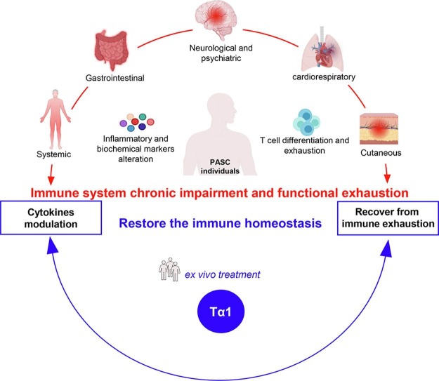
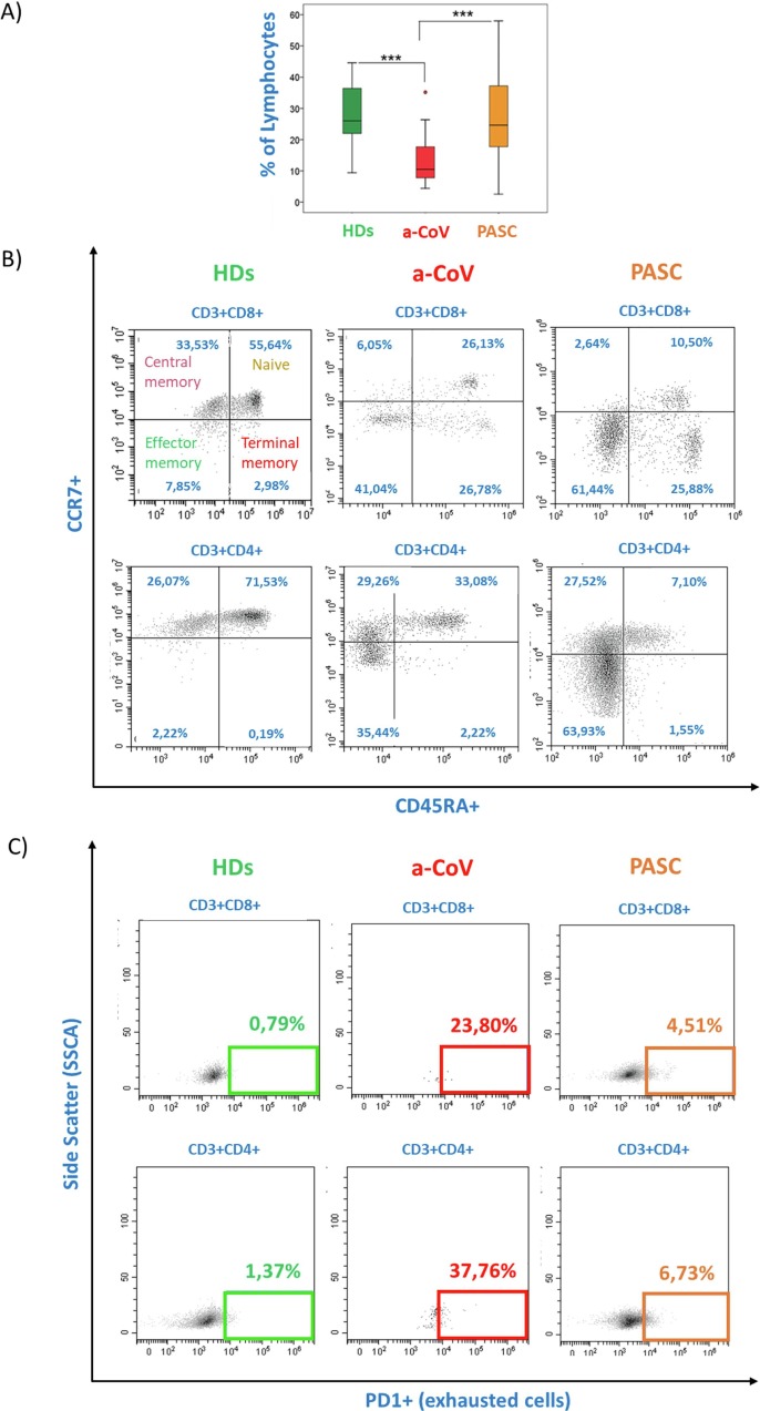
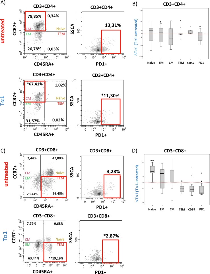
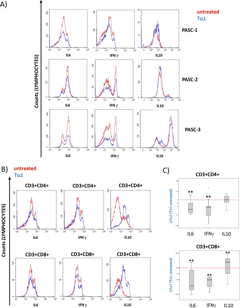
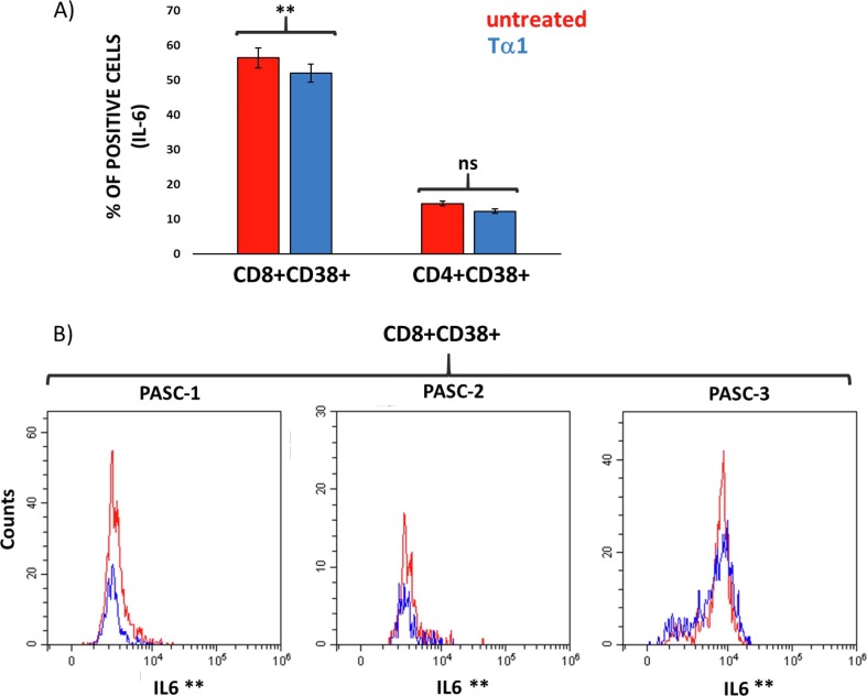
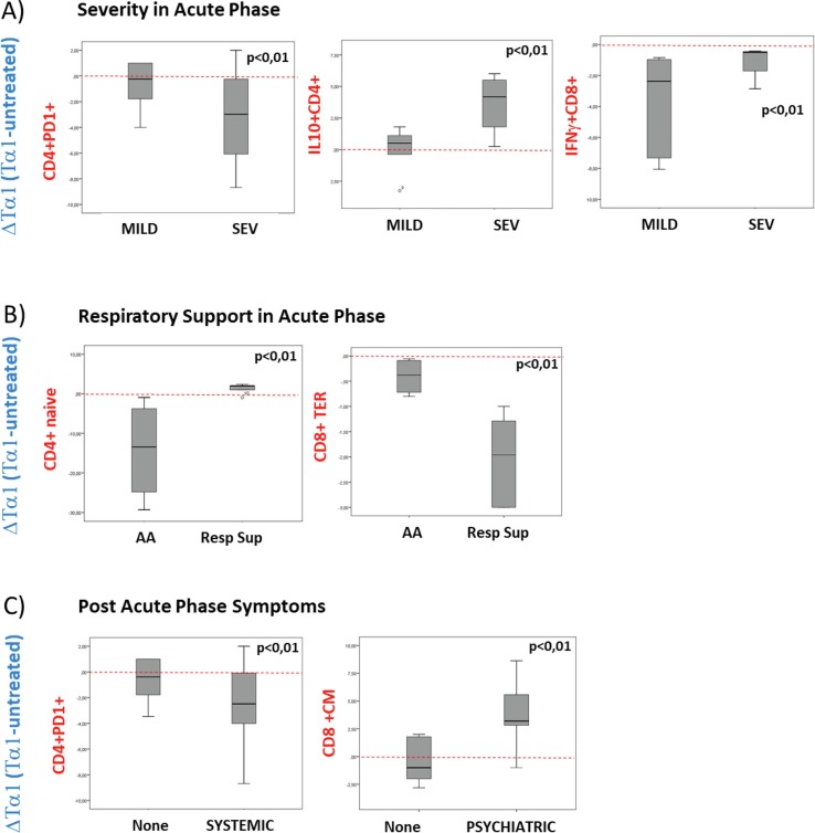
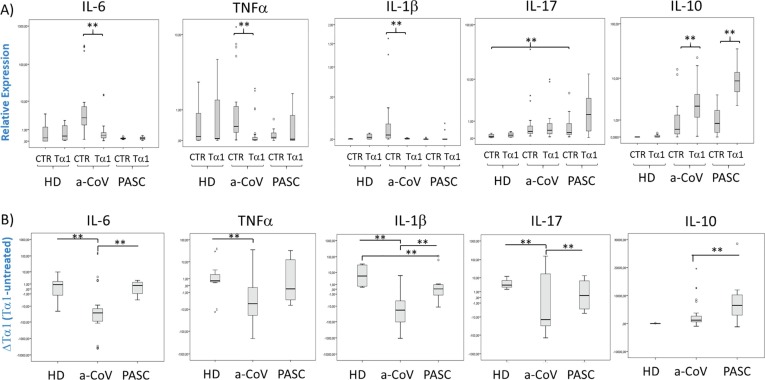
The complex alterations of the immune system and the immune-mediated multiorgan injury plays a key role in host response to SARS-CoV-2 infection and in the pathogenesis of COVID-19, being also associated with adverse outcomes. Thymosin alpha 1 (Tα1) is one of the molecules used in the treatment of COVID-19, as it is known to restore the homeostasis of the immune system during infections and cancer. The use of Tα1 in COVID-19 patients had been widely used in China and in COVID-19 patients, it has been shown to decrease hospitalization rate, especially in those with greater disease severity, and reduce mortality by restoring lymphocytopenia and more specifically, depleted T cells. Persistent dysregulation with depletion of naive B and T cell subpopulations and expansion of memory T cells suggest a chronic stimulation of the immune response in individuals with post-acute sequelae of SARS-CoV-2 infection (PASC). Our data obtained from an ex vivo study, showed that in PASC individuals with a chronically altered immune response, Tα1 improve the restoration of an appropriate response, most evident in those with more severe illness and who need respiratory support during acute phase, and in those with specific systemic and psychiatric symptoms of PASC, confirming Tα1 treatment being more effective in compromised patients. The results obtained, along with promising reports on recent trials on Tα1 administration in patients with COVID-19, offer new insights into intervention also for those patients with long-lasting inflammation with post-infectious symptoms, some of which have a delayed onset.
Keywords: Anti-inflammatory response; Immune regulation; Post-acute SARS-CoV-2 symptoms; Thymosin alpha 1.
Copyright © 2023 Elsevier B.V. All rights reserved.
Conflict of interest statement
Declaration of Competing Interest The authors declare that they have no known competing financial interests or personal relationships that could have appeared to influence the work reported in this paper.
Figures
Similar articles
-
Novel evidence of Thymosin α1 immunomodulatory properties in SARS-CoV-2 infection: Effect on innate inflammatory response in a peripheral blood mononuclear cell-based in vitro model.Int Immunopharmacol. 2023 Apr;117:109996. doi: 10.1016/j.intimp.2023.109996. Epub 2023 Mar 13. Int Immunopharmacol. 2023. PMID: 36933449 Free PMC article.
-
Thymosin Alpha-1 Has no Beneficial Effect on Restoring CD4+ and CD8+ T Lymphocyte Counts in COVID-19 Patients.Front Immunol. 2021 Jun 3;12:568789. doi: 10.3389/fimmu.2021.568789. eCollection 2021. Front Immunol. 2021. PMID: 34149679 Free PMC article.
-
Thymosin Alpha 1 Reduces the Mortality of Severe Coronavirus Disease 2019 by Restoration of Lymphocytopenia and Reversion of Exhausted T Cells.Clin Infect Dis. 2020 Nov 19;71(16):2150-2157. doi: 10.1093/cid/ciaa630. Clin Infect Dis. 2020. PMID: 32442287 Free PMC article.
-
Tissue injury and leukocyte changes in post-acute sequelae of SARS-CoV-2: review of 2833 post-acute patient outcomes per immune dysregulation and microbial translocation in long COVID.J Leukoc Biol. 2023 Mar 1;113(3):236-254. doi: 10.1093/jleuko/qiac001. J Leukoc Biol. 2023. PMID: 36807444 Review.
-
Thymosin alpha 1 treatment for patients with sepsis.Expert Opin Biol Ther. 2018 Jul;18(sup1):71-76. doi: 10.1080/14712598.2018.1484104. Expert Opin Biol Ther. 2018. PMID: 30063866 Review.
Cited by
-
Persistence of circulating CD169+monocytes and HLA-DR downregulation underline the immune response impairment in PASC individuals: the potential contribution of different COVID-19 pandemic waves.Curr Res Microb Sci. 2023 Dec 12;6:100215. doi: 10.1016/j.crmicr.2023.100215. eCollection 2024. Curr Res Microb Sci. 2023. PMID: 38187999 Free PMC article.
-
Mucosal immune response in biology, disease prevention and treatment.Signal Transduct Target Ther. 2025 Jan 8;10(1):7. doi: 10.1038/s41392-024-02043-4. Signal Transduct Target Ther. 2025. PMID: 39774607 Free PMC article.
-
Flow cytometry for extracellular vesicle characterization in COVID-19 and post-acute sequelae of SARS-CoV-2 infection.Extracell Vesicles Circ Nucl Acids. 2024 Aug 9;5(3):417-437. doi: 10.20517/evcna.2024.20. eCollection 2024. Extracell Vesicles Circ Nucl Acids. 2024. PMID: 39697632 Free PMC article. Review.
-
Effect of thymosin α1 on Immune response and organ function in acute aortic dissection surgery: PANDA II trial protocol.Future Cardiol. 2025 Jun;21(7):447-454. doi: 10.1080/14796678.2025.2505401. Epub 2025 May 14. Future Cardiol. 2025. PMID: 40367062 Clinical Trial.
-
Phenotypic drug discovery: a case for thymosin alpha-1.Front Med (Lausanne). 2024 Jun 6;11:1388959. doi: 10.3389/fmed.2024.1388959. eCollection 2024. Front Med (Lausanne). 2024. PMID: 38903817 Free PMC article. Review.
References
MeSH terms
Substances
LinkOut - more resources
Full Text Sources
Medical
Miscellaneous
