. 2023 Mar;11(3):e006721.
doi: 10.1136/jitc-2023-006721.

Identification of HPV16 E1 and E2-specific T cells in the oropharyngeal cancer tumor microenvironment

Affiliations

Identification of HPV16 E1 and E2-specific T cells in the oropharyngeal cancer tumor microenvironment

Christine McInnis et al. J Immunother Cancer. 2023 Mar.

Abstract

Background: High-risk human papillomavirus (HPV) is a primary cause of an increasing number of oropharyngeal squamous cell carcinomas (OPSCCs). The viral etiology of these cancers provides the opportunity for antigen-directed therapies that are restricted in scope compared with cancers without viral components. However, specific virally-encoded epitopes and their corresponding immune responses are not fully defined.

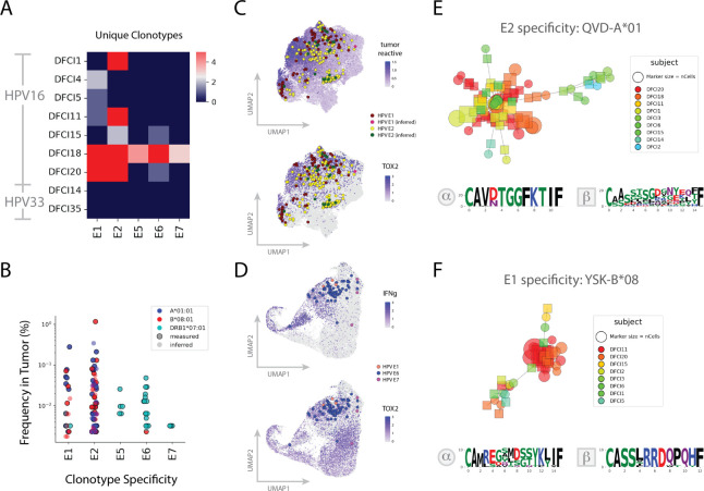
Methods: To understand the OPSCC immune landscape, we conducted a comprehensive single-cell analysis of HPV16+ and HPV33+ primary tumors and metastatic lymph nodes. We used single-cell analysis with encoded peptide-human leukocyte antigen (HLA) tetramers to analyze HPV16+ and HPV33+ OPSCC tumors, characterizing the ex vivo cellular responses to HPV-derived antigens presented in major Class I and Class II HLA alleles.

Results: We identified robust cytotoxic T-cell responses to HPV16 proteins E1 and E2 that were shared across multiple patients, particularly in HLA-A*01:01 and HLA-B*08:01. Responses to E2 were associated with loss of E2 expression in at least one tumor, indicating the functional capacity of these E2-recognizing T cells and many of these interactions validated in a functional assay. Conversely, cellular responses to E6 and E7 were limited in quantity and cytotoxic capacity, and tumor E6 and E7 expression persisted.

Conclusions: These data highlight antigenicity beyond HPV16 E6 and E7 and nominate candidates for antigen-directed therapies.

Keywords: Adaptive Immunity; Head and Neck Neoplasms; Lymphocytes, Tumor-Infiltrating; T-Lymphocytes; Tumor Microenvironment.


Conflict of interest statement

Competing interests: CM, BV, QT, DL-E, DCP and AJC are employees and/or stockholders of Repertoire Immune Medicine. GJH reports grants and institutional support from ASCO CCF, Bicara, BMS, Gateway for Cancer Research, GSK, Kite, KSQ, Kura Oncology, ImmunityBio, Regeneron. Consulting/honoraria from Bicara, BMS, Coherus, Exicure, Kura, Maverick, Merck, Naveris, Regeneron, and SIRPant. AME reports support from NIH/NIDCR U0 DE029188. RU serves on a Merck advisory board.

Figures

Figure 1
Figure 1
Experimental workflow. Biopsies were dissociated and subjected to scRNA-seq. Separately, tumor-infiltrating lymphocytes were isolated and probed with DNA-encoded peptide-MHC tetramers, sorted using flow cytometry and subjected to scRNA-seq. Readout of TCR, GEX, and tetramer sequencing data allowed for the characterization of TCRs, cellular phenotype, and tetramer binding. GEX, gene expression; HLA, human leukocyte antigen; HPV, human papillomavirus; MHC, major histocompatibility complex; OPSCC, oropharyngeal squamous cell carcinoma; scRNA-seq, single-cell RNA sequencing; TCR, T-cell receptor; UMAP, uniform manifold approximation and projection.
Figure 2
Figure 2
Heterogeneity in HPV gene expression influences T cell infiltrates. (A) The percentage of HPV+ tumor cells expressing each HPV gene assessed is heterogenous. (B) UMAP showing cell classifications of T cell subtypes in TME. (C). HPV E1 expression is positively correlated with CD8 memory infiltration. HPV E1 expression is positively associated with CD8 effector T cell infiltration. HPV E1 expression is negatively associated with CD4 Treg infiltration. adj., adjusted; HPV, human papillomavirus; Th1, type 1 T helper cells; TME, tumor microenvironment; Treg, regulatory T cells; UMAP, uniform manifold approximation and projection.
Figure 3
Figure 3
Cellular response to HPV antigens in OPSCC. (A) Number of clonotypes by subject measured and inferred with high confidence to be specific to epitopes derived from HPV antigens. (B) Frequency of HPV-reactive clonotypes in dissociated tumor samples. Measured clonotypes are displayed with filled dots and inferred clonotypes are displayed with open dots. (C) UMAP of CD8+ TIL showing HPV-specific clonotypes overlaid on tumor reactive and TOX2 gene scores. (D) UMAP of CD4+ TIL showing HPV-specific clonotypes overlaid on IFN-y and TOX2 gene scores. (E) HPV16 E2 QVDYYGLYY-A*01:01-specific T cells show strong clonotype sharing within the TCR-alpha chain across patients. We observed T cells from several different patients appearing in this cluster, with heaviest contributions from DFCI1, DFCI11, DFCI20 and DFCI18, all patients that were HLA-A*01:01+and HPV16+. (F) HPV16 E2 YSKNKVWEV-B*08:01-specific T cells show strong clonotype sharing within the TCR-beta chain across patients. We observed T cells from several different patients appearing in this cluster, with heaviest contributions from DFCI1, DFCI11, DFCI20 and DFCI15, all patients that were HLA-B*08:01+ and HPV16+. HLA, human leukocyte antigen; HPV, human papillomavirus; IFN, interferon; OPSCC, oropharyngeal squamous cell carcinoma; TCR, T-cell receptor; TIL, tumor-infiltrating lymphocytes; TOX2, TOX high mobility group box family member 2; UMAP, uniform manifold approximation and projection.
Figure 4
Figure 4
Functional validation of TCR-epitope pairs. (A) TCR-epitope interactions across HPV16 genes were tested using this workflow. (B) The TCRs derived from patient TIL tested signaled when presented with cognate peptide as measured by CD69 induction. Peptides tested included WPYLHNRLV-B*08:01, QVDYYGLYY-A*01:01, YSKNKVWEV-B*08:01, KFKELYGVSFSELVR-DRB1*07:01, KQRHLDKKQRFHNIRGRWTGR-DRB1*07:01 and LDKKQRFHNIRGRWTGRCMSC-DRB1*07:01. (C) Three TCRs derived from tumor of patient DFCI11 tested with beta chain homology to YSKNKVWEV-B*08:01-reactive T cells signal when presented with cognate peptide and make up 1.2%, 0.13% and 0.025% of TIL. (D) TCRs with beta chains homologous to QVDYYGLYY-reactive T cells fail to signal but both TCRs with similar alphas signal when presented with cognate peptide. APC, allophycocyanin; DMSO, dimethylsulfoxide; TCR, T-cell receptor; TIL, tumor-infiltrating lymphocytes.

References

    1. Mork J, Lie AK, Glattre E, et al. . Human papillomavirus infection as a risk factor for squamous-cell carcinoma of the head and neck. N Engl J Med 2001;344:1125–31. 10.1056/NEJM200104123441503 - DOI - PubMed
    1. Mroz EA, Rocco JW. Math, a novel measure of intratumor genetic heterogeneity, is high in poor-outcome classes of head and neck squamous cell carcinoma. Oral Oncol 2013;49:211–5. 10.1016/j.oraloncology.2012.09.007 - DOI - PMC - PubMed
    1. Kürten CHL, Kulkarni A, Cillo AR, et al. . Investigating immune and non-immune cell interactions in head and neck tumors by single-cell RNA sequencing. Nat Commun 2021;12:7338. 10.1038/s41467-021-27619-4 - DOI - PMC - PubMed
    1. Powell SF, Vu L, Spanos WC, et al. . The key differences between human papillomavirus-positive and -negative head and neck cancers: biological and clinical implications. Cancers (Basel) 2021;13:5206. 10.3390/cancers13205206 - DOI - PMC - PubMed
    1. Gillison ML, Akagi K, Xiao W, et al. . Human papillomavirus and the landscape of secondary genetic alterations in oral cancers. Genome Res 2019;29:1–17. 10.1101/gr.241141.118 - DOI - PMC - PubMed

Publication types

Supplementary concepts