Benefit of prednisolone alone in nodal peripheral T-cell lymphoma with T follicular helper phenotype
- PMID: 36990775
- PMCID: PMC10158724
- DOI: 10.3960/jslrt.22038
Benefit of prednisolone alone in nodal peripheral T-cell lymphoma with T follicular helper phenotype
Abstract
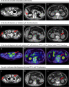
A 71-year-old Japanese man presented with severe thrombocytopenia. A whole-body CT at presentation showed small cervical, axillary, and para-aortic lymphadenopathy, leading to suspicion of immune thrombocytopenia due to lymphoma. Biopsy was difficult to perform because of severe thrombocytopenia. Thus, he received prednisolone (PSL) therapy and his platelet count gradually recovered. Two and a half years after PSL therapy initiation, his cervical lymphadenopathy slightly progressed without other clinical symptoms. Hence, a biopsy from the left cervical lymph node was performed, and he was diagnosed with nodal peripheral T-cell lymphoma (PTCL) with T follicular helper (TFH) phenotype. Due to various complications, we continued treatment with prednisolone alone after the diagnosis of lymphoma; however, there was no further increase in lymph node enlargement and no other lymphoma-related symptoms for one and a half years after diagnosis. Although immunosuppressive therapy has been reported to produce a response in some patients with angioimmunoblastic T-cell lymphoma, our experience suggests that a similar subset may exist in patients with nodal PTCL with TFH phenotype, which has the same cellular origin. Immunosuppressive therapies may constitute an alternative treatment option even in the era of novel molecular-targeted therapies, especially for elderly patients who are ineligible for chemotherapy.
Keywords: immune thrombocytopenia; nodal peripheral T-cell lymphoma with T follicular helper phenotype; prednisolone.
Conflict of interest statement
CONFLICT OF INTEREST
The authors declare that they have no conflict of interest.
Figures


References
-
- Rodríguez-Pinilla SM, Atienza L, Murillo C, et al. Peripheral T-cell lymphoma with follicular T-cell markers. Am J Surg Pathol. 2008; 32: 1787-1799. - PubMed
-
- Watatani Y, Sato Y, Miyoshi H, et al. Molecular heterogeneity in peripheral T-cell lymphoma, not otherwise specified revealed by comprehensive genetic profiling. Leukemia. 2019; 33: 2867-2883. - PubMed

