. 2023 Jul;44(7):1380-1390.
doi: 10.1038/s41401-023-01076-9. Epub 2023 Mar 29.

Cell-autonomous effect of cardiomyocyte branched-chain amino acid catabolism in heart failure in mice

Affiliations

Cell-autonomous effect of cardiomyocyte branched-chain amino acid catabolism in heart failure in mice

Jia-Yu Yu et al. Acta Pharmacol Sin. 2023 Jul.

Abstract

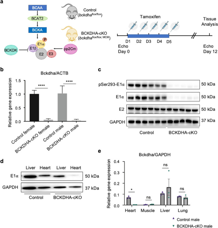
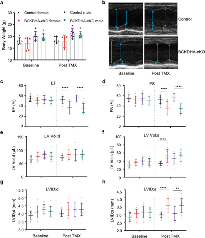
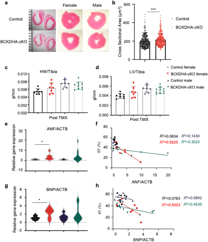
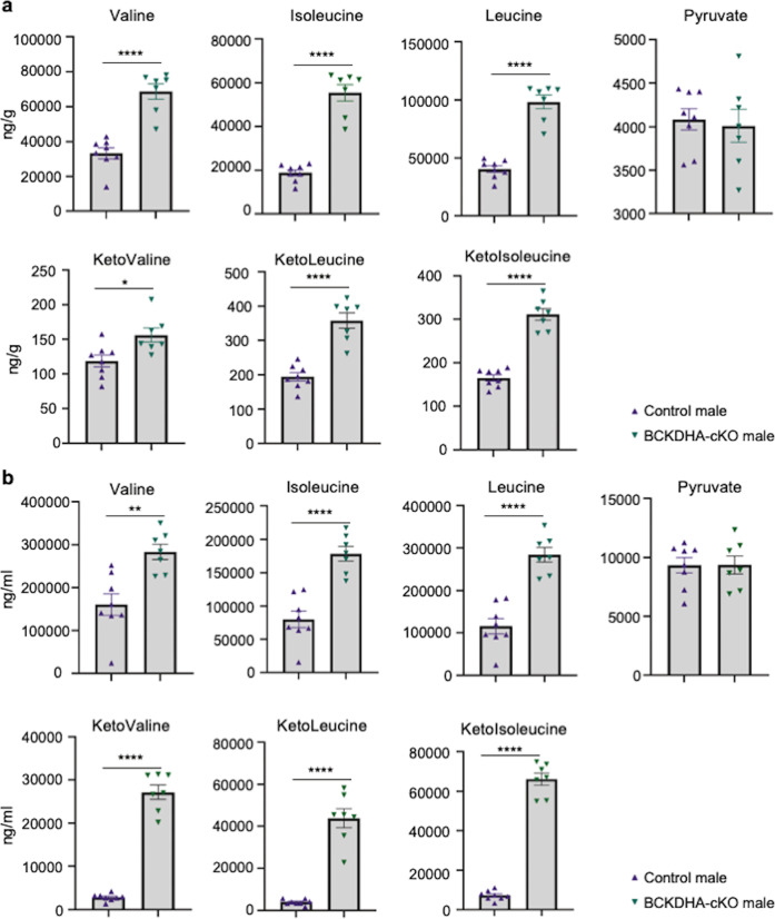
Parallel to major changes in fatty acid and glucose metabolism, defect in branched-chain amino acid (BCAA) catabolism has also been recognized as a metabolic hallmark and potential therapeutic target for heart failure. However, BCAA catabolic enzymes are ubiquitously expressed in all cell types and a systemic BCAA catabolic defect is also manifested in metabolic disorder associated with obesity and diabetes. Therefore, it remains to be determined the cell-autonomous impact of BCAA catabolic defect in cardiomyocytes in intact hearts independent from its potential global effects. In this study, we developed two mouse models. One is cardiomyocyte and temporal-specific inactivation of the E1α subunit (BCKDHA-cKO) of the branched-chain α-ketoacid dehydrogenase (BCKDH) complex, which blocks BCAA catabolism. Another model is cardiomyocyte specific inactivation of the BCKDH kinase (BCKDK-cKO), which promotes BCAA catabolism by constitutively activating BCKDH activity in adult cardiomyocytes. Functional and molecular characterizations showed E1α inactivation in cardiomyocytes was sufficient to induce loss of cardiac function, systolic chamber dilation and pathological transcriptome reprogramming. On the other hand, inactivation of BCKDK in intact heart does not have an impact on baseline cardiac function or cardiac dysfunction under pressure overload. Our results for the first time established the cardiomyocyte cell autonomous role of BCAA catabolism in cardiac physiology. These mouse lines will serve as valuable model systems to investigate the underlying mechanisms of BCAA catabolic defect induced heart failure and to provide potential insights for BCAA targeted therapy.

Keywords: branched-chain amino acid; heart failure; metabolism.


Conflict of interest statement

Rachel J Roth Flach is a Pfizer employee.

Figures

Fig. 1
Fig. 1. Generation of cardiac specific BCAA metabolism defective mouse model using BCKDHA-cKO mice.
a Schematic view of BCKDH complex in BCAA catabolic pathway and the experimental design. b Bckdha mRNA levels in the Control and the BCKDHA-cKO mouse hearts measured by real-time RT-PCR, n = 7–10 each group, ****P < 0.001. c Representative immunoblot data of total and phosphorylated E1α protein, and E2 protein in the BCKDHA-cKO and the Control mouse hearts. d Representative immunoblot data of E1α expression in liver and heart tissues from the Control and the BCKDHA-cKO mice. e Real-time PCR analysis of BCKDHA normalized to GAPDH in different tissues as indicated. n = 3 each group, *P < 0.05.
Fig. 2
Fig. 2. Impaired cardiac function following BCAA catabolic defect in heart.
a Body weight of the male and the female Control and BCKDHA-cKO mice at baseline (Day 0) and one week post tamoxifen injection (Day 12). b Representative echo M-mode images for the Control and the BCKDHA-cKO mice at baseline and post tamoxifen mediated gene inactivation. ch Echocardiography parameters, including (c) ejection fraction (EF%); (d) fractional shortening (FS%); (e) left ventricle end-diastolic volume (LV Vol;d); (f) left ventricle end-systolic volume (LV Vol;s); (g) left ventricle end-diastolic internal diameter (LVID;d) and (h) left ventricle end-systolic internal diameter (LVID;s) in the Control and the BCKDHA-cKO male and female animals at baseline (Day 0) and one week post-tamoxifen induced gene inactivation (Day 12). **P < 0.01; ****P < 0.001.
Fig. 3
Fig. 3. Pathological remodeling following BCAA catabolic defect in heart.
a Representative H&E staining of the Control and the BCKDHA-cKO male and female mice cardiac section. Magnification: 10×. b Cardiomyocytes cross-section area in the Control and the BCKDHA-cKO hearts, ***P < 0.005. c, d Heart weight vs. tibial length (HW/TL), Left ventricle weight vs. tibial length (LV/TL) in the Control and the BCKDHA-cKO animals. eh mRNA levels of ANF (e, f) and BNP relative to ACTB (g, h) in the Control and the BCKDHA-cKO male and female mouse hearts (e, g) as well as their quantitative correlation with cardiac function in each cohort (f, h).
Fig. 4
Fig. 4. Impaired cardiac BCAA metabolism led to changes of systemic BCAA/BCKA.
a BCAA, pyruvate and BCKA concentrations in the Control and the BCKDHA-cKO mouse cardiac tissues collected at one week post tamoxifen administration. b BCAA, pyruvate and BCKA concentrations in the Control and BCKDHA-cKO mouse serum samples collected at one week post tamoxifen administration. *P < 0.05, **P < 0.01, ****P < 0.001.
Fig. 5
Fig. 5. Generation of cardiac specific BCAA metabolism active mouse via BCKDK-cKO mouse line.
a Schematic view of the BCKDK-cKO mouse line generation and experimental design. b Real-time PCR analysis of BCKDK mRNA expression level in the Control and the BCDK-cKO male and female mouse left ventricle tissues. n = 4–5 each group, ****P < 0.001. c Western Blot analysis of BCKDK, E1α, phosphor-E1α (S293) and E2 expression level in the Control and the BCKDK-cKO mouse left ventricle tissues, GAPDH was used as control.
Fig. 6
Fig. 6. Inactivation of BCKDK in heart does not prevent heart failure post pressure overload.
af Echocardiography parameters including Ejection Fraction (a), Fraction Shortening (b), LVID;s (c), LVID;d (d), LV Vol;s (e) and LV Vol; d(f) in the Control and the BCKDK-cKO male and female mice, at baseline and 6 weeks post pressure overload injury. g Representative H&E staining for the Control and the BCKDK-cKO female and male mouse hearts post pressure overload surgery. Magnification: 10×. h Heart weight/tibia length ratio in the Control and the BCKDK-cKO male and female mice 6 weeks post TAC. i Left ventricle weight/tibia length ratio in the Control and the BCKDK-cKO male and female mice 6 weeks post TAC. jk: Survival curve for the Control and the BCKDK-cKO female (j) and male (k) mice post TAC at indicated time points. **P < 0.01.
Fig. 7
Fig. 7. Transcriptome changes in BCKDHA-cKO and BCKDK-cKO mouse hearts.
a Heatmap of selected genes detected in the Control vs the BCKDHA-cKO hearts from RNA-seq dataset ranked based on their expression levels in the BCKDHA-cKO hearts. b Integrated biological pathway and Gene-Ontology analysis for the top 500 differentially regulated genes ranked by significance in P values were determined by ClueGO in the Control and the BCKDHA-cKO hearts. c Heatmap of selected genes detected in the Control vs the BCKDK-cKO hearts from RNA-seq dataset ranked based on their expression levels in BCKDK-cKO hearts. d Integrated biological pathway and Gene-Ontology analysis for the top 500 differentially regulated genes ranked by significance in P values were determined by ClueGO in the Control and the BCKDK-cKO hearts. Colors represent related GO term families and only GO terms with q < 1E-20 are included.

References

    1. Neinast M, Murashige D, Arany Z. Branched chain amino acids. Annu Rev Physiol. 2019;81:139–64. doi: 10.1146/annurev-physiol-020518-114455. - DOI - PMC - PubMed
    1. Lu G, Sun H, She P, Youn JY, Warburton S, Ping P, et al. Protein phosphatase 2Cm is a critical regulator of branched-chain amino acid catabolism in mice and cultured cells. J Clin Invest. 2009;119:1678–87. doi: 10.1172/JCI38151. - DOI - PMC - PubMed
    1. Neinast MD, Jang C, Hui S, Murashige DS, Chu Q, Morscher RJ, et al. Quantitative analysis of the whole-body metabolic fate of branched-chain amino acids. Cell Metab. 2019;29:417–29. doi: 10.1016/j.cmet.2018.10.013. - DOI - PMC - PubMed
    1. Sun H, Olson KC, Gao C, Prosdocimo DA, Zhou M, Wang Z, et al. Catabolic defect of branched-chain amino acids promotes heart failure. Circulation. 2016;133:2038–49. doi: 10.1161/CIRCULATIONAHA.115.020226. - DOI - PMC - PubMed
    1. Bloomgarden Z. Diabetes and branched-chain amino acids: What is the link? J Diabetes. 2018;10:350–2. doi: 10.1111/1753-0407.12645. - DOI - PubMed

Substances