

Magnetic resonance brain volumetry biomarkers of CLN2 Batten disease identified with miniswine model
- PMID: 36991106
- PMCID: PMC10060411
- DOI: 10.1038/s41598-023-32071-z
Magnetic resonance brain volumetry biomarkers of CLN2 Batten disease identified with miniswine model
Abstract
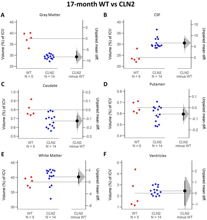
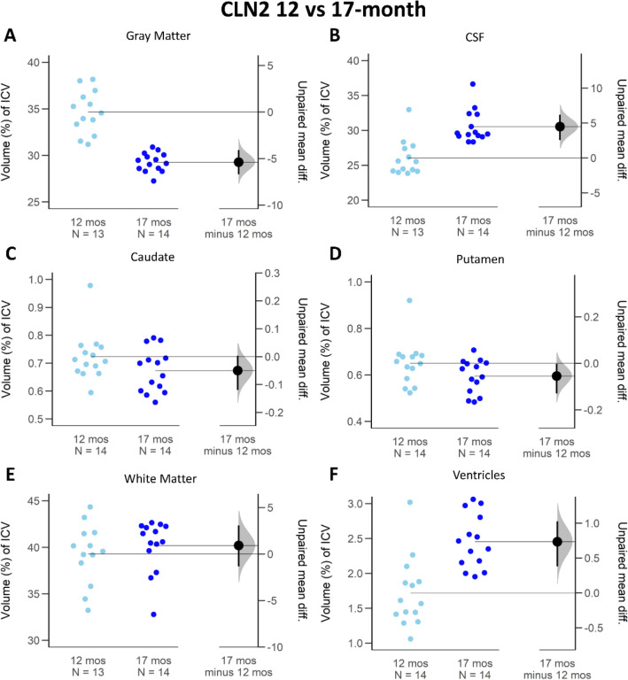
Late-infantile neuronal ceroid lipofuscinosis type 2 (CLN2) disease (Batten disease) is a rare pediatric disease, with symptom development leading to clinical diagnosis. Early diagnosis and effective tracking of disease progression are required for treatment. We hypothesize that brain volumetry is valuable in identifying CLN2 disease at an early stage and tracking disease progression in a genetically modified miniswine model. CLN2R208X/R208X miniswine and wild type controls were evaluated at 12- and 17-months of age, correlating to early and late stages of disease progression. Magnetic resonance imaging (MRI) T1- and T2-weighted data were acquired. Total intercranial, gray matter, cerebrospinal fluid, white matter, caudate, putamen, and ventricle volumes were calculated and expressed as proportions of the intracranial volume. The brain regions were compared between timepoints and cohorts using Gardner-Altman plots, mean differences, and confidence intervals. At an early stage of disease, the total intracranial volume (- 9.06 cm3), gray matter (- 4.37% 95 CI - 7.41; - 1.83), caudate (- 0.16%, 95 CI - 0.24; - 0.08) and putamen (- 0.11% 95 CI - 0.23; - 0.02) were all notably smaller in CLN2R208X/R208X miniswines versus WT, while cerebrospinal fluid was larger (+ 3.42%, 95 CI 2.54; 6.18). As the disease progressed to a later stage, the difference between the gray matter (- 8.27%, 95 CI - 10.1; - 5.56) and cerebrospinal fluid (+ 6.88%, 95 CI 4.31; 8.51) continued to become more pronounced, while others remained stable. MRI brain volumetry in this miniswine model of CLN2 disease is sensitive to early disease detection and longitudinal change monitoring, providing a valuable tool for pre-clinical treatment development and evaluation.
© 2023. The Author(s).
Conflict of interest statement
C.S.R. is an employee of Precigen Exemplar, a company commercializing the CLN2 porcine model. J.M.W. has equity in Amicus Therapeutics, a biotechnology company focused on developing medicines for people with rare metabolic diseases. All other authors have no competing interests.
Figures


Similar articles
-
A Novel Porcine Model of CLN2 Batten Disease that Recapitulates Patient Phenotypes.Neurotherapeutics. 2022 Oct;19(6):1905-1919. doi: 10.1007/s13311-022-01296-7. Epub 2022 Sep 13. Neurotherapeutics. 2022. PMID: 36100791 Free PMC article.
-
Enzyme replacement therapy attenuates disease progression in a canine model of late-infantile neuronal ceroid lipofuscinosis (CLN2 disease).J Neurosci Res. 2014 Nov;92(11):1591-8. doi: 10.1002/jnr.23423. Epub 2014 Jun 17. J Neurosci Res. 2014. PMID: 24938720 Free PMC article.
-
Untargeted Metabolite Profiling of Cerebrospinal Fluid Uncovers Biomarkers for Severity of Late Infantile Neuronal Ceroid Lipofuscinosis (CLN2, Batten Disease).Sci Rep. 2018 Oct 15;8(1):15229. doi: 10.1038/s41598-018-33449-0. Sci Rep. 2018. PMID: 30323181 Free PMC article.
-
CLN2 Disease (Classic Late Infantile Neuronal Ceroid Lipofuscinosis).Pediatr Endocrinol Rev. 2016 Jun;13 Suppl 1:682-8. Pediatr Endocrinol Rev. 2016. PMID: 27491216 Review.
-
The neuronal ceroid-lipofuscinoses.Dev Disabil Res Rev. 2013;17(3):254-9. doi: 10.1002/ddrr.1118. Dev Disabil Res Rev. 2013. PMID: 23798013 Review.
Cited by
-
Enzyme Replacement Therapy for CLN2 Disease: MRI Volumetry Shows Significantly Slower Volume Loss Compared with a Natural History Cohort.AJNR Am J Neuroradiol. 2024 Nov 7;45(11):1791-1797. doi: 10.3174/ajnr.A8408. AJNR Am J Neuroradiol. 2024. PMID: 38977290
-
A Case for Automated Segmentation of MRI Data in Neurodegenerative Diseases: Type II GM1 Gangliosidosis.NeuroSci. 2025 Apr 3;6(2):31. doi: 10.3390/neurosci6020031. NeuroSci. 2025. PMID: 40265361 Free PMC article.
References
-
- Mole S, Williams R, Goebel H. The Neuronal Ceroid Lipofuscinoses (Batten Disease) Oxford University Press; 2012.
-
- Nickel M, Simonati A, Jacoby D, Lezius S, Kilian D, Van de Graaf B, Pagovich OE, Kosofsky B, Yohay K, Downs M, et al. Disease characteristics and progression in patients with late-infantile neuronal ceroid lipofuscinosis type 2 (CLN2) disease: An observational cohort study. Lancet Child Adolesc. Health. 2018;2(8):582–590. doi: 10.1016/S2352-4642(18)30179-2. - DOI - PMC - PubMed
-
- Kovacs KD, Patel S, Orlin A, Kim K, Van Everen S, Conner T, Sondhi D, Kaminsky SM, D'Amico DJ, Crystal RG, et al. Symmetric age association of retinal degeneration in patients with CLN2-associated batten disease. Ophthalmol. Retina. 2020;4(7):728–736. doi: 10.1016/j.oret.2020.01.011. - DOI - PMC - PubMed
Publication types
MeSH terms
Substances
Grants and funding
LinkOut - more resources
Full Text Sources
Research Materials
