. 2023 Apr;616(7958):806-813.
doi: 10.1038/s41586-023-05880-5. Epub 2023 Mar 29.

STING inhibits the reactivation of dormant metastasis in lung adenocarcinoma

Affiliations

STING inhibits the reactivation of dormant metastasis in lung adenocarcinoma

Jing Hu et al. Nature. 2023 Apr.

Abstract

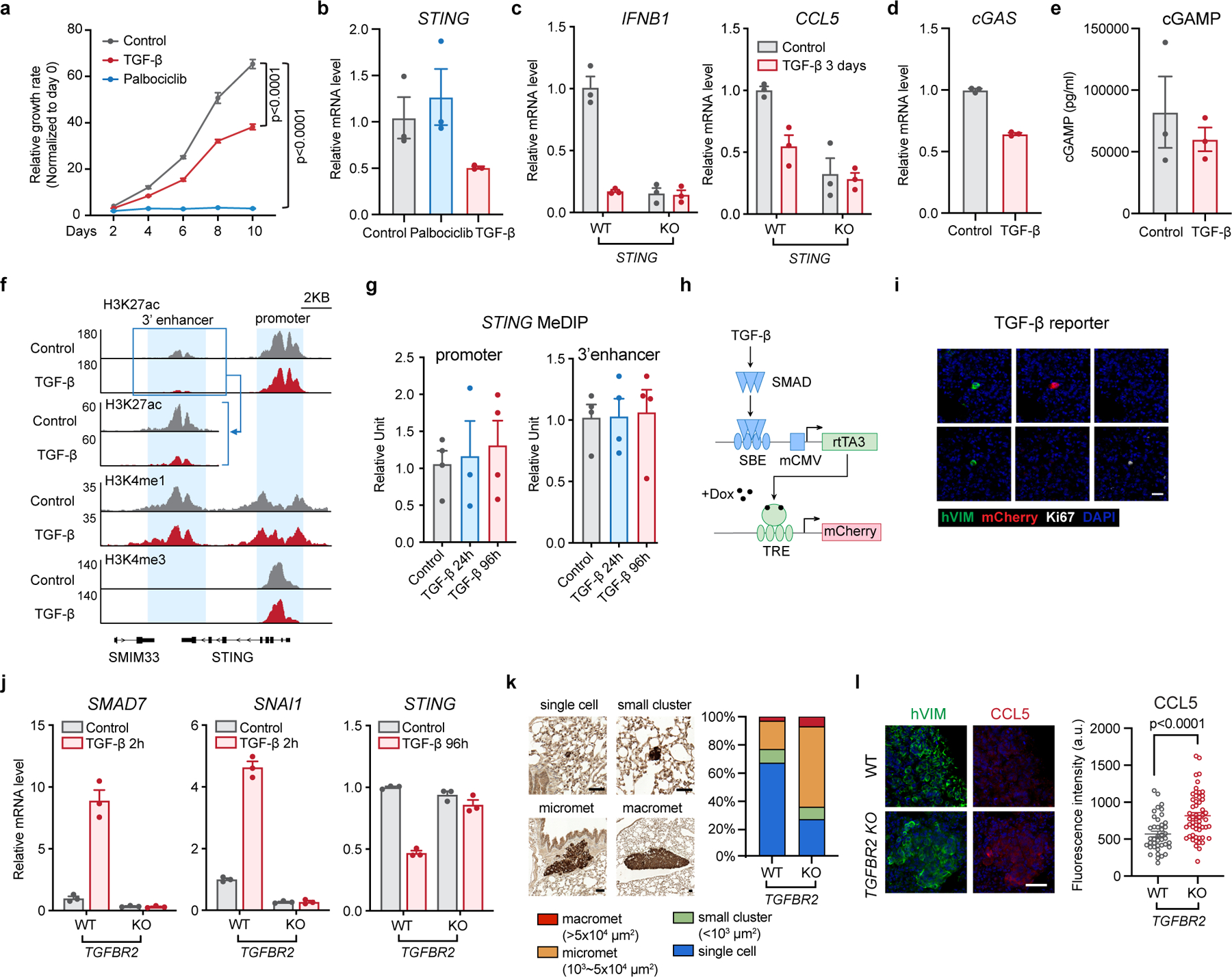
Metastasis frequently develops from disseminated cancer cells that remain dormant after the apparently successful treatment of a primary tumour. These cells fluctuate between an immune-evasive quiescent state and a proliferative state liable to immune-mediated elimination1-6. Little is known about the clearing of reawakened metastatic cells and how this process could be therapeutically activated to eliminate residual disease in patients. Here we use models of indolent lung adenocarcinoma metastasis to identify cancer cell-intrinsic determinants of immune reactivity during exit from dormancy. Genetic screens of tumour-intrinsic immune regulators identified the stimulator of interferon genes (STING) pathway as a suppressor of metastatic outbreak. STING activity increases in metastatic progenitors that re-enter the cell cycle and is dampened by hypermethylation of the STING promoter and enhancer in breakthrough metastases or by chromatin repression in cells re-entering dormancy in response to TGFβ. STING expression in cancer cells derived from spontaneous metastases suppresses their outgrowth. Systemic treatment of mice with STING agonists eliminates dormant metastasis and prevents spontaneous outbreaks in a T cell- and natural killer cell-dependent manner-these effects require cancer cell STING function. Thus, STING provides a checkpoint against the progression of dormant metastasis and a therapeutically actionable strategy for the prevention of disease relapse.


Conflict of interest statement

Competing interests

SWL received funding and research support from Agilent Technologies for the purposes of massively parallel oligo synthesis to generate the sgRNA libraries described herein.

Figures

Extended Data Figure 1.
Extended Data Figure 1.. In vivo CRISPR screen for cell-autonomous immune regulators of dormant metastasis
(a) Metastasis-free survival Kaplan-Meier plots of athymic mice intracardially inoculated with 1×10⁵ H2030-BrM or H2087-LCC cells. One set of mice inoculated with H2087-LCC cells was treated with anti-asialo-GM1 antibody to deplete NK cells 30 days after cell inoculation. n=9 (H2030-BrM) or 10 (H2087-LCC) mice per group, log-rank test. (b) Flow cytometry analysis of LineageCD45+NKP46+ NK cells in blood from athymic mice that were treated with IgG control or anti-asialo-GM1 antibody for 4 days. Lineage : TCRβCD3CD19B220CD11cLy6GF4/80. n=3 mice per group. Mean ± s.e.m., two-sided unpaired t-test. (c) Petal charts of metastasis incidence in specific organs of 10 athymic mice intracardially inoculated with 1×10⁵ H2087-LCC cells, treated with or without anti-asialo-GM1 antibody. Metastasis progression was monitored by BLI for 160 days after cell inoculation. (d) Petal charts of metastasis incidence in specific organs after intracardiac inoculation of 2×10⁴ KPad1 cells into 8–9 B6-albino mice or NSG mice. Metastasis progression was monitored by BLI for 60 days (B6-albino mice) or 28 days (NSG mice) after cell inoculation. (e) Flow cytometry analysis of LineageCD45+NKP46+ NK cells, CD45+CD3+CD4+, and CD45+CD3+CD8+ T cells in blood from B6-albino mice that were treated for 3 days with IgG control, anti-NK1.1, anti-CD4, or anti-CD8 antibodies. n=3 mice per group. Mean ± s.e.m., two-sided unpaired t-test. (f) Metastasis-free survival Kaplan-Meier plots of NSG mice inoculated intracardially with KPad1 cells, or B6-albino mice inoculated intracardially with KPad1 cells or KP-482T1 cells, and treated with the indicated antibodies to deplete specific types of immune cells. 1×10⁵ cells were inoculated per mouse. Antibody treatments started 12 days after inoculation. n=8 (KPad1/control, KPad1/αCD4), 7 (KPad1/αNK1.1, KPad1/αCD8, KP-482T1), or 9 (NSG) mice per group, log-rank test. *, p=0.03 (KPad1/αNK1.1 vs. KPad1/control), *, p=0.0146 (KPad1/αCD8 vs. KPad1/control), **, p=0.0094 (KPad1/αCD4 vs. KPad1/control), ***, p=0.0002 (KP-482T1 vs. KPad1/control), ****, p<0.0001 (KPad1/NSG vs. KPad1/control in B6-albino). (g) Gene set enrichment analysis (GSEA) showing pathways enriched in dormant H2087-LCC cells that were induced to proliferate in culture compared to cells from spontaneous metastatic outbreaks, analyzed from previous scRNA-seq data sets. (h-i) Metastasis-free survival of athymic mice (h) or B6-albino mice (i) intracardially inoculated with 2.5×10⁵ H2087-LCC (h) or KPad1 cells (i), expressing a scrambled RNA control or various sgRNA pools. n=9 (Scrambled), 10 (NK ligands and MHC class I), 12 (STING pathway), 10 (LPS and RNA-sensing pathways), or 11 (Interferon response, Complement pathway) mice per group (h); n=7 (Scrambled, LPS and RNA-sensing pathways), 8 (Complement pathway), or 9 (other groups) mice per group (i). (j) BLI quantification of lung metastatic colonies after intravenous inoculation of 2.5×10⁵ KPad1 cells expressing a scrambled control or different sgRNA pools in B6-albino mice. Tissues above the dotted line were harvested 57 days after cell inoculation, then subjected to sgRNA recovery and analysis. n=9 (Additional MHC class I) or 10 (other groups) mice per group. (k-l) Gene rank based on the average fold enrichment of sgRNAs in multi-organ metastases from H2087-LCC (k) or KPad1 (l) cells. (m) Gene rank based on average fold change of sgRNAs enriched in the lung colonies formed after tail-vein inoculation of 2.5×10⁵ KPad1 cells into B6-albino mice. For k-m, genes with average log2FC >0 are plotted and genes targeted by the top 10 enriched sgRNAs are listed.
Extended Data Figure 2.
Extended Data Figure 2.. sgRNAs enriched in metastases of specific organs
(a-b) Dot plots showing genes targeted by the enriched sgRNAs in metastases of the indicated organs after intracardiac inoculation of 2.5×10⁵ H2087-LCC cells into athymic mice (a) or 2.5×10⁵ KPad1 cells into B6-albino mice (b). Genes with average log2FC >0 are plotted.
Extended Data Figure 3.
Extended Data Figure 3.. STING in dormant and reactivated metastatic progenitors and progenies
(a) As in Fig. 2a, representative IF images of single quiescent (Ki67low) and proliferative (Ki67high) cells during dormancy, and spontaneous outbreaks (SO) from H2087-LCC, stained for CCL5 IF (red), human vimentin (green), Ki67 IF (white), and DAPI (blue). The staining signal intensity of CCL5 in vimentin-positive cells was quantified and plotted (a.u., arbitrary units). Scale bar: 10 μm. n=89 cells (Ki67low), 24 cells (Ki67high), or 45 regions (outbreaks), from 5 mice per group. Mean ± s.e.m, two-sided unpaired t-test. (b-c) Representative IF staining images of KPad1 cells (GFP, green) in brains of B6-albino mice that were intracardially inoculated with 1×10⁵ KPad1 cells. Organs were harvested 25 days after inoculation to capture the dormant state, and 43–61 days after inoculation to capture spontaneous outbreaks. Tissue sections were stained for STING (b, red), CCL5 (c, red), Ki67 (b and c, white), and DAPI (b and c, blue), and the staining signal intensity of STING (b) and CCL5 (c) in GFP-positive cells was quantified and plotted (a.u., arbitrary units). Scale bar: 20 μm. n=16 cells (Ki67low), 7 cells (Ki67high), or 22 lesions (outbreaks), from 4 mice per group in (b); n=15 cells (Ki67low), 10 cells (Ki67high), or 33 lesions (outbreaks), from 4 mice per group in (c). Mean ± s.e.m, two-sided unpaired t-test. (d) Representative images of micrometastatic cells (cytokeratin AE1/AE3, green) in lymph nodes from stage II and stage III LUAD patients, stained for CCL5 IF (red), Ki67 (white), and DAPI (blue). The signal intensity of CCL5 in cytokeratin-positive cells was quantified and plotted. Scale bar: 20 μm. n=24 (Ki67low) or 44 (Ki67high) regions, from 14 lymph nodes of 9 patients. Mean ± s.e.m., two-sided unpaired t-test. (e) Representative images of STING IHC intensity levels (0, none; 1, weak, 2, moderate; 3, strong) in patient-derived lung adenocarcinoma tissue samples. STING expression H-scores were calculated based on the intensity levels, representative of 2 independent experiments. Scale bar: 50 μm. (f-g) IF staining (f) and quantification (g) of CCL5 expression in patient-derived LUAD metastases compared to matched primary tumors. Tissue sections were stained for cytokeratin AE1/AE3 IF (green), CCL5 IF (red), and DAPI (blue), and the staining signal intensity of CCL5 in cytokeratin-positive areas was quantified and plotted (a.u., arbitrary units). Scale bar: 50 μm. n=4 matched pairs, two-tailed paired t-test. (h) Expression of SOX transcription factors specifying early (SOX2) and late (SOX9) lung epithelial progenitor states, STING, canonical STING pathway targets, hallmark_interferon α response genes (GSEA), canonical and non-canonical NF-kB target genes (rows) across H2087-LCC single cells isolated from dormant metastases or spontaneous macrometastases in athymic mice, and cultured under growth-promoting conditions. STING pathway targets refers to the mean expression of the genes shown in Extended Data Fig. 4. For each gene, imputed expression was z-normalized across all cells. In the top row, cells (columns) are colored and labeled by source. n=2245 cells isolated from 3 mice. (i) Violin plots showing imputed and z-normalized expression of STING in H2087-LCC cells from the data set used in (h). n=2245 cells isolated from 3 mice. Two-sided Mann-Whitney U-test. (j) Expression of SOX transcription factors, STING, canonical STING pathway targets, hallmark interferon α response genes (GSEA), canonical and non-canonical NF-kB target genes (rows) across patient-derived metastatic tumor cells assigned to early or late-stage progenitor states. STING pathway targets refers to the mean expression of the genes shown in Extended Data Fig. 4. For each gene, imputed expression was z-normalized across all cells and smoothed using a 20-cell moving average window. Individual cells (columns) are ranked left to right by average lung epithelial development score. n=991 cells isolated from 5 patients. (k) Violin plots showing imputed and z-normalized expression of STING across patient-derived tumor cells assigned to early or late-stage progenitor states from the data set used in (j). n=991 cells isolated from 5 patients. Two-sided Mann-Whitney U-test. (l) Clustered heatmap showing expression of SOX2, SOX9, and STING in H2087-LCC cells isolated from metastases formed after NK cell depletion by anti-asialo-GM1 antibody treatment in athymic mice. For each gene, imputed expression was z-normalized across all cells. n= 6073 cells isolated from 5 mice. (m) qRT-PCR analysis of STING mRNA levels in H2087-LCC cell cultures derived from spontaneous outbreaks (SO) compared to those derived from outbreaks following depletion of NK cells. NK cells were depleted by treating mice with anti-asialo-GM1 antibody 30 days after cancer cell inoculation. n=9 H2087-LCC cell lines from 4 mice after NK cell depletion or n=17 cell lines from 8 mice with spontaneous outbreaks. Mean ± s.e.m., two-sided unpaired t-test. (n) IHC staining and quantification of STING expression (H-score) in H2087-LCC spontaneous outbreaks and outbreaks following NK cell depletion in multiple organs from athymic mice. Scale bar: 50 μm. n=3 organs from 3 mice (NK depletion) or 7 organs from 3 mice (spontaneous). Mean ± s.e.m., two-sided unpaired t-test.
Extended Data Figure 4.
Extended Data Figure 4.. Expression of canonical STING pathway targets from scRNA-seq datasets
(a) Expression of SOX transcription factors specifying early (SOX2) and late (SOX9) lung epithelial progenitor states, STING and canonical STING pathway targets (rows) across H2087-LCC single cells isolated from dormant metastases, or spontaneous macrometastases in athymic nude mice, and cultured under growth-promoting conditions. STING pathway targets refers to the mean expression of the genes shown in the bottom heatmap panel. For each gene, imputed expression was z-normalized across all cells. In the top row, cells (columns) are colored and labeled by source. n=2245 cells isolated from 3 mice. (b) Expression of SOX transcription factors, STING and canonical STING pathway targets (rows) across patient-derived metastatic tumor cells assigned to early or late-stage progenitor states. STING pathway targets refers to the mean expression of the genes shown in the bottom heatmap panel. For each gene, imputed expression was z-normalized across all cells and smoothed using a 20-cell moving average window. Individual cells (columns) are ranked left to right by average lung epithelial development score, as in Laughney et al. n=991 cells isolated from 5 patients.
Extended Data Figure 5.
Extended Data Figure 5.. Perturbation of STING activity in human LUAD models
(a) Western immunoblot analysis of STING and β-actin in WT and STING knockout H2087-LCC cells, representative of 2 independent experiments. (b-c) Immunoblotting (b) and qRT-PCR analyses (c) of STING expression in parental H2087-LCC cells and the spontaneous outbreak derivatives SO1 and SO2. Mean ± s.e.m, representative of 2 independent experiments. In (c), each dot represents a technical replicate of the assay. (d) qRT-PCR analysis of IFNB1 expression after cGAMP treatment of H2087-LCC, SO1, and SO2 cells. Mean ± s.e.m, representative of 2 independent experiments. Each dot represents a technical replicate of the assay. (e) Western immunoblot analysis of STING (endogenous STING and HA-tagged STING) and β-actin in H2087-LCC-SO cells inducibly expressing STING-HA under doxycycline (Dox) treatment for 24 h. Representative of 2 independent experiments. (f) Metastasis-free survival plots of athymic mice inoculated intracardially with 1×10⁵ H2087-LCC-SO cells. Cells were transduced with a vector control or a Dox-inducible STING expression vector. Dox treatment started 7 days after cell inoculation. n=13 (Control, STING), 10 (Control+Dox), 11 (STING+Dox) mice per group, log-rank test. (g) qRT-PCR analysis of STING mRNA levels in human primary bronchial epithelial cells, H2087, A549, and H2030 LUAD cell lines. Mean ± s.e.m, representative of 3 independent experiments. Each dot represents a technical replicate of the assay. (h-i) qRT-PCR analysis of IFNB1 mRNA expression in human primary bronchial epithelial cells and lung adenocarcinoma cell lines treated with cGAMP (h) or poly(I:C) (i). Mean ± s.e.m, representative of 3 independent experiments. Each dot represents a technical replicate of the assay. (j) BLI and quantification of metastases formed by H2030-BrM cells inducibly expressing activated STING-V155M, starting the day after intracardiac inoculation of 1×10⁵ cells in athymic mice. Metastases were quantified 21 days after inoculation. n=9 mice per group. Mean ± s.e.m., two-sided Mann-Whitney U-test. For gel source data, see Supplemental Figure 1.
Extended Data Figure 6.
Extended Data Figure 6.. STING activity in mouse lung adenocarcinoma cells
(a-b) qRT-PCR analysis of Sting (a) and Cxcl10 (b) mRNA levels after cGAMP treatment in KPad1 and KP-482T1 cells. Mean ± s.e.m, representative of 2 independent experiments. Each dot represents a technical replicate of the assay. (c) Western immunoblotting of STING and β-actin in parental and Sting knockout KPad1 cells. Representative of 2 independent experiments. For gel source data, see Supplemental Figure 1. (d) RFP-positive WT KPad1 cells and GFP-positive Sting KO KPad1 cells were mixed at the indicated ratios and intracardially inoculated (1×10⁵ cells) in B6-albino mice. Metastases-bearing organs were harvested 4–17 weeks after inoculation and the percentage of GFP-positive cells and RFP-positive cells was determined by flow cytometry. n=9 (1:1, 9:1) or 5 (3:1) lesions from 3–5 mice per group. Mean ± s.e.m. (e) Percentage of EdU positive cells of WT or Sting knockout KPad1 cells incubated with EdU for 2h. n=3 per group. Mean ± s.e.m. (f) Percentage of EdU positive cells in KP-482T1 Tet-On Sting cultures treated with Dox for 24h and then incubated with EdU for 2h. n=3 per group. Mean ± s.e.m. (g-h) BLI signal intensity at the indicated time points after intracardiac inoculation of 1×10⁵ WT or Sting knockout KPad1 cells (g) or KP-482T1 cells with induced overexpression of STING (h) in B6-albino mice. n=16 (WT) or 15 (KO) mice (g); n=7 (control) or 6 (Dox) mice (h). Mean ± s.e.m. (i) Heatmap representation of cytokine array analysis of culture supernatant from KP-482T1 cells transduced with a vector control or a Sting overexpression vector. n=3 for each condition.
Extended Data Figure 7.
Extended Data Figure 7.. STING knockout and overexpression effects on growth and differentiation of metastatic progenitors
(a) Percentage of EdU positive cells of WT or STING knockout H2087-LCC cells incubated with EdU for 2h. n=3 per group. Mean ± s.e.m. (b) Percentage of EdU positive cells in H2087-LCC-SO Tet-On STING or H2030-BrM Tet-On STINGV155M cultures treated with Dox for 24h, and then incubated with EdU for 2h. n=3 per group. Mean ± s.e.m. (c-d) BLI signal intensity at the indicated time points after intracardiac inoculation of 1×10⁵ WT or STING knockout H2087-LCC cells (c) or H2030-BrM cells with Dox-induced overexpression of STINGV155M (d) in athymic mice. n=7 (WT, KO1) or 6 (KO2) mice per group (c); n=9 (7 days) or 7 (other groups) mice per group (d). Mean ± s.e.m. (e) IF staining analysis showing percentage of SOX2high and SOX9high cells in spontaneous metastases formed after intracardiac inoculation of 1×10⁵ WT or STING knockout (KO) H2087-LCC cells in athymic mice. Organs were harvested 12 weeks after inoculation. n=10 lesions (WT) or 23 lesions (STING KO). Mean ± s.e.m. (f) IF staining analysis showing percentage of SOX2high and SOX9high cells in metastases formed after intracardiac inoculation of 1×10⁵ H2087-LCC-SO cells inducibly overexpressing STING in athymic mice. Organs were harvested 7–13 weeks after inoculation for the control cells and 18 weeks after inoculation for the STING overexpressing cells. n=48 images from 5 mice (control) or 13 images from 3 mice (Dox). Mean ± s.e.m.
Extended Data Figure 8.
Extended Data Figure 8.. Cancer cell STING increases NK and T cell levels in metastases
(a) Schematic of flow cytometry or immunofluorescence analysis of immune cells in bone metastasis. (b) Flow cytometry analysis and quantification of CD45+ leukocytes, CD45+CD19TCRβXCR1+CD11c+ cDC1 cells, CD45+NK1.1+TCRβCD49b+NKp46+ NK cells, CD45+TCRβ+NK1.1CD8+ T cells, and CD45+TCRβ+NK1.1CD4+ T cells in KP-482T1 metastasis-bearing femurs after Dox-induced expression of STING for 2 days or 7 days. Cell numbers per gram of femur tissue were counted and normalized to control (-Dox). Metastasis-bearing femurs were collected 2 weeks after intracardiac inoculation of 2×10⁴ KP-482T1 cells in B6-albino mice. n=7 mice per group. Mean ± s.e.m., two-sided unpaired t-test. p values comparing each Dox treatment group with control (-Dox). (c-f) Percentage of NK cells degranulating (CD107a+) (c) or producing IFNγ (d), and percentage of CD44+CD8+ T cells producing IFNγ (e) or TNF (f), isolated from KP-482T1 metastasis-bearing femurs after Dox-induced expression of STING for 2 days or 7 days, and then cultured ex vivo with phorbol 12-myristate 13-acetate (PMA) and ionomycin for 4h. n=5 (+Dox 7 days) or 6 (other groups) mice per group. Mean ± s.e.m. (g) Schematic of the NK cell-mediated killing assay, and the percentage of WT or STING KO H2087-LCC cells killed by incubation with naïve NK cells for 4h at the indicated effector:target ratios. n=3 per group. Mean ± s.e.m. (h) Schematic of the trans-well migration assay, and the number of NK cells migrated into cell culture media conditioned by WT or STING knockout H2087-LCC cells. n=3 per group. Mean ± s.e.m., two-sided unpaired t-test.
Extended Data Figure 9.
Extended Data Figure 9.. STING locus regulation during metastatic progression
(a) Gene track view for H3K27ac, H3K4me1, and H3K4me3 ChIP-Seq tags at the STING locus in H2087-LCC, spontaneous outbreak derivative H2087-LCC-SO1, and H2030 cells. (b) ChIP-PCR analysis of DNMT3B binding to the STING promoter or 3’ enhancer regions in parental H2087-LCC, H2087-LCC-SO1, H2087-LCC-SO2, and in A549 and H2030 cells. Mean ± s.e.m., representative of 2 independent experiments. Each dot represents a technical replicate of the assay. (c) qRT-PCR analysis of STING mRNA levels in H2030 and A549 cells after a 3-day treatment with 100nM 5-aza-2’deoxycytidine (5-azadC) in culture. Mean ± s.e.m., representative of 3 independent experiments. Each dot represents a technical replicate of the assay. (d) Schematic of targeted bisulfite sequencing analysis of the STING locus in metastatic cells of interest.
Extended Data Figure 10.
Extended Data Figure 10.. TGF-β mediated suppression of STING expression
(a) Growth rate of H2087-LCC cells treated with TGF-β or CDK4/6 inhibitor palbociclib for the indicated times. n=4. Mean ± s.e.m., two-way ANOVA analysis. (b) qRT-PCR analysis of STING mRNA levels in H2087-LCC cells treated with TGF-β or palbociclib for 7 days. Mean ± s.e.m., representative of 3 independent experiments. Each dot represents a technical replicate of the assay. (c) qRT-PCR analysis of IFNB1 and CCL5 expression in WT or STING knockout H2087-LCC cells treated with TGF-β for 3 days. Mean ± s.e.m., representative of 2 independent experiments. Each dot represents a technical replicate of the assay. (d) qRT-PCR analysis of cGAS mRNA levels in H2087-LCC cells treated with TGF-β for 7 days. Mean ± s.e.m., representative of 3 independent experiments. Each dot represents a technical replicate of the assay. (e) ELISA analysis of cGAMP levels in H2087-LCC cells treated with TGF-β for 5 days. n=3 per group. Mean ± s.e.m. (f) Gene track view of H3K27ac, H3K4me1, and H3K4me3 ChIP-Seq tags at the STING locus in H2087-LCC cells treated with TGF-β for 4 days. (g) PCR based quantitation of methylated STING promoter and 3’ enhancer sequences in MeDIP samples from H2087-LCC cells that were treated with TGF-β for the indicated times. Mean ± s.e.m., representative of 3 independent experiments. Each dot represents a technical replicate of the assay. (h) Schematic of the TGF-β and SMAD-dependent transcriptional reporter. SBE: Smad Binding Element. mCMV: minimal CMV promoter. TRE: Tetracycline-Responsive promoter Element. (i) Representative IF staining images of H2087-LCC cells (human vimentin, green) expressing SMAD-responsive mCherry reporter in the lungs of athymic mice intravenously inoculated with 1×10⁵ cells. Organs were harvested 8 weeks after inoculation. Disseminated cancer cells were present as single quiescent (Ki67low) or proliferative cells (Ki67high, white). Representative of 2 independent experiments. Scale bar: 20 μm. (j) qRT-PCR analysis of SMAD7, SNAI1 and STING expression in H2087-LCC cells treated with TGF-β or no additions for 2 h or 96 h, respectively. Mean ± s.e.m., representative of 2 independent experiments. Each dot represents a technical replicate of the assay. (k) Representative IHC images (left) and quantification (right) of the proportion of H2087-LCC single cells, small clusters, micrometastases, and macrometastases in the lungs of NSG mice 7 weeks after intravenous injection of 1×10⁵ cells. Scale bar: 50 μm. n=102 (WT) or 33 (KO) single cells, 17 (WT) or 12 (KO) small clusters, 31 (WT) or 71 (KO) micrometastases, and 5 (WT) or 9 (KO) macrometastases. (l) IF staining of human vimentin (green), DAPI (blue), and CCL5 (red), in lung metastases generated 7 weeks after intravenous inoculation of 1×10⁵ WT or TGFBR2 knockout H2087-LCC cells in NSG mice. The IF staining intensity in vimentin-positive areas was quantified. Scale bar: 50 μm. n=41 (WT) or 55 (KO) lesions from 5 mice. Mean ± s.e.m., two-sided unpaired t-test.
Extended Data Figure 11.
Extended Data Figure 11.. Immune cell dependence of STING agonist-mediated suppression of metastasis reactivation
(a) Rationale for experimental design. (b-c) qRT-PCR analysis of CXCL10 and CCL5 mRNA levels in WT or STING knockout KPad1 (b) or H2087-LCC (c) cells treated with 33 μM MSA-2 for 4h. Mean ± s.e.m., representative of 2 independent experiments. Each dot represents a technical replicate of the assay. (d-e) Treatment was performed as in (b, c) but with 10 μM (KPad1) or 50 μM (H2087-LCC) ADU-S100. Mean ± s.e.m., representative of 2 independent experiments. Each dot represents a technical replicate of the assay. (f-j) Metastasis-free survival plots of B6-albino mice intracardially inoculated with 2.5×10⁵ KPad1 cells, treated with both MSA-2 and IgG control (f, n=10 mice) or individual antibodies to deplete NK cells (g, n=10 mice for vehicle or 8 mice for MSA-2), CD4+ T cells (h, n=10 mice), CD8+ T cells (i, n=10 mice), or a combination of these antibodies (j, n=10 mice for vehicle or 9 mice for MSA-2). Mice were administered antibodies (200 μg/mouse) once weekly for 3 weeks starting 6 days after cell inoculation, and vehicle or MSA-2 (50 mg/kg of body weight) once weekly for 2 weeks starting 9 days after cell inoculation. Log-rank test. (k) Metastasis-free survival plots of B6-albino mice intracardially inoculated with 2.5×10⁵ KPad1 cells pre-treated with 33 μM MSA-2 for 24h before inoculation. n=10 mice per group. (l) Metastasis-free survival plots of C57BL/6J mice intravenously inoculated with 2.5×10⁵ WT or Sting knockout KPad1 cells and treated with vehicle or ADU-S100 (1.25 mg/kg of body weight) once weekly by intratracheal delivery until the study endpoint. ADU-S100 treatment started 5 days after cell inoculation. n=15 mice (control) or 8 mice (ADU-S100). Log-rank test. (m) Quantification of WT or STING knockout H2087-LCC cell numbers in lungs from athymic mice treated with vehicle or ADU-S100 (6.25 mg/kg of body weight) once weekly for 4 weeks by intratracheal delivery. ADU-S100 treatment started 1 week after intravenous inoculation of 1×10⁵ cells. Lungs were harvested 5 weeks after inoculation. n=5 mice per group. Mean ± s.e.m., two-sided unpaired t-test.
Figure 1.
Figure 1.. Genetic screens identify cell-autonomous immune regulators of dormant metastasis
(a) Schematic of experimental models of metastatic dormancy in which disseminated cancer cells fluctuate between an immune evasive quiescent state and a proliferative state eliminated by the immune system. (b) Metastasis-free survival Kaplan-Meier plots of immunocompetent B6-albino or immunodeficient NSG mice intracardially inoculated with 2×10⁴ KPad1 or KP-482T1 cells. n=9 (KP-482T1) or 10 (KPad1) mice per group, log-rank test. (c) Representative immunofluorescence (IF) images (left) and quantification (right) of the proportion of KPad1 single cells and clusters in the brains of B6-albino mice 25 or 75 days after intracardiac inoculation of 2×10⁴ cells. Scale bar, 50 μm. n=23 (25 days) or 17 (75 days) single cells or clusters. (d) Schematic of CRISPR screen design. (e-f) Petal plots of metastasis incidence in specific organs after intracardiac inoculation of 2.5×10⁵ H2087-LCC cells in athymic mice (e) or KPad1 cells in B6-albino mice (f). Cells were transduced with the indicated sgRNA pools. Metastasis progression was monitored by bioluminescence imaging (BLI) for 106 (e) or 62 days (f) after inoculation. The radius of each black petal represents the proportion of mice developing metastases in the indicated organ from 8–12 mice per pool (e) or 7–9 mice per pool (f). (g) Enrichment of sgRNAs in multi-organ metastases from H2087-LCC and KPad1 cells after intracardiac inoculation into athymic mice or B6-albino mice, respectively. Genes with average log2 fold change (log2FC) >0 are plotted. Genes targeted by the top enriched sgRNAs in both screens are listed. Due to non-homology of genes encoding NK activating ligands and MHC class I in human and mouse, these genes scored in the H2087-LCC screen or the KPad1 screen but not both. See also Extended Data Figures 1–2.
Figure 2.
Figure 2.. STING activation in metastatic progenitors reentering the cell cycle
(a) Representative IF staining images of H2087-LCC cells (human vimentin, green) in lungs of athymic mice inoculated with 1×10⁵ cells. Organs were harvested 4 weeks after inoculation to capture the dormant state, and 10–13 weeks after inoculation to capture spontaneous outbreaks (SO). Tissue sections were stained for STING IF (red), Ki67 IF (white), and DAPI (blue). Disseminated cancer cells were present as single quiescent (Ki67low) and proliferative cells (Ki67high) during dormancy. The signal intensity of STING in vimentin-positive cells was quantified and plotted (a.u., arbitrary units). Scale bar: 10 μm. n=70 cells (Ki67low), 40 cells (Ki67high), or 50 regions (outbreaks), from 5 mice per group. Mean ± s.e.m., two-sided unpaired t-test. (b) Representative images of micrometastatic cells (cytokeratin AE1/AE3, green) in lymph nodes from stage II and stage III LUAD patients, stained for STING IF (red), Ki67 (white), and DAPI (blue). The signal intensity of STING in cytokeratin-positive cells was quantified and plotted. Scale bar: 20 μm. n=32 (Ki67low) or 40 (Ki67high) regions from 14 lymph nodes of 9 patients. Mean ± s.e.m., two-sided unpaired t-test. (c-d) Immunohistochemistry (IHC) staining (c) and quantification (d) of STING expression (H-score) in patient-derived LUAD metastases compared to matched primary tumors. Scale bar: 50 μm. n=7 matched pairs, two-tailed paired t-test. (e) UMAP plots showing imputed and z-normalized expression of STING, SOX2, and SOX9 in H2087-LCC cells isolated from dormant metastases or spontaneous macrometastases in athymic mice and placed in growth-promoting culture conditions. n=2245 cells isolated from 3 mice. See also Extended Data Figure 3–4.
Figure 3.
Figure 3.. Metastasis suppression by tumor-intrinsic STING
(a) Metastasis-free survival plots of athymic mice intracardially inoculated with 1×10⁵ wild-type (WT) or STING knockout H2087-LCC cells. n=11 (WT, KO2) or 12 (KO1) mice per group, log-rank test. (b) Metastasis-free survival of athymic mice intracardially inoculated with 1×10⁵ H2087-LCC cells derived from spontaneous outbreaks and inducibly overexpressing STING by doxycycline (Dox). n=15 (Control) or 14 (Dox) mice, log-rank test. (c) Metastases 26 days after intracardiac inoculation of 2.5×10⁵ WT or Sting knockout KPad1 in B6-albino mice. Mean ± s.e.m. n=10 (WT) or 8 (KO) mice. Two-sided Mann-Whitney U-test. (d) Metastases 21 days after inoculation of 5×10⁴ KP-482T1 cells with Dox-inducible STING expression in C57BL/6J mice. n=7 mice per group. Mean ± s.e.m., two-sided Mann-Whitney U-test. (e) Metastasis incidence in specific organs after intracardiac inoculation of 1×10⁵ WT or STING knockout H2087-LCC cells into 11 athymic mice (left), or 2.5×10⁵ KPad1 cells into 8–10 B6-albino mice (right). Metastasis monitored for 12 (H2087-LCC) or 7 (KPad1) weeks after inoculation. (f) Metastases by KP-482T1 cells with Dox-inducible expression of STING after inoculation of 2×10⁴ cells into B6-albino mice. Mice were treated with the indicated antibodies. BLI results are plotted relative to values without Dox per treatment group. n=16 (control/IgG), 14 (Dox/IgG), 10 (control/αCD8) or 9 (other groups) mice per group. Mean ± s.e.m., two-sided Mann-Whitney U-test. (g-h) Representative IF images (g) and quantification (h) of NKp46+ NK cells (white) or CD3+ T cells (red) in femoral metastases in C57BL/6J mice. Mice were intracardially inoculated with 1×10⁵ KP-482T1-Tet-On Sting cells and placed on Dox diet one week later for 2 days or 7 days. Femurs were harvested 14 days after cell inoculation. Scale bar: 100 μm. n=15 (control), 11 (2 days), 12 (7 days) lesions, from 4–8 mice per group. Mean ± s.e.m., two-sided unpaired t-test. See also Extended Data Figure 5–8.
Figure 4.
Figure 4.. STING downregulation during dormancy and metastatic progression
(a) PCR based quantitation of STING promoter (left) or 3’ enhancer (right) reads in methylated DNA immunoprecipitation (MeDIP) samples from parental H2087-LCC, two spontaneous metastasis outbreaks (SO1 and SO2), and the aggressive cell lines A549 and H2030. Mean ± s.e.m., representative of 2 independent experiments. Each dot represents a technical replicate of the assay. (b) qRT-PCR analysis of STING mRNA levels after 3-day treatment of the indicated cell lines with 100nM 5-aza-2’deoxycytidine (5-azadC). Mean ± s.e.m., representative of 2 independent experiments. Each dot represents a technical replicate of the assay. (c-d) Schematic of the STING locus and the locations of 30 CpG sites analyzed in the STING promoter (c) and 14 sites in the STING 3’ enhancer (d), and percentage of methylation on each CpG site as determined by bisulfite sequencing of H2087-LCC cells freshly sorted from 4 metastatic outbreaks in 2 athymic mice or 2 outbreaks in 2 NSG mice, and H2087-LCC and H2030 cells in culture. Organs were harvested 5–7 weeks after intracardiac inoculation of 1×10⁵ H2087-LCC cells. n=4 (athymic) or 2 (NSG) outbreaks. Mean ± s.e.m., two-way ANOVA analysis. (e) Western immunoblot analysis of STING in H2087-LCC cells treated with TGF-β or CDK4/6 inhibitor palbociclib for the indicated time periods, representative of 2 independent experiments. For gel source data, see Supplemental Figure 1. (f) IF staining of human vimentin (green), DAPI (blue), and STING (red), in lung metastases generated 7 weeks after intravenous inoculation of 1×10⁵ wild-type (WT) or TGFBR2 knockout H2087-LCC cells in NSG mice. The IF staining intensity in vimentin-positive areas was quantified. Scale bar: 50 μm. n=28 (WT) or 34 (KO) lesions from 6 mice. Mean ± s.e.m., two-sided unpaired t-test. (g) Schematic summary of epigenetic mechanisms regulating STING expression during dormancy and metastasis progression. See also Extended Data Figure 9–10.
Figure 5.
Figure 5.. STING agonist depletion of disseminated cancer cells to prevent metastasis
(a) Schematic of experimental design. DTE, disseminated tumor entities. (b) B6-albino mice were intracardially inoculated with 1×10⁵ wild-type (WT) or Sting knockout KPad1 cells and treated with vehicle or MSA-2 (50 mg/kg of body weight) once weekly at weeks 2 and 3. Brains were harvested 5 weeks after inoculation and the number of DTEs were counted. DTEs included single cells and small cell clusters, except in the case of Sting knockout KPad1 cells, which included mostly micro and macrometastases. n=8 (WT/Vehicle), 7 (WT/MSA-2), or 6 (KO/Vehicle, KO/MSA-2) mice per group. Mean ± s.e.m., two-sided unpaired t-test. (c-d) Kaplan-Meier plots of metastasis-free survival (c) or overall survival (d) of B6-albino mice that were intracardially inoculated with 2.5×10⁵ WT or Sting knockout KPad1 cells, treated as in (b) and tracked for metastasis formation and for survival. n=10 mice per group. Log-rank test. (e) Athymic mice were inoculated with 1×10⁵ WT or STING knockout H2087-LCC cells via the tail vein and treated with vehicle or MSA-2 (50 mg/kg) once weekly for 4 weeks starting two weeks after inoculation. Lungs were harvested 6 weeks after inoculation and the number of DTEs were counted. DTEs included single cells and small cell clusters. n=5 mice per group. Mean ± s.e.m., two-sided unpaired t-test. (f-g) Kaplan-Meier plots of metastasis-free survival (f) and overall survival (g) of athymic mice intracardially inoculated with 1×10⁵ WT or STING knockout H2087-LCC cell and treated as in (e). Anti-asialo-GM1 treatment started 6 weeks after cell inoculation. n=11 (WT/Vehicle, WT/MSA-2, KO/Vehicle), or 10 (KO/MSA-2) mice per group. Log-rank test. See also Extended Data Figure 11.

References

    1. Goddard ET, Bozic I, Riddell SR & Ghajar CM Dormant tumour cells, their niches and the influence of immunity. Nat Cell Biol 20, 1240–1249, doi:10.1038/s41556-018-0214-0 (2018). - DOI - PubMed
    1. Malladi S et al. Metastatic Latency and Immune Evasion through Autocrine Inhibition of WNT. Cell 165, 45–60, doi:10.1016/j.cell.2016.02.025 (2016). - DOI - PMC - PubMed
    1. Pommier A et al. Unresolved endoplasmic reticulum stress engenders immune-resistant, latent pancreatic cancer metastases. Science 360, doi:10.1126/science.aao4908 (2018). - DOI - PMC - PubMed
    1. Pantel K et al. Frequent down-regulation of major histocompatibility class I antigen expression on individual micrometastatic carcinoma cells. Cancer Res 51, 4712–4715 (1991). - PubMed
    1. Eyles J et al. Tumor cells disseminate early, but immunosurveillance limits metastatic outgrowth, in a mouse model of melanoma. J Clin Invest 120, 2030–2039, doi:10.1172/JCI42002 (2010). - DOI - PMC - PubMed

Publication types

MeSH terms

Substances