. 2023 Mar 9;23(6):2982.
doi: 10.3390/s23062982.

Conformable Electrode Arrays for Wearable Neuroprostheses

Affiliations

Conformable Electrode Arrays for Wearable Neuroprostheses

Narrendar RaviChandran et al. Sensors (Basel). .

Abstract

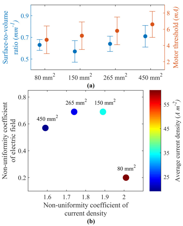
Wearable electrode arrays can selectively stimulate muscle groups by modulating their shape, size, and position over a targeted region. They can potentially revolutionize personalized rehabilitation by being noninvasive and allowing easy donning and doffing. Nevertheless, users should feel comfortable using such arrays, as they are typically worn for an extended time period. Additionally, to deliver safe and selective stimulation, these arrays must be tailored to a user's physiology. Fabricating customizable electrode arrays needs a rapid and economical technique that accommodates scalability. By leveraging a multilayer screen-printing technique, this study aims to develop personalizable electrode arrays by embedding conductive materials into silicone-based elastomers. Accordingly, the conductivity of a silicone-based elastomer was altered by adding carbonaceous material. The 1:8 and 1:9 weight ratio percentages of carbon black (CB) to elastomer achieved conductivities between 0.0021-0.0030 S cm-1 and were suitable for transcutaneous stimulation. Moreover, these ratios maintained their stimulation performance after several stretching cycles of up to 200%. Thus, a soft, conformable electrode array with a customizable design was demonstrated. Lastly, the efficacy of the proposed electrode arrays to stimulate hand function tasks was evaluated by in vivo experiments. The demonstration of such arrays encourages the realization of cost-effective, wearable stimulation systems for hand function restoration.

Keywords: carbon black; elastomer; electrode array; functional electrical stimulation; hand function.


Conflict of interest statement

The authors declare no conflict of interest.

Figures

Figure 1
Figure 1
Multilayered screen printing of an electrode array. (a) Pristine Ecoflex forming the substrate layer. (b) The trace layer was screen-printed over the substrate layer using a patterned stencil. Here the base mixture (carbon black and Ecoflex) was evenly poured and smeared using a squeegee. (c) Exposed trace layer over the substrate layer. (d) 3D-printed masking structures were used to create cavities, while another layer of pristine Ecoflex concealed the trace layer. (e) Trace layer concealed by the second layer of pristine Ecoflex with cavities exposed. (f) The last layer, i.e., the electrode layer, was screen printed using a patterned stencil. The base mixture was again poured and smeared to fill the exposed cavities from the previous step. This process also bridged the electrodes to their respective traces in the trace layer. (g) Wearable sleeve array with exposed electrode layer. Insert shows the cross-section of an individual electrode. (h) Experimental setup to evaluate hand function during transcutaneous stimulation. Figure depicts initial scanning for motor point locations. Activating respective elements in the array (Ei,j) with appropriate stimulation waveform (It ) elicited the desired outcome.
Figure 2
Figure 2
(a) Motor thresholds and normalized surface-to-volume ratios for respective activation volume. The error bars represent values for motor nerve stimulation at depths of 12.4, 14.6, and 16.8 mm. (b) Plot comparing model-predicted nonuniformity coefficients of current density and electric fields for electrodes with different surface areas. The color bar represents the average current density.
Figure 3
Figure 3
Conductivity measurements across sixteen samples of CB:Ecoflex grouped by different weight ratio percentages.
Figure 4
Figure 4
Comparison of model-predicted nonuniformity coefficients of current density and electric fields for electrodes with different weight ratio percentages. The color bar represents normalized surface-to-volume ratio.
Figure 5
Figure 5
Change in resistance observed across twelve samples of CB:Ecoflex grouped by different weight ratio percentages of 1:8 (a), 1:9 (b), 1:10 (c), and 1:11 (d). Resistance changes were reported while the samples were subjected to three stretching cycles.
Figure 6
Figure 6
Average surface thickness across twelve samples of CB:Ecoflex grouped by different weight ratio percentages.
Figure 7
Figure 7
Scanning electron microscope images for 1:8 and 1:9 weight ratios of CB:Ecoflex. Images for 1:8 weight ratio magnified at ×250 (a) and ×450 (b), and 1:9 weight ratio magnified at ×450 (c).
Figure 8
Figure 8
(a) Previous attempt of an electrode-array-based sleeve with disposable electrodes having a bulky construction. (b) Computational model of a forearm under transcutaneous stimulation. (c) Electrode-array-based sleeve fabricated using multilayered screen-printing process (in this study). (d,e) demonstrate that the fabrication process can allow for customizing the electrode elements in terms of shape and different materials.

References

    1. Marquez-Chin C., Popovic M.R. Functional electrical stimulation therapy for restoration of motor function after spinal cord injury and stroke: A review. Biomed. Eng. Online. 2020;19:34. doi: 10.1186/s12938-020-00773-4. - DOI - PMC - PubMed
    1. Kenney L.P., Heller B.W., Barker A.T., Reeves M.L., Healey J., Good T.R., Cooper G., Sha N., Prenton S., Liu A., et al. A review of the design and clinical evaluation of the ShefStim array-based functional electrical stimulation system. Med. Eng. Phys. 2016;38:1159–1165. doi: 10.1016/j.medengphy.2016.08.005. - DOI - PubMed
    1. Koutsou A.D., Moreno J.C., del Ama A.J., Rocon E., Pons J.L. Advances in selective activation of muscles for non-invasive motor neuroprostheses. J. Neuroeng. Rehabil. 2016;13:56. doi: 10.1186/s12984-016-0165-2. - DOI - PMC - PubMed
    1. Hwang K., Jin S., Hwang S.-H., Lee K.-M., Han S.-H. Location of nerve entry points of flexor digitorum profundus. Surg. Radiol. Anat. 2007;29:617–621. doi: 10.1007/s00276-007-0260-y. - DOI - PubMed
    1. RaviChandran N., Aw K.C., McDaid A. Characterizing the Motor Points of Forearm Muscles for Dexterous Neuroprostheses. IEEE Trans. Biomed. Eng. 2020;67:50–59. doi: 10.1109/TBME.2019.2907926. - DOI - PubMed

LinkOut - more resources