Review
. 2023 Mar 21;23(6):3313.
doi: 10.3390/s23063313.

Programmable Nanostructures Based on Framework-DNA for Applications in Biosensing

Affiliations
Review

Programmable Nanostructures Based on Framework-DNA for Applications in Biosensing

Bing Liu et al. Sensors (Basel). .

Abstract

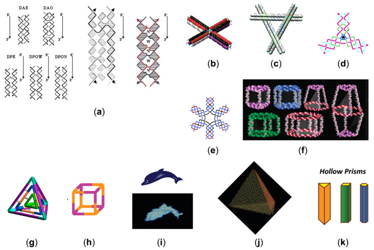
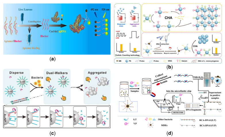
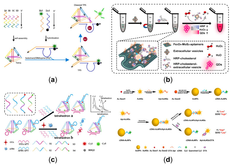
DNA has been actively utilized as bricks to construct exquisite nanostructures due to their unparalleled programmability. Particularly, nanostructures based on framework DNA (F-DNA) with controllable size, tailorable functionality, and precise addressability hold excellent promise for molecular biology studies and versatile tools for biosensor applications. In this review, we provide an overview of the current development of F-DNA-enabled biosensors. Firstly, we summarize the design and working principle of F-DNA-based nanodevices. Then, recent advances in their use in different kinds of target sensing with effectiveness have been exhibited. Finally, we envision potential perspectives on the future opportunities and challenges of biosensing platforms.

Keywords: DNA nanostructures; bacteria; biosensing; nucleic acids; protein; virus.


Conflict of interest statement

The authors declare no conflict of interest.

Figures

Figure 1
Figure 1
(a) The left is the five models of DNA double crossover molecules [25]. (Reprinted from Biochemistry, T. J. Fu, and N. C. Seeman, DNA double-crossover molecules, 3211–3220, Copyright (1993), with permission from the American Chemical Society). The middle one is TX [26], which contains three helices and four strands. (Reprinted from the Journal of the American Chemical Society, LaBean, T.H.; Yan, H.; Kopatsch, J.; Liu, F.; Winfree, E.; Reif, J.H.; Seeman, N.C. Construction, Analysis, Ligation, and Self-Assembly of DNA Triple Crossover Complexes, 2000, 122, 1848–1860, Copyright (2000), with permission from the American Chemical Society). The last one is PX [27], which contains four strands, arranged in two double-helical domains related by a central dyad axis. (Reprinted from the Journal of the American Chemical Society, Shen, Z.; Yan, H.; Wang, T.; Seeman, N.C. Paranemic Crossover DNA: A Generalized Holliday Structure with Applications in Nanotechnology, 2004, 126, 1666–1674, Copyright (2004), with permission from the American Chemical Society). (b) Cross-shaped double-decker tile [28]. (Reprinted from the Journal of the American Chemical Society, Majumder, U.; Rangnekar, A.; Gothelf, K.V.; Reif, J.H.; LaBean, T.H. Design and construction of double-decker tile as a route to three-dimensional periodic assembly of DNA, 2011, 133, 3843–3845, Copyright (2011), with permission from the American Chemical Society). (c) Tensegrity triangle with double-crossover edges [29]. (Reprinted from Nano Letters, Zheng, J.; Constantinou, P.E.; Micheel, C.; Alivisatos, A.P.; Kiehl, R.A.; Seeman, N.C. Two-Dimensional Nanoparticle Arrays Show the Organizational Power of Robust DNA Motifs, 2006, 6, 1502–1504, Copyright (2006), with permission from the American Chemical Society). (d) Three-point star motif [30]. (Reprinted from the Journal of the American Chemical Society, He, Y.; Chen, Y.; Liu, H.; Ribbe, A.E.; Mao, C. Self-Assembly of Hexagonal DNA Two-Dimensional (2D) Arrays, 2005, 127, 12202–12203, with permission from the American Chemical Society). (e) Six-point star motif [31]. (Reprinted from the Journal of the American Chemical Society, He, Y.; Tian, Y.; Ribbe, A.E.; Mao, C. Highly Connected Two-Dimensional Crystals of DNA Six-Point-Stars, 2006, 128, 15978–15979, Copyright (2006), with permission from the American Chemical Society). (f) A large number of three-dimensional discrete DNA assemblies [33]. (Reprinted from the Journal of the American Chemical Society, Aldaye, F.A.; Sleiman, H.F. Modular Access to Structurally Switchable 3D Discrete DNA Assemblies, 2007, 129, 13376–13377, Copyright (2007), with permission from the American Chemical Society). (g) Self-assembly of a multilayered DNA tetrahedron [35]. (Reprinted from the Journal of the American Chemical Society, Liu, Z.; Tian, C.; Yu, J.; Li, Y.; Jiang, W.; Mao, C. Self-assembly of responsive multilayered DNA nanocages, 2015, 137, 1730–1733, Copyright (2015), with permission from the American Chemical Society). (h) DNA tubes of self-assembly from eight copies of identical three-point star tiles [36]. (Reprinted from the Journal of the American Chemical Society, Zhang, C.; Ko, S.H.; Su, M.; Leng, Y.; Ribbe, A.E.; Jiang, W.; Mao, C. Symmetry Controls the Face Geometry of DNA Polyhedra, 2009, 131, 1413–1415, Copyright (2009), with permission from the American Chemical Society). (i) The origami of dolphin shape [42]. (Reprinted from ACS Nano, Andersen, E.S.; Dong, M.; Nielsen, M.M.; Jahn, K.; Lind-Thomsen, A.; Mamdouh, W.; Gothelf, K.V.; Besenbacher, F.; Kjems, J. DNA Origami Design of Dolphin-Shaped Structures with Flexible Tails, 2008, 2, 1213–1218, Copyright (2008), with permission from the American Chemical Society). (j) A DNA tetrahedron [45]. (Reprinted from Nano Letters, Ke, Y.; Sharma, J.; Liu, M.; Jahn, K.; Liu, Y.; Yan, H. Scaffolded DNA Origami of a DNA Tetrahedron Molecular Container, 2009, 9, 2445–2447, Copyright (2009), with permission from the American Chemical Society). (k) Hollow prism of folding multiple-arm DNA structures [47]. (Reprinted from the Journal of the American Chemical Society, Endo, M.; Hidaka, K.; Kato, T.; Namba, K.; Sugiyama, H. DNA Prism Structures Constructed by Folding of Multiple Rectangular Arms, 2009, 131, 15570–15571, Copyright (2009), with permission from the American Chemical Society).
Figure 2
Figure 2
(a) Electrochemical DNA biosensor based on Mg2+-dependent DNAzyme cleavage recycling amplification and a regenerated DNA tweezer’s dynamical regulation of the enzyme cascade reaction [59]. (Reprinted from Analytical Chemistry, Kou, B.; Chai, Y.; Yuan, Y.; Yuan, R. Dynamical Regulation of Enzyme Cascade Amplification by a Regenerated DNA Nanotweezer for Ultrasensitive Electrochemical DNA Detection, 2018, 90, 10701–10706, Copyright (2018), with permission from the American Chemical Society). (b) The sensing of Cas13a-bHCR utilizing cascade signal amplification for detection of miRNA [65]. (Reprinted from Biosensors and Bioelectronics, Zhang, J.; Song, C.; Zhu, Y.; Gan, H.; Fang, X.; Peng, Q.; Xiong, J.; Dong, C.; Han, C.; Wang, L. A novel cascade signal amplification strategy integrating CRISPR/Cas13a and branched hybridization chain reaction for ultra-sensitive and specific SERS detection of disease-related nucleic acids, 2023, 219, Copyright (2023), with permission from Elsevier). (c) Hairpin-mediated fluorescence changes triggered by the target circRNA [82]. (Reprinted from Biosensors and Bioelectronics, Yang, M.; Li, H.; Li, X.; Huang, K.; Xu, W.; Zhu, L. Catalytic hairpin self-assembly regulated chameleon silver nanoclusters for the ratiometric detection of CircRNA, 2022, 209, 114258, Copyright (2022), with permission from Elsevier). (d) The process of electrochemical detection based on AuNFs-CFME [83]. (Reprinted from Biosensors and Bioelectronics, Zhang, B.; Chen, M.; Cao, J.; Liang, Y.; Tu, T.; Hu, J.; Li, T.; Cai, Y.; Li, S.; Liu, B.; et al. An integrated electrochemical POCT platform for ultrasensitive circRNA detection towards hepatocellular carcinoma diagnosis, 2021, 192, 113500, Copyright (2021), with permission from Elsevier).
Figure 3
Figure 3
(a) Molecular nanostructures with electrochemical signal enhancement to detect SARS-CoV-2 by automated microfluidics [88]. (Reprinted from Biosensors and Bioelectronics, Zhao, H.; Zhang, Y.; Chen, Y.; Ho, N.R.Y.; Sundah, N.R.; Natalia, A.; Liu, Y.; Miow, Q.H.; Wang, Y.; Tambyah, P.A.; et al. Accessible detection of SARS-CoV-2 through molecular nanostructures and automated microfluidics, 2021, 194, 113629, Copyright (2021), with permission from Elsevier). (b) Aptamer-mediated target recognition with the DHCR process for the detection of the virus [92]. (Reprinted from Analytical Chemistry, Jiang, Y.; Chen, X.; Feng, N.; Miao, P. Electrochemical Aptasensing of SARS-CoV-2 Based on Triangular Prism DNA Nanostructures and Dumbbell Hybridization Chain Reaction, 2022, 94, 14755–14760, Copyright (2022), with permission from the American Chemical Society). (c) Using the DNA Net-aptamer for the viral capture and reading/inhibition [94]. (Reprinted from the Journal of the American Chemical Society, Chauhan, N.; Xiong, Y.; Ren, S.; Dwivedy, A.; Magazine, N.; Zhou, L.; Jin, X.; Zhang, T.; Cunningham, B.T.; Yao, S.; et al. Net-Shaped DNA Nanostructures Designed for Rapid/Sensitive Detection and Potential Inhibition of the SARS-CoV-2 Virus, 2022, Copyright (2022), with permission from the American Chemical Society). (d) Complementarity of SARS-CoV-2 trimer spike proteins with trimer aptamers for molecular recognition [95]. (Reprinted from the Journal of the American Chemical Society, Li, J.; Zhang, Z.; Gu, J.; Amini, R.; Mansfield, A.G.; Xia, J.; White, D.; Stacey, H.D.; Ang, J.C.; Panesar, G.; et al. Three on Three: Universal and High-Affinity Molecular Recognition of the Symmetric Homotrimeric Spike Protein of SARS-CoV-2 with a Symmetric Homotrimeric Aptamer, 2022, 144, 23465–23473, Copyright (2022), with permission from the American Chemical Society).
Figure 4
Figure 4
(a) Dual CHA system, with the released individual aptamer for the simultaneous analysis of AFP and GPC3 [101]. (Reprinted from ACS Applied Materials and Interfaces, Chen, P.; Jiang, P.; Lin, Q.; Zeng, X.; Liu, T.; Li, M.; Yuan, Y.; Song, S.; Zhang, J.; Huang, J.; et al. Simultaneous Homogeneous Fluorescence Detection of AFP and GPC3 in Hepatocellular Carcinoma Clinical Samples Assisted by Enzyme-Free Catalytic Hairpin Assembly, 2022, 14, 28697–28705, Copyright (2022), with permission from the American Chemical Society). (b) Due to the steric hindrance effects, a highly selective electrochemical sensor based on DNA to detect the presence of macromolecules [103]. (Reprinted from the Journal of the American Chemical Society, Mahshid, S.S.; Camiré, S.; Ricci, F.; Vallée-Bélisle, A. A Highly Selective Electrochemical DNA-Based Sensor That Employs Steric Hindrance Effects to Detect Proteins Directly in Whole Blood, 2015, 137, 15596–15599, Copyright (2015), with permission from the American Chemical Society). (c) The DNA tetrahedral structure forms a switchable scaffold. The cleavage reaction of the Ago2/miR-21 complex opens the hairpin structure, causing the PET pairs to separate in the spatial state [106]. (Reprinted from Analytical Chemistry, Zhang, K.; Huang, W.; Huang, Y.; Li, H.; Wang, K.; Zhu, X.; Xie, M. DNA Tetrahedron Based Biosensor for Argonaute2 Assay in Single Cells and Human Immunodeficiency Virus Type-1 Related Ribonuclease H Detection in Vitro, 2019, 91, 7086–7096, Copyright (2019), with permission from the American Chemical Society). (d) Ratio-type ECL immunosensor for transcription factor (NF-κB p50) determination [112]. (Reprinted from ACS Applied Materials and Interfaces, Fan, Z.; Lin, Z.; Wang, Z.; Wang, J.; Xie, M.; Zhao, J.; Zhang, K.; Huang, W. Dual-Wavelength Electrochemiluminescence Ratiometric Biosensor for NF-kappaB p50 Detection with Dimethylthiodiaminoterephthalate Fluorophore and Self-Assembled DNA Tetrahedron Nanostructures Probe, 2020, 12, 11409–11418, Copyright (2020), with permission from the American Chemical Society).
Figure 5
Figure 5
(a) The released blocker from the dsDNA triggered by the live S. aureus, combined with Cas14a1/sgRNA to cleave the FQ probe and produce an intense fluorescent signal [114]. (Reprinted from Biosensors and Bioelectronics, Wei, Y.; Tao, Z.; Wan, L.; Zong, C.; Wu, J.; Tan, X.; Wang, B.; Guo, Z.; Zhang, L.; Yuan, H.; et al. Aptamer-based Cas14a1 biosensor for amplification-free live pathogenic detection, 2022, 211, 114282, Copyright (2022), with permission from Elsevier). (b) The PS microspheres in different states based on the CHA-mediated micro-orifice resistance assay using DNA hybridization reaction to detect L. monocytogenes [116]. (Reprinted from Biosensors and Bioelectronics, Ren, L.; Hong, F.; Chen, Y. Enzyme-free catalytic hairpin assembly reaction-mediated micro-orifice resistance assay for the ultrasensitive and low-cost detection of Listeria monocytogenes, 2022, 214, 114490, Copyright (2022), with permission from Elsevier) (c) A stochastic DNA dual walker with two kinds of released multiple walking strands as a colorimetric biosensor for bacterial detection [123]. (Reprinted from Analytical Chemistry, Yang, H.; Xiao, M.; Lai, W.; Wan, Y.; Li, L.; Pei, H. Stochastic DNA Dual-Walkers for Ultrafast Colorimetric Bacteria Detection, 2020, 92, 4990–4995, Copyright (2020), with permission from the American Chemical Society) (d) A dual-mode aptasensor for multiple bacterial tests either by naked eyes or microfluidic chips [125]. (Reprinted from Talanta, Yu, J.; Wu, H.; He, L.; Tan, L.; Jia, Z.; Gan, N. The universal dual-mode aptasensor for simultaneous determination of different bacteria based on naked eyes and microfluidic-chip together with magnetic DNA encoded probes, 2021, 225, 122062, Copyright (2021), with permission from Elsevier).
Figure 6
Figure 6
(a) Tetrahedron-based Pb2+-sensitive DNAzyme sensor (TPS) that combines GR-5 DNAzyme with DNA tetrahedral [131]. (Reprinted from Talanta, Guan, H.; Yang, S.; Zheng, C.; Zhu, L.; Sun, S.; Guo, M.; Hu, X.; Huang, X.; Wang, L.; Shen, Z. DNAzyme-based sensing probe protected by DNA tetrahedron from nuclease degradation for the detection of lead ions, 2021, 233, 122543, Copyright (2021), with permission from Elsevier). (b) The 2D Fe3O4-MoS2 platform for efficient enrichment and test of extracellular vesicles based on peroxidase-coupled cholesterol [135]. (Reprinted from Analytical Chemistry, Li, Z.; Ma, D.; Zhang, Y.; Luo, Z.; Weng, L.; Ding, X.; Wang, L. Biomimetic 3D Recognition with 2D Flexible Nanoarchitectures for Ultrasensitive and Visual Extracellular Vesicle Detection, 2022, 94, 14794–14800, Copyright (2022), with permission from the American Chemical Society). (c) A signal on-off fluorescence sensor based on the self-assembly DNA tetrahedron for the rapid simultaneous detection of ochratoxin A (OTA) and aflatoxin B1 (AFB1) [141]. (Reprinted from Analytica Chimica Acta, Ren, W.; Pang, J.; Ma, R.; Liang, X.; Wei, M.; Suo, Z.; He, B.; Liu, Y. A signal on-off fluorescence sensor based on the self-assembly DNA tetrahedron for simultaneous detection of ochratoxin A and aflatoxin B1, 2022, 1198, 339566, Copyright (2022), with permission from Elsevier). (d) The FL-SERS dual-mode mechanism involving the Apt-AuNSs and the cDNA-AuNPs to detect OTA [142]. (Reprinted from Biosensors and Bioelectronics, Wang, H.; Zhao, B.; Ye, Y.; Qi, X.; Zhang, Y.; Xia, X.; Wang, X.; Zhou, N. A fluorescence and surface-enhanced Raman scattering dual-mode aptasensor for rapid and sensitive detection of ochratoxin A, 2022, 207, 114164, Copyright (2022), with permission from Elsevier).

Similar articles

Cited by

References

    1. Watson J.D., Crick F.H.C. Molecular Structure of Nucleic Acids A Structure. Nature. 1953;171:737–738. doi: 10.1038/171737a0. - DOI - PubMed
    1. Ranallo S., Porchetta A., Ricci F. DNA-Based Scaffolds for Sensing Applications. Anal. Chem. 2019;91:44–59. doi: 10.1021/acs.analchem.8b05009. - DOI - PubMed
    1. Pei H., Zuo X., Zhu D., Huang Q., Fan C. Functional DNA Nanostructures for Theranostic Applications. Acc. Chem. Res. 2014;47:550–559. doi: 10.1021/ar400195t. - DOI - PubMed
    1. Charoenphol P., Bermudez H. Design and application of multifunctional DNA nanocarriers for therapeutic delivery. Acta Biomater. 2014;10:1683–1691. doi: 10.1016/j.actbio.2013.07.021. - DOI - PMC - PubMed
    1. Chen Y.-J., Groves B., Muscat R.A., Seelig G. DNA nanotechnology from the test tube to the cell. Nat. Nanotechnol. 2015;10:748–760. doi: 10.1038/nnano.2015.195. - DOI - PubMed

LinkOut - more resources