. 2023 Feb 21;11(3):495.
doi: 10.3390/vaccines11030495.

Astragalus Saponins, Astragaloside VII and Newly Synthesized Derivatives, Induce Dendritic Cell Maturation and T Cell Activation

Affiliations

Astragalus Saponins, Astragaloside VII and Newly Synthesized Derivatives, Induce Dendritic Cell Maturation and T Cell Activation

Nilgun Yakubogullari et al. Vaccines (Basel). .

Abstract

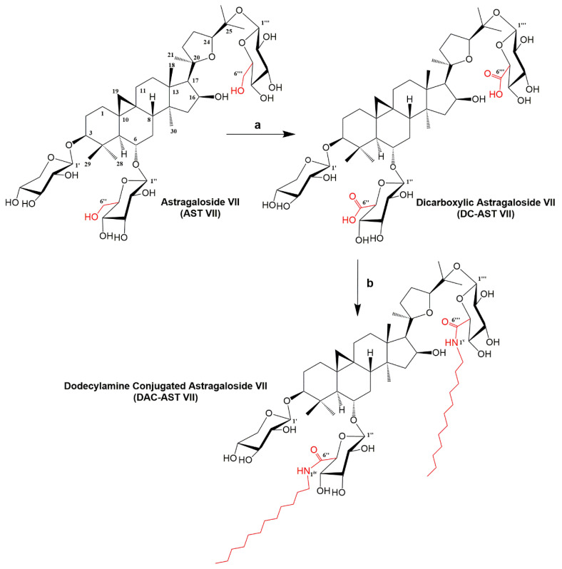
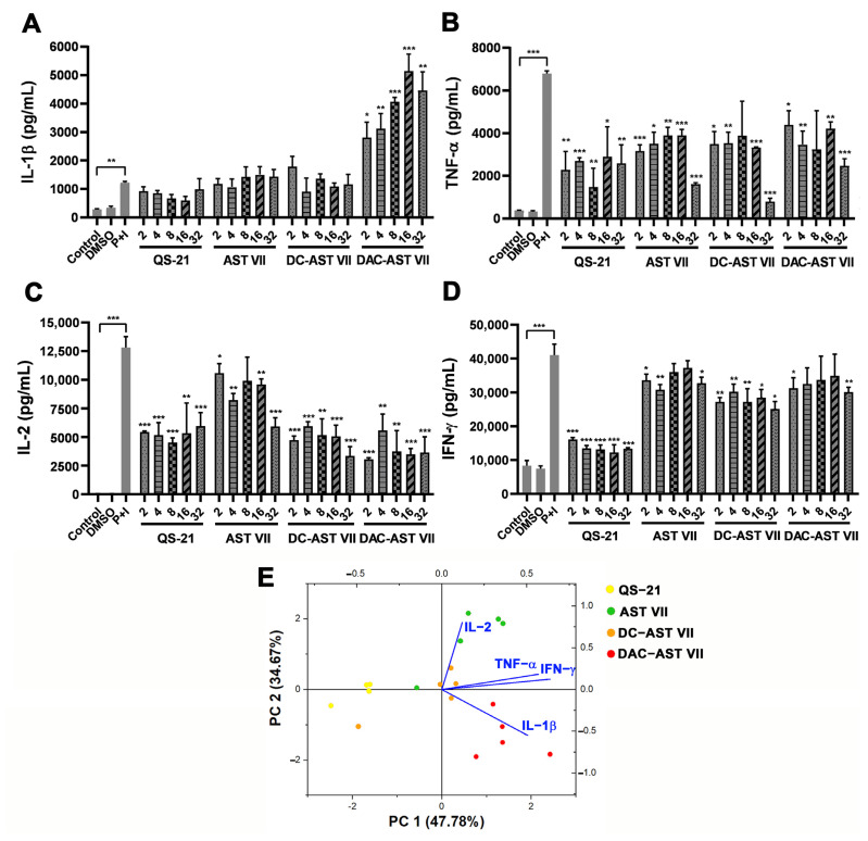
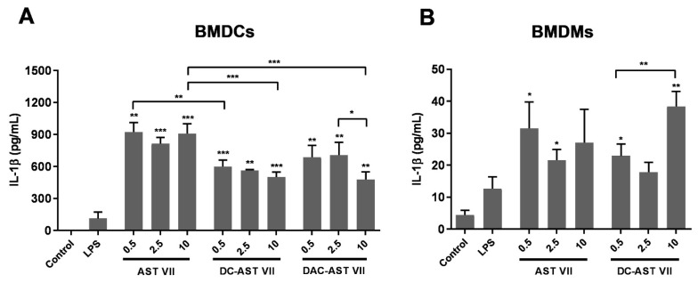
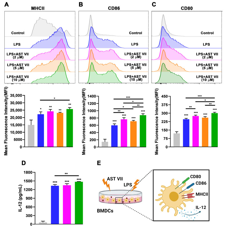
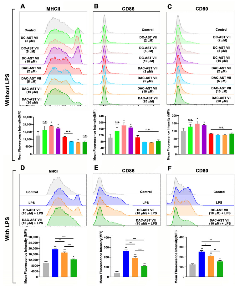
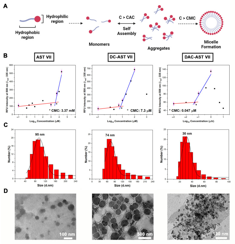
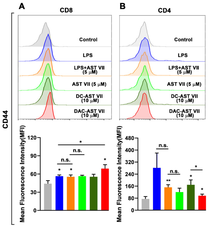
Astragaloside VII (AST VII), a triterpenic saponin isolated from Astragalus species, shows promise as a vaccine adjuvant, as it supported a balanced Th1/Th2 immune response in previous in vivo studies. However, the underlying mechanisms of its adjuvant activity have not been defined. Here, we investigated the impact of AST VII and its newly synthesized semi-synthetic analogs on human whole blood cells, as well as on mouse bone marrow-derived dendritic cells (BMDCs). Cells were stimulated with AST VII and its derivatives in the presence or absence of LPS or PMA/ionomycin and the secretion of cytokines and the expression of activation markers were analyzed using ELISA and flow cytometry, respectively. AST VII and its analogs increased the production of IL-1β in PMA/ionomycin-stimulated human whole blood cells. In LPS-treated mouse BMDCs, AST VII increased the production of IL-1β and IL-12, and the expression of MHC II, CD86, and CD80. In mixed leukocyte reaction, AST VII and derivatives increased the expression of the activation marker CD44 on mouse CD4+ and CD8+ T cells. In conclusion, AST VII and its derivatives strengthen pro-inflammatory responses and support dendritic cell maturation and T cell activation in vitro. Our results provide insights into the mechanisms of the adjuvant activities of AST VII and its analogs, which will be instrumental to improve their utility as a vaccine adjuvant.

Keywords: immunological evaluation; immunomodulation; semi-synthesis; triterpenoid saponin; vaccine adjuvant.


Conflict of interest statement

The authors declare no conflict of interest.

Figures

Figure 1
Figure 1
Semi-synthesis of AST VII derivatives. Reagents and conditions: (a) TEMPO, NaBr, NaOCl, H2O, 0 °C (57%) and (b) EDC/HOBt, DIPEA, Pyridine, 60 °C (28%).
Figure 2
Figure 2
AST VII and its derivatives (DC-AST VII and DAC-AST VII) alter the production of pro-inflammatory and Th- mediated cytokines in hWB cells. Diluted hWB was co-treated with PMA (50 ng/mL)/ionomycin (400 ng/mL) and the following compounds: QS-21, AST VII, DC-AST VII, DAC-AST VII at the concentration of 2–32 µg/mL for 48 h. The supernatants were collected for the detection of cytokines using ELISA. (A) IL-1β, (B) TNF-α, (C) IL-2, and (D) IFN-γ in 1/20 diluted hWB. (E) Principal component analysis (PCA) biplot illustrating the cytokine responses and corresponding compound treatments in hWB. Data are projected onto the plane of the first two principal components (PCs) and colored by the different compounds. DMSO was used as vehicle control. Data shown are mean ± SD of triplicate determinations and representative of two independent experiments with similar results. Statistical analyses were performed between the control and P+I (PMA+Ionomycin) using Student’s t-test and P+I and treated groups using One-way ANOVA and Tukey’s multiple comparison tests. * p < 0.05, ** p < 0.01, *** p < 0.001, not statistically significant (ns).
Figure 3
Figure 3
AST VII and its derivatives induce IL-1β secretion in BMDCs and BMDMs. (A) BMDCs and (B) BMDMs generated from bone marrow cells of C57BL/6 mice were treated with LPS (10 ng/mL) alone or LPS with AST VII, DC-AST VII, or DAC-AST VII at the concentrations of 0.5, 2.5, or 10 µM for 6 h. Unstimulated cells were used as the control. The cell culture supernatants were collected to analyze IL-1β concentrations using ELISA. Data shown are the mean ± SD of triplicates and representative of two independent experiments with similar results. Statistically significant differences in the intragroup were analyzed compared to LPS. Intragroup and intergroup comparisons were performed with one-way ANOVA and Tukey’s multiple comparisons test. * p < 0.05, ** p < 0.01, *** p < 0.001.
Figure 4
Figure 4
AST VII induces the maturation and activation of BMDCs in the presence of LPS. BMDCs generated from C57BL/6 mice bone marrow cells were co-treated with LPS (10 ng/mL) and AST VII at the concentrations of 2, 5, or 10 µM for 24 h. The expression of (A) MHCII, (B) CD86, and (C) CD80 on CD11c+MHCII+ BMDCs was determined using flow cytometry. For (AC), representative histogram (top panel) and summary graphs (bottom panel) are shown. (D) IL-12 titers were measured using ELISA. (E) Schematic illustration of DC maturation/activation after the treatment with AST VII and LPS. Data shown are mean ± SD of triplicates and representative of three independent experiments with similar results. Statistically significant differences in the treated group were analyzed compared to the control (untreated cells). Intragroup and intergroup comparisons were performed using one-way ANOVA and Tukey’s multiple comparison test. * p < 0.05, ** p ≤ 0.01, *** p ≤ 0.001. Created with BioRender.com.
Figure 5
Figure 5
The effects of AST VII derivatives (DC-AST VII and DAC-AST VII) on the maturation and activation of BMDCs in the absence/presence of LPS. BMDCs generated from C57BL/6 mice bone marrow cells were treated with DC-AST VII or DAC-AST VII at the concentrations of 2, 5, 10, or 20 µM for 24 h. The expression of (A) MHCII, (B) CD86, and (C) CD80 on CD11c+MHCII+ BMDCs without LPS treatment and (D) MHCII, (E) CD86, and (F) CD80 on CD11c+MHCII+ BMDCs with LPS treatment was determined using flow cytometry. Representative histograms (top panel) and summary graphs (bottom panel) are shown. Data are shown as the mean and SD of triplicates and are representative of two independent experiments with similar results. Statistically significant differences between the treated group were analyzed compared to the control (untreated cells). Intragroup and intergroup comparisons were performed using one-way ANOVA and Tukey’s multiple comparison test. * p < 0.05, ** p ≤ 0.01, *** p ≤ 0.001, n.s. (not statistically significant).
Figure 6
Figure 6
Self-assembling nanoparticles based on Astragalus saponins. (A) A general illustration for self-assembling particle formation. C: Concentration, CAC: Critical aggregation concentration, CMC: Critical micelle concentration. (B) Relative fluorescence intensity at 508 nm vs. logarithm of Astragalus saponins concentrations to determine CMC. (C) Representative particle size distribution of self-assembling Astragalus saponins that were measured using dynamic light scattering (DLS) analysis. (D) Transmission electron microscopy (TEM) images of self-assembled nanoparticles based on AST VII (4000 μM), DC-AST VII (10 μM), and DAC-AST VII (10 μM) in dH2O. Data represent two or three independent experiments. Created with BioRender.com. *: the intersection of two linear lines, showing CMC value.
Figure 7
Figure 7
AST VII and its derivatives (DC-AST VII and DAC-AST VII) activated T cells in MLR. BMDCs derived from BALB/c mice were treated with LPS, AST VII, DC-AST VII, or DAC-AST VII for 24 h. The next day, BMDCs were co-cultured with naive CD4+ and CD8+ T cells isolated from the spleens of C57BL/6 mice for 3 days. The expression of CD44 on (A) CD8+ T cells and on (B) CD4+ T cells was determined using flow cytometry. Representative histograms (top panel) and summary graphs (bottom panel) are shown. Data are shown as the mean and SD of triplicates and represent three independent experiments with similar results. Statistically significant differences in the treated group were analyzed compared to the control (untreated cells). Intragroup and intergroup comparisons were performed using one-way ANOVA and Tukey’s multiple comparison test. * p < 0.05, ** p ≤ 0.01, n.s. (not statistically significant).

Similar articles

Cited by

References

    1. Reed S.G., Orr M.T., Fox C.B. Key Roles of Adjuvants in Modern Vaccines. Nat. Med. 2013;19:1597–1608. doi: 10.1038/nm.3409. - DOI - PubMed
    1. Pifferi C., Fuentes R., Fernández-Tejada A. Natural and Synthetic Carbohydratebased Vaccine Adjuvants and Their Mechanisms of Action. Nat. Rev. Chem. 2021;5:197–216. doi: 10.1038/s41570-020-00244-3. - DOI - PMC - PubMed
    1. Mirotti L., Alberca Custódio R.W.A., Gomes E., Rammauro F., de Araujo E.F., Garcia Calich V.L.G., Russo M. CpG-ODN Shapes Alum Adjuvant Activity Signaling via MyD88 and IL-10. Front. Immunol. 2017;8:47. doi: 10.3389/fimmu.2017.00047. - DOI - PMC - PubMed
    1. Bonam S.R., Partidos C.D., Halmuthur S.K.M., Muller S. An Overview of Novel Adjuvants Designed for Improving Vaccine Efficacy. Trends Pharmacol. Sci. 2017;38:771–793. doi: 10.1016/j.tips.2017.06.002. - DOI - PubMed
    1. Laupèze B., Hervé C., Di Pasquale A., Da Silva F.D. Adjuvant Systems for Vaccines: 13 Years of Post-Licensure Experience in Diverse Populations Have Progressed the Way Adjuvanted Vaccine Safety Is Investigated and Understood. Vaccine. 2019;37:5670–5680. doi: 10.1016/j.vaccine.2019.07.098. - DOI - PubMed