. 2023 Mar 16;11(3):672.
doi: 10.3390/vaccines11030672.

RSV A2-Based Prefusion F Vaccine Candidates Induce RSV A and RSV B Cross Binding and Neutralizing Antibodies and Provide Protection against RSV A and RSV B Challenge in Preclinical Models

Affiliations

RSV A2-Based Prefusion F Vaccine Candidates Induce RSV A and RSV B Cross Binding and Neutralizing Antibodies and Provide Protection against RSV A and RSV B Challenge in Preclinical Models

Freek Cox et al. Vaccines (Basel). .

Abstract

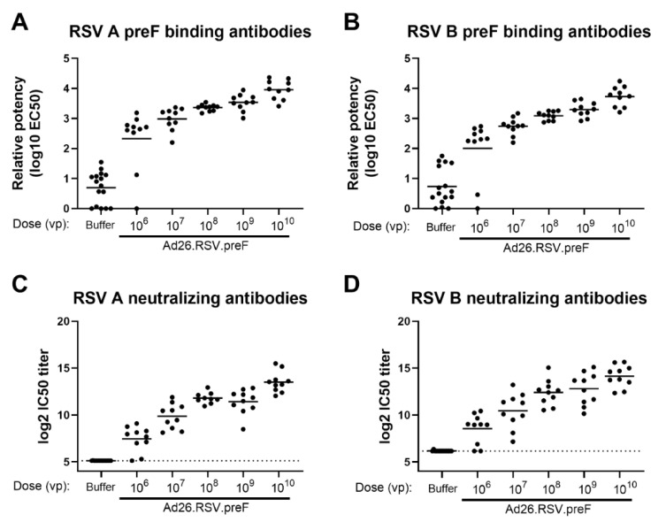
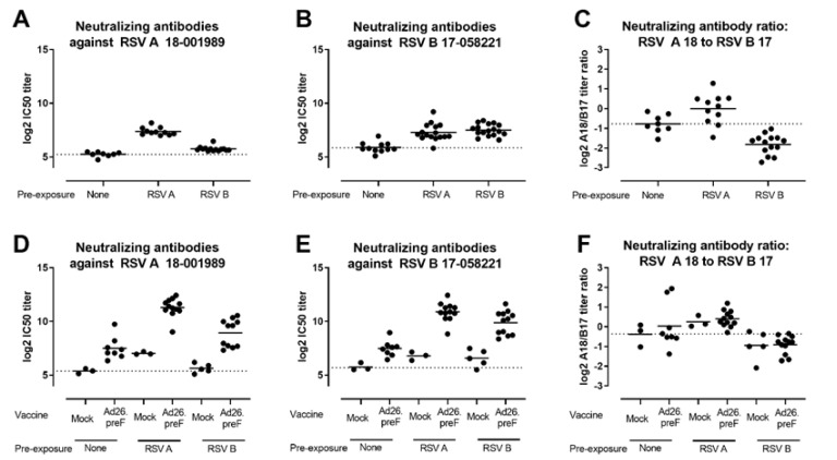
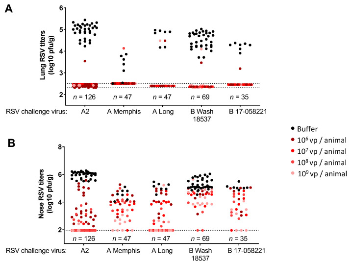
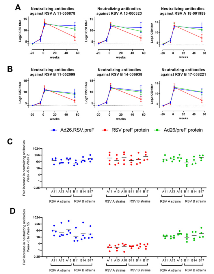
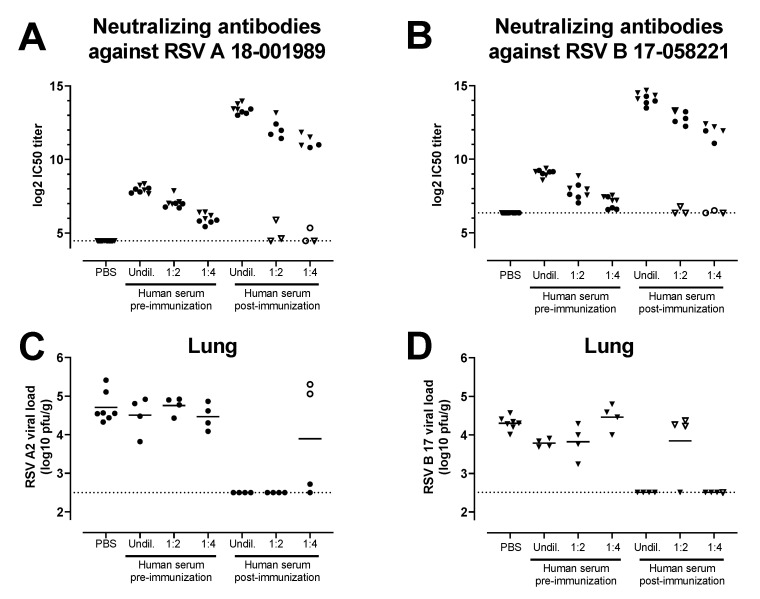
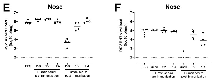
RSV is divided into two antigenic subtypes, RSV A and RSV B, which is largely based on the variation in the G protein, while the fusion protein F is more conserved and a target for antibody-mediated neutralization. Here we evaluate the breadth of the protective immune responses across RSV A and RSV B subtypes, induced by vaccines based on the RSV A-based fusion protein, stabilized in the prefusion conformation (preF) in preclinical models. Immunization of naïve cotton rats with preF subunit or preF encoded by a replication incompetent Adenoviral 26, induced antibodies capable of neutralizing recent RSV A and RSV B clinical isolates, as well as protective efficacy against a challenge with RSV A and RSV B strains. Similarly, induction of cross-neutralizing antibodies was observed after immunization with Ad26-encoded preF, preF protein or a mix of both (Ad26/preF protein) in RSV pre-exposed mice and African Green Monkeys. Transfer of serum of human subjects immunized with Ad26/preF protein into cotton rats provide protection against challenges with both RSV A and RSV B, with complete protection against both strains observed in the lower respiratory tract. In contrast, almost no protection against RSV A and B infection was observed after the transfer of a human serum pool isolated pre-vaccination. These results collectively show that the RSV A-based monovalent Ad26/preF protein vaccine induced neutralizing antibodies, as well as protection against both RSV A and RSV B subtypes in animals, including by passive transfer of human antibodies alone, suggesting that clinical efficacy against both subtypes can be achieved.

Keywords: Adenoviral vector 26; RSV prefusion protein; RSV subtypes; cotton rat model; respiratory syncytial virus.


Conflict of interest statement

All authors are employees of Janssen Infectious Diseases & Vaccines and may own stock or stock options in Johnson & Johnson, its parent company.

Figures

Figure 1
Figure 1
Ad26.RSV.preF dose-dependently induces antibodies binding to preF from RSV A and RSV B, which are capable of neutralizing both RSV A and RSV B strains in naïve cotton rats. Cotton rats were immunized with 106 to 1010 vp Ad26.RSV.preF (n = 10 per group) or formulation buffer (n = 16) at day 0, and serum isolated at day 49 was analyzed. Binding IgG was determined by ELISA using RSV A preF (A) or RSV B preF (B). Neutralizing antibody titers were determined against RSV A 18-001989 (C) or RSV B 17-058221 (D) by microneutralization virus neutralization assay (MN VNA). The lower limit of detection for MN VNA was defined as the mean plus 3× the standard deviation of the formulation buffer group, indicated with a dotted line. Means per group are indicated with horizontal lines. Note: MN VNA titers against RSV A 18-001989 from one animal in the 108 vp dose group were unexpectedly and unexplainably high (24.2 log2 IC50), and this data point was excluded.
Figure 2
Figure 2
Ad26.RSV.preF induces RSV A and RSV B subtype-specific neutralizing antibodies in both RSV A and RSV B pre-exposed mice. Mice were pre-exposed to RSV A 18-001989 or RSV B 17-058221 12 weeks prior to immunization with 108 vp Ad26.RSV.preF (n = 12), or empty Ad26 vector (mock, n = 5). As controls, naïve mice were immunized with 109 vp Ad26.RSV.preF (n = 8), or empty Ad26 vector (mock, n = 3). Neutralizing antibodies titers against RSV A 18-001989 (A,D) or RSV B 17-058221 (B,E) were determined by MN VNA on serum isolated at week 12, prior to immunization (a,b), and at week 16, 4 weeks posts immunization (D,E). Log2 ratios between RSV A and RSV B VNT are depicted (C,F). The average responses in mock immunized naïve animals are indicated with dotted lines. Mean responses per group are indicated with horizontal lines. Note some data points are missing due to insufficient serum availability, dropout of animals from the study or experimental failure.
Figure 3
Figure 3
Immunization with Ad26.RSV.preF confers protection against challenges with RSV A and RSV B strains in naïve cotton rats. Cotton rats were immunized with 106 to 109 vp of Ad26.RSV.preF at day 0, and challenged with RSV A2, RSV A Memphis, RSV A Long, RSV B Wash 18537 or RSV B 17-058221 at week 7. RSV viral load was determined by plaque assay in homogenates of the lung (A) or nose tissue (B) isolated at day 5 post-challenge and expressed as log10 plaque forming units (pfu) per gram of tissue. Compiled results from multiple challenge studies are shown (five studies for RSV A2, one study for RSV A Memphis, one study for RSV A Long, four studies for RSV B Wash 18537 and one study for RSV B 17-058221), with total numbers of animals per challenge strain shown in the figure, and number of animals per Ad26.RSV.preF dose level (indicated with different colored dots) ranging from seven to forty-two. Pooling of results of several different studies is considered justified, as viral loads in the control groups were within acceptable ranges between the separate studies that use the same challenge strain. The limit of detection of the plaque assays is indicated with dotted lines.
Figure 4
Figure 4
Ad26.RSV.preF, RSV preF protein and their combination induce antibodies that can neutralize various RSV A and RSV B strains in RSV A Memphis pre-exposed African Green Monkeys. African Green Monkeys were intranasally pre-exposed with RSV A Memphis 37 at week −19 and immunized intramuscularly at Week 0 with 1011 vp Ad26.RSV.preF (in blue), 150 μg preF protein (in red), or a mixture of both components (in green). MN VNT against 3 RSV A strains (A) and 3 RSV B strains (B) were determined in serum samples (n = 4 per group at week −5, week 2 and week 55; one sample per group at week −19). The 50% inhibitory concentration (IC50) titers were calculated, and the limit of detection (LOD) was set at 4 log2 (dotted line), which is the lowest serum dilution used in the assay. Mean ± standard deviations are shown. Fold increase from week −5 to week 2 (C) or to week 55 (D) was calculated per animal, with geometric means per group per RSV strain indicated.
Figure 5
Figure 5
Serum of human subjects that received vaccination with the combination of Ad26.RSV.preF and RSV preF protein can provide protection against RSV A and RSV B strains in the cotton rat model. Cotton rats received different amounts (2 mL of undiluted (undil.), 1:2 or 1:4 diluted serum in PBS) of a human serum pool generated by pooling serum isolated pre-vaccination or at 14 days post-vaccination with a mixture of 1011 vp Ad26.RSV.preF and 150 μg preF protein (n = 39) by intraperitoneal injection (n = 4 per group) or with PBS (n = 7). The next day, pre-challenge serum samples of the recipient animals were isolated and assayed for VNT against RSV A 18-001989 (A) or for RSV B 17-058221 (B). Animals were challenged with RSV A2 (C,E) or RSV B 17-058221 (D,F). RSV viral load was determined five days after the challenge using plaque assay on homogenates of the lung (C,D) or nose tissue (E,F) and expressed as pfu per gram of tissue. Samples of animals that were (to be) challenged with RSV A2 are indicated with dots, whereas samples of animals that were (to be) challenged with RSV B 17-058221 are indicated with triangles. Animals in which serum transfer was not successful, based on low VNT pre-challenge, are indicated with open symbols. LODs for MN VNA were determined as the mean plus 3× standard deviations of the PBS control group. LODs are indicated with dotted lines.
Figure 5
Figure 5
Serum of human subjects that received vaccination with the combination of Ad26.RSV.preF and RSV preF protein can provide protection against RSV A and RSV B strains in the cotton rat model. Cotton rats received different amounts (2 mL of undiluted (undil.), 1:2 or 1:4 diluted serum in PBS) of a human serum pool generated by pooling serum isolated pre-vaccination or at 14 days post-vaccination with a mixture of 1011 vp Ad26.RSV.preF and 150 μg preF protein (n = 39) by intraperitoneal injection (n = 4 per group) or with PBS (n = 7). The next day, pre-challenge serum samples of the recipient animals were isolated and assayed for VNT against RSV A 18-001989 (A) or for RSV B 17-058221 (B). Animals were challenged with RSV A2 (C,E) or RSV B 17-058221 (D,F). RSV viral load was determined five days after the challenge using plaque assay on homogenates of the lung (C,D) or nose tissue (E,F) and expressed as pfu per gram of tissue. Samples of animals that were (to be) challenged with RSV A2 are indicated with dots, whereas samples of animals that were (to be) challenged with RSV B 17-058221 are indicated with triangles. Animals in which serum transfer was not successful, based on low VNT pre-challenge, are indicated with open symbols. LODs for MN VNA were determined as the mean plus 3× standard deviations of the PBS control group. LODs are indicated with dotted lines.

References

    1. Ackerson B., Tseng H.F., Sy L.S., Solano Z., Slezak J., Luo Y., Fischetti C.A., Shinde V. Severe Morbidity and Mortality Associated with Respiratory Syncytial Virus Versus Influenza Infection in Hospitalized Older Adults. Clin. Infect. Dis. 2019;69:197–203. doi: 10.1093/cid/ciy991. - DOI - PMC - PubMed
    1. Shi T., Denouel A., Tietjen A.K., Campbell I., Moran E., Li X., Campbell H., Demont C., Nyawanda B.O., Chu H.Y., et al. Global disease burden estimates of respiratory syncytial virus-associated acute respiratory infection in older adults in 2015: A systematic review and meta-analysis. J. Infect. Dis. 2020;222:S577–S583. doi: 10.1093/infdis/jiz059. - DOI - PubMed
    1. Hall C.B., Walsh E.E., Long C.E., Schnabel K.C. Immunity to and frequency of re-infection with respiratory syncytial virus. J. Infect. Dis. 1991;163:693–698. doi: 10.1093/infdis/163.4.693. - DOI - PubMed
    1. Capella C., Chaiwatpongsakorn S., Gorrell E., Risch Z.A., Ye F., Mertz S.E., Johnson S.M., Moore-Clingenpeel M., Ramilo O., Mejias A., et al. Prefusion F, Postfusion F, G Antibodies, and Disease Severity in Infants and Young Children with Acute Respiratory Syncytial Virus Infection. J. Infect. Dis. 2017;216:1398–1406. doi: 10.1093/infdis/jix489. - DOI - PMC - PubMed
    1. Ngwuta J.O., Chen M., Modjarrad K., Joyce M.G., Kanekiyo M., Kumar A., Yassine H.M., Moin S.M., Killikelly A.M., Chuang G.Y., et al. Prefusion F-specific antibodies determine the magnitude of RSV neutralizing activity in human sera. Sci. Transl. Med. 2015;7:309ra162. doi: 10.1126/scitranslmed.aac4241. - DOI - PMC - PubMed

LinkOut - more resources