. 2023 Feb 24;15(3):627.
doi: 10.3390/v15030627.

Impact of Previous Common Human Coronavirus Exposure on SARS-CoV-2-Specific T-Cell and Memory B-Cell Response after mRNA-Based Vaccination

Affiliations

Impact of Previous Common Human Coronavirus Exposure on SARS-CoV-2-Specific T-Cell and Memory B-Cell Response after mRNA-Based Vaccination

José L Casado et al. Viruses. .

Abstract

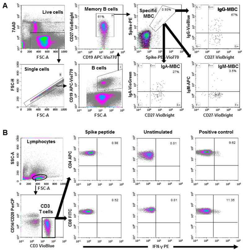
Objective: T-cell responses against SARS-CoV-2 are observed in unexposed individuals, attributed to previous common human coronavirus (HCoV) infections. We evaluated the evolution of this T-cell cross-reactive response and the specific memory B-cells (MBCs) after the SARS-CoV-2 mRNA-based vaccination and its impact on incident SARS-CoV-2 infections.

Methods: This was a longitudinal study of 149 healthcare workers (HCWs) that included 85 unexposed individuals that were subdivided according to previous T-cell cross-reactivity, who were compared to 64 convalescent HCWs. Changes in specific T-cell response and memory B-cell (MBC) levels were compared at baseline and after two doses of the SARS-CoV-2 mRNA-based vaccine.

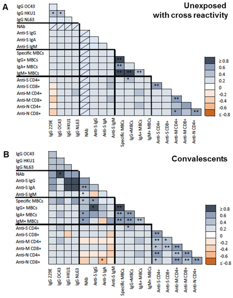
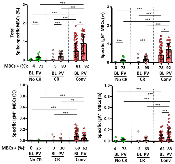
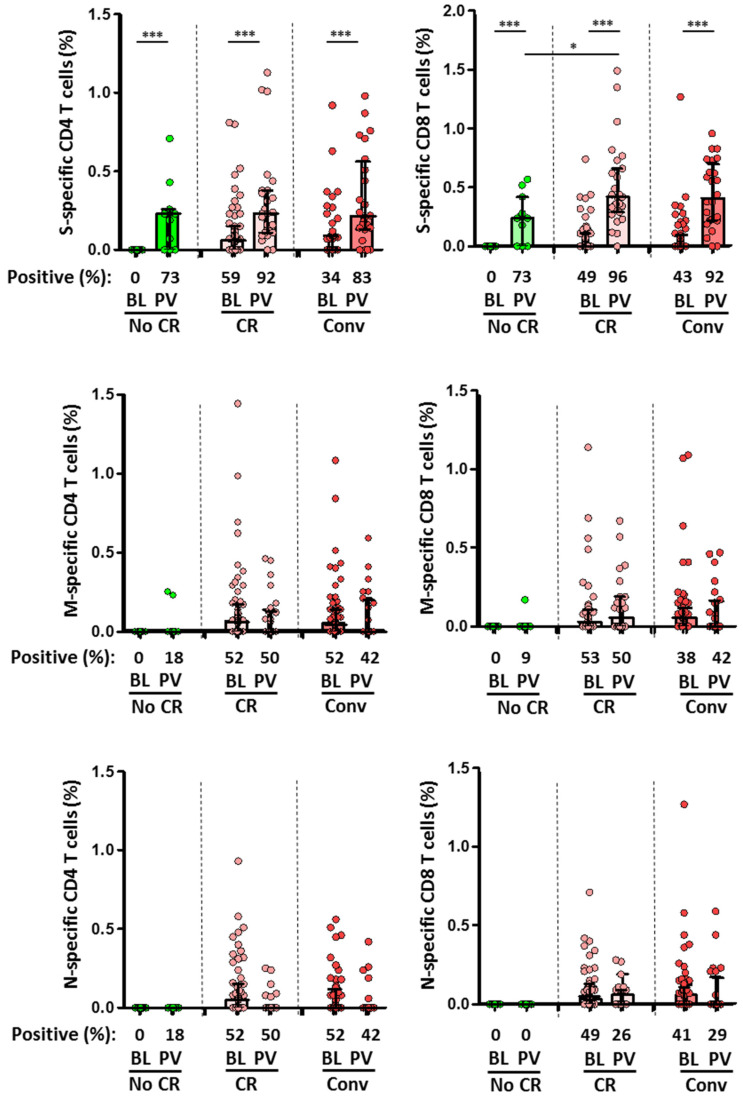
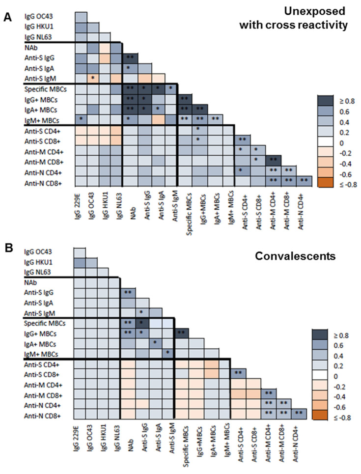
Results: A cross-reactive T-cell response was found in 59% of unexposed individuals before vaccination. Antibodies against HKU1 positively correlated with OC43 and 229E antibodies. Spike-specific MBCs was scarce in unexposed HCWs regardless of the presence of baseline T-cell cross-reactivity. After vaccination, 92% and 96% of unexposed HCWs with cross-reactive T-cells had CD4+ and CD8+ T-cell responses to the spike protein, respectively. Similar results to that were found in convalescents (83% and 92%, respectively). Contrarily, higher than that which was observed in unexposed individuals without T-cell cross-reactivity showed lower CD4+ and CD8+ T-cell responses (73% in both cases, p = 0.03). Nevertheless, previous cross-reactive T-cell response was not associated with higher levels of MBCs after vaccination in unexposed HCWs. During a follow-up of 434 days (IQR, 339-495) after vaccination, 49 HCWs (33%) became infected, with a significant positive correlation between spike-specific MBC levels and isotypes IgG+ and IgA+ after vaccination and a longer time to get infected. Interestingly, T-cell cross-reactivity did not reduce the time to vaccine breakthrough infections.

Conclusion: While pre-existing T-cell cross-reactivity enhances the T-cell response after vaccination, it does not increase SARS-CoV-2-specific MBC levels in the absence of previous infection. Overall, the level of specific MBCs determines the time to breakthrough infections, regardless of the presence of T-cell cross-reactivity.

Keywords: SARS-CoV-2; coronavirus; cross-reactive; mRNA vaccine; memory B-cells.


Conflict of interest statement

The authors declare no conflict of interest.

Figures

Figure 1
Figure 1
(A) Flow cytometry strategy for the quantification of SARS-CoV-2-specific memory B-cells (MBC). Viable cells (FSC-A/7AAA plot) were plotted with FSC-H/FSC-A parameters to exclude doublets. Single cells were then gated using CD19-APC-Vio770 and CD27-Vio-Bright-FITC to identify memory B-cells. Spike-specific B-cells were then identified with a double staining with the two spike-tetramer on the diagonal of the dot plot. Finally, the use of IgG-VioBlue, IgA-VioGreen, and IgM-APC were used to quantify each specific isotype of spike-specific memory B-cells. The results are recorded as percentage among the total memory B-cells and isotypes among specific memory B-cells. (B) Flow cytometry gating strategy for the quantification of SARS-CoV-2 spike-specific T-cells. After stimulation with the spike peptide, staining of the cells was carried out with the following fluorochrome-conjugated antibodies: CD3-VioBlue, CD4-APC, CD8-FITC, CD14-PerCP, CD20-PerCP, IFN-γ-PE, and FcR blocking reagent. To exclude dead cells, viability 405/520 fixable dye staining was added for the last 10 min of incubation. The samples were measured and analyzed by flow cytometry on a MACSQuant Analyzer 10 using MACSQuantify software. At least 105 cells were analyzed and gated with the following strategy: Single (FSC-A/FSC-H dot plot) and live cells were first selected. Cell debris, monocytes, and B-cells were excluded from the analysis with CD14-and CD20-PerCP antibodies. Then, lymphocytes were selected with an FSC-A/SSC-A dot plot, and CD3 T-cells were gated. IFN-γ expression was finally analyzed separately for CD4+ and CD8+ T-cells. A representative sample of negative control (without stimulation), positive control (stimulated with SEB), and with SARS-CoV-2 spike peptide are shown.
Figure 2
Figure 2
Baseline correlations between antibodies against common human coronavirus (HCoVs), SARS-CoV-2 spike-specific (IgG, IgA, and IgM) antibodies, neutralizing antibodies (NAB), specific memory B-cells (MBCs) and IgG+, IgA+, and IgM+ MBCs isotypes, CD4+ and CD8+ T-cell responses to SARS-CoV-2 spike (S), membrane (M), and nucleocapsid (N) proteins. (A) Spearman test heatmap analysis of the different variables in unexposed subjects with cross-reactivity. (B) Spearman test heatmap of the different variables in convalescent individuals. The grey color represents a positive correlation and the red color a negative correlation. The intensity of the color indicates the R2 coefficient. Statistically significant when p < 0.05. * = p < 0.05, and ** = p < 0.01.
Figure 3
Figure 3
Comparison of specific memory B-cells (MBCs, top left) and IgG+ MBC (top right), IgM+ MBCs (bottom left), and IgA+ MBCs (bottom right) isotypes between the different subgroups at baseline (BL) and after two doses of vaccine (PV, post-vaccination). No CR (unexposed without cross-reactivity, green dots and bars), CR (unexposed with cross-reactivity, rose dots and bars), and Conv (convalescent, red dots and bars). Frequencies are indicated at the X axis. Statistically significant when p < 0.05. * = p < 0.05, ** = p < 0.01, and *** = p < 0.001.
Figure 4
Figure 4
Comparison of the magnitude of CD4+ (left) and CD8+ (right) T-cell responses against SARS-CoV-2 S, M, and N proteins between the different subgroups at baseline (BL) and after two doses of vaccine (PV, post-vaccination). No CR (unexposed without cross-reactivity, green dots and bars), CR (unexposed with cross-reactivity, rose dots and bars), and Conv (convalescent, red dots and bars). Frequencies are indicated at the X axis. Statistically significant when p < 0.05. * = p < 0.05, and *** = p < 0.001.
Figure 5
Figure 5
Post-vaccination correlations between antibodies against common human coronavirus (HCoVs), SARS-CoV-2 spike-specific (IgG, IgA, and IgM) antibodies, neutralizing antibodies (NAB), specific memory B-cells (MBCs) and IgG+, IgA+, and IgM+ MBCs isotypes, CD4+ and CD8+ T-cell responses to SARS-CoV-2 spike (S), membrane (M), and nucleocapsid (N) proteins. (A), Spearman test heatmap analysis of the different variables in unexposed subjects with cross-reactivity. (B), Spearman test heatmap of the different variables in convalescent individuals. The grey color represents a positive correlation and the red color a negative correlation. The intensity of the color indicates the R2 coefficient. Statistically significant when p < 0.05. * = p < 0.05, and ** = p < 0.01.

References

    1. Ibarrondo F.J., Hofmann C., Fulcher J.A., Goodman-Meza D., Mu W., Hausner M.A., Ali A., Balamurugan A., Taus E., Elliott J., et al. Primary, Recall, and Decay Kinetics of SARS-CoV-2 Vaccine Antibody Responses. ACS Nano. 2021;15:11180–11191. doi: 10.1021/acsnano.1c03972. - DOI - PubMed
    1. Long Q.X., Tang X.J., Shi Q.L., Li Q., Deng H.J., Yuan J., Hu J.L., Xu W., Zhang Y., Lv F.J., et al. Clinical and immunological assessment of asymptomatic SARS-CoV-2 infections. Nat. Med. 2020;26:1200–1204. doi: 10.1038/s41591-020-0965-6. - DOI - PubMed
    1. Goel R.R., Apostolidis S.A., Painter M.M., Mathew D., Pattekar A., Kuthuru O., Gouma S., Hicks P., Meng W., Rosenfeld A.M., et al. Distinct antibody and memory B cell responses in SARS-CoV-2 naive and recovered individuals following mRNA vaccination. Sci. Immunol. 2021;6:eabi6950. doi: 10.1126/sciimmunol.abi6950. - DOI - PMC - PubMed
    1. Dan J.M., Mateus J., Kato Y., Hastie K.M., Yu E.D., Faliti C.E., Grifoni A., Ramirez S.I., Haupt S., Frazier A., et al. Immunological memory to SARS-CoV-2 assessed for up to 8 months after infection. Science. 2021;371:abf4063. doi: 10.1126/science.abf4063. - DOI - PMC - PubMed
    1. Grifoni A., Weiskopf D., Ramirez S.I., Mateus J., Dan J.M., Moderbacher C.R., Rawlings S.A., Sutherland A., Premkumar L., Jadi R.S., et al. Targets of T Cell Responses to SARS-CoV-2 Coronavirus in Humans with COVID-19 Disease and Unexposed Individuals. Cell. 2020;181:1489–1501.E15. doi: 10.1016/j.cell.2020.05.015. - DOI - PMC - PubMed