Review
. 2023 Mar 14;15(3):744.
doi: 10.3390/v15030744.

Manipulation of the Cellular Membrane-Cytoskeleton Network for RNA Virus Replication and Movement in Plants

Affiliations
Review

Manipulation of the Cellular Membrane-Cytoskeleton Network for RNA Virus Replication and Movement in Plants

Rongrong He et al. Viruses. .

Abstract

Viruses infect all cellular life forms and cause various diseases and significant economic losses worldwide. The majority of viruses are positive-sense RNA viruses. A common feature of infection by diverse RNA viruses is to induce the formation of altered membrane structures in infected host cells. Indeed, upon entry into host cells, plant-infecting RNA viruses target preferred organelles of the cellular endomembrane system and remodel organellar membranes to form organelle-like structures for virus genome replication, termed as the viral replication organelle (VRO) or the viral replication complex (VRC). Different viruses may recruit different host factors for membrane modifications. These membrane-enclosed virus-induced replication factories provide an optimum, protective microenvironment to concentrate viral and host components for robust viral replication. Although different viruses prefer specific organelles to build VROs, at least some of them have the ability to exploit alternative organellar membranes for replication. Besides being responsible for viral replication, VROs of some viruses can be mobile to reach plasmodesmata (PD) via the endomembrane system, as well as the cytoskeleton machinery. Viral movement protein (MP) and/or MP-associated viral movement complexes also exploit the endomembrane-cytoskeleton network for trafficking to PD where progeny viruses pass through the cell-wall barrier to enter neighboring cells.

Keywords: cytoskeleton; endomembrane; plant virus; positive-sense RNA virus; virus intercellular movement; virus replication; virus-plant interaction.


Conflict of interest statement

The authors declare no conflict of interest.

Figures
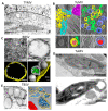
Figure 1
Figure 1
Virus-induced membranous structures derived from organellar membranes. (A) Electron micrograph of an ultrathin section of part of a chloroplast from TYMV-infected Chinese cabbage leaf cell. The arrow points to a vesicle in which an open channel is connecting the interior to the cytoplasm. Adapted from [69]. (B) Three-dimensional reconstruction of TuMV-induced membrane rearrangement. Overview of tomogram slice from TuMV-infected vascular parenchymal cell at the mid stage (upper left) and late stage (upper right) of infection. Three-dimensional model of a single-membrane tubule (SMT) with fibrillary material inside and with an adjacent intermediate tubular structure (lower left). Three-dimensional model of a double-membrane tubule (DMT) with a core of electron-dense materials inside (lower right). Yellow, SMTs; light red, electron-dense materials; green, intermediate tubular structures; light blue, outer membranes of DMTs; dark blue, inner membranes of DMTs; dark red, the electron-dense materials inside DMTs; sky blue, rough ER; magenta, cytoplasmic inclusion body; red arrow, connection between the rough ER membrane and an SMT. Adapted with permission from [47]. (C) Transmission electron microcopy (TEM) analysis and 3D model of BBSV-induced ER membrane rearrangements. (upper left) BBSV-induced vesiculation of the ER. Vesicle packets were observed in the aggregates of branched ER cisternae (*). Black arrowheads, ER membrane; Vi, virus crystal. (upper right) Tomographic slice of BBSV-induced vesicle packets and ER-derived spherules. I II and III represent different units of VPs. (Lower left) Three-dimensional model of BBSV-induced vesicle packets and ER-derived spherules. (Lower right) Enlargement of the connections between the spherules and the outer ER membrane. Gold, outer ER membrane; gray, BBSV-induced spherules; green, fibrillary materials inside the spherules. Adapted with permission from [45]. (D) TEM analysis of abnormally distorted chloroplasts in TuMV-infected leaves. (Upper panel) Chloroplast with membrane-bound extrusion. (Lower panel) Amoeboid shaping of chloroplasts, showing the extrusion of chloroplast encircling a large vesicle. Adapted with permission from [71]. (E) Electron analysis and 3D model of TBSV replication platform. (Left) Transmission electron micrograph of a TBSV-infected N. benthamiana leaf cell. Arrowheads, TBSV-induced individual peroxisomal multivesicular bodies (MVBs). Adapted with permission from [62]. (Right) Three-dimensional model of TBSV replication platform in yeast showing peripheral MVB with characteristic spherules. Yellow, peroxisome membrane; blue, spherules; red, mitochondrion. Adapted with permission from [46].
Figure 2
Figure 2
Schematic representation of the involvement of endomembrane system and cytoskeleton in viral replication and movement. Early in the infection process, translation of viral RNAs occurs at the rough ER (A). The viral membrane-targeting protein(s) target specific organelles such as the ER (Ba), peroxisomes (Bb), chloroplasts (Bc), multivesicular bodies (MVBs) (Bd) and vacuole (Be) and remodel organellar membranes to initiate the formation of viral replication organelles (VROs), and various virus-specific host proteins (host factors) are recruited to VROs. Dependent on virus, different cellular endomembrane-cytoskeleton network components, such as the ER, ER-Golgi, microtubules, actin filaments and endosomes, are hijacked to play roles in VRO biogenesis. VROs, viral movement protein(s) (MPs) and MP-associated viral movement complexes traffic to the plasmodesmata (PD) via the cytoskeleton machinery (mainly the actomyson mobility system) (Ca), secretory pathway (Cb), or the ER membrane (Cc). To pass through the PD, some viral MPs may form tubules within the PD to displace the desmotubule (Da) and others may increase the size exclusion limit (SEL) of PD (Db). Different viruses may pass through the PD via different MP-associated complexes: VRO (Dc), virion (Dd), or the viral ribonucleoprotein (vRNP) complex (De).

References

    1. Wolf Y.I., Kazlauskas D., Iranzo J., Lucia-Sanz A., Kuhn J.H., Krupovic M., Dolja V.V., Koonin E.V. Origins and Evolution of the Global RNA Virome. mBio. 2018;9:e02329-18. doi: 10.1128/mBio.02329-18. - DOI - PMC - PubMed
    1. Wang A. Dissecting the molecular network of virus-plant interactions: The complex roles of host factors. Annu. Rev. Phytopathol. 2015;53:45–66. doi: 10.1146/annurev-phyto-080614-120001. - DOI - PubMed
    1. Nagy P.D., Strating J.R., van Kuppeveld F.J. Building viral replication organelles: Close encounters of the membrane types. PLoS Pathog. 2016;12:e1005912. doi: 10.1371/journal.ppat.1005912. - DOI - PMC - PubMed
    1. Hyodo K., Okuno T. Hijacking of host cellular components as proviral factors by plant-infecting viruses. Adv. Virus Res. 2020;107:37–86. doi: 10.1016/bs.aivir.2020.04.002. - DOI - PubMed
    1. Nagy P.D., Feng Z. Tombusviruses orchestrate the host endomembrane system to create elaborate membranous replication organelles. Curr. Opin. Virol. 2021;48:30–41. doi: 10.1016/j.coviro.2021.03.007. - DOI - PubMed

Publication types

LinkOut - more resources