. 2023 Mar 14;15(3):745.
doi: 10.3390/v15030745.

Human Brain Microvascular Endothelial Cells Exposure to SARS-CoV-2 Leads to Inflammatory Activation through NF-κB Non-Canonical Pathway and Mitochondrial Remodeling

Affiliations

Human Brain Microvascular Endothelial Cells Exposure to SARS-CoV-2 Leads to Inflammatory Activation through NF-κB Non-Canonical Pathway and Mitochondrial Remodeling

Carolline Soares Motta et al. Viruses. .

Abstract

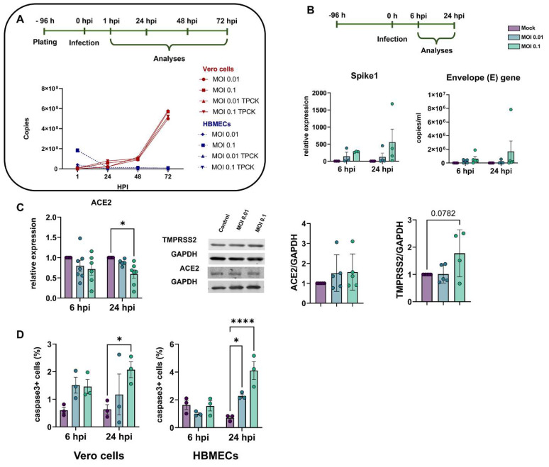
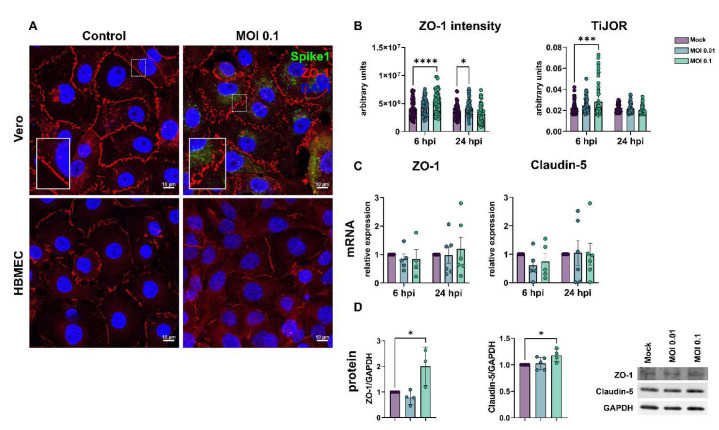
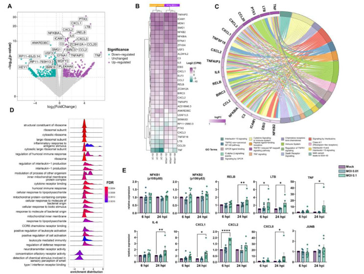
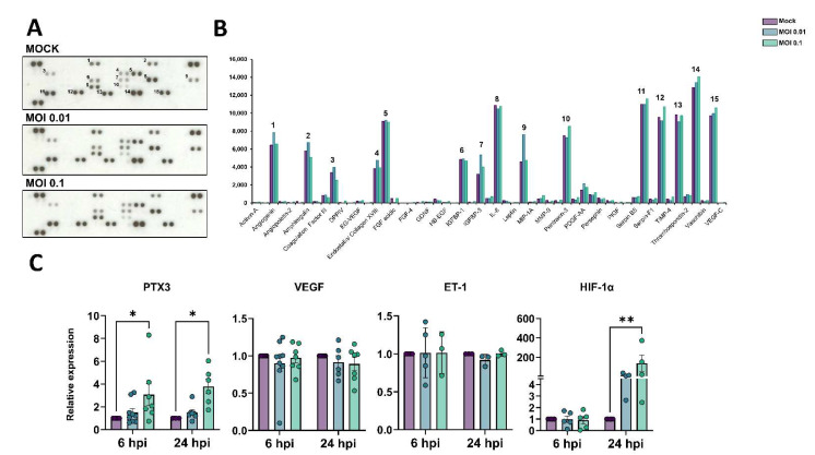
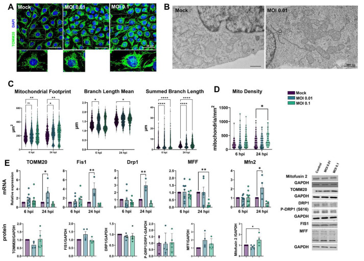
Neurological effects of COVID-19 and long-COVID-19, as well as neuroinvasion by SARS-CoV-2, still pose several questions and are of both clinical and scientific relevance. We described the cellular and molecular effects of the human brain microvascular endothelial cells (HBMECs) in vitro exposure by SARS-CoV-2 to understand the underlying mechanisms of viral transmigration through the blood-brain barrier. Despite the low to non-productive viral replication, SARS-CoV-2-exposed cultures displayed increased immunoreactivity for cleaved caspase-3, an indicator of apoptotic cell death, tight junction protein expression, and immunolocalization. Transcriptomic profiling of SARS-CoV-2-challenged cultures revealed endothelial activation via NF-κB non-canonical pathway, including RELB overexpression and mitochondrial dysfunction. Additionally, SARS-CoV-2 led to altered secretion of key angiogenic factors and to significant changes in mitochondrial dynamics, with increased mitofusin-2 expression and increased mitochondrial networks. Endothelial activation and remodeling can further contribute to neuroinflammatory processes and lead to further BBB permeability in COVID-19.

Keywords: COVID-19; NF-κB signaling pathway; blood–brain barrier; endothelial activation; mitochondrial dynamics.


Conflict of interest statement

The authors declare no conflict of interest.

Figures

Figure 1
Figure 1
Characterization of infectivity profile of HBMECs and Vero cells by SARS-CoV-2. (A) Cells were exposed to different MOIs of SARS-CoV-2 (variant D614G) and viral production, and release to supernatant was analyzed by RT-qPCR for Envelope (E) gene from 0 to 72 h post-infection (hpi). As compared to Vero cells, HBMECs showed a non-productive infection. (B) At desired time points (6 and 24 hpi), total RNA from HBMEC cultures and expression of Spike1 and E genes were analyzed by RT-qPCR. HBMECs exposed to MOI 0.1 showed an increase in the expression of these two transcripts at 24 hpi (p > 0.05). (C) Evaluation of SARS-CoV-2 receptors expression in HBMECs after SARS-CoV-2 challenge. ACE2 mRNA had a significant decrease at 24 hpi with the MOI 0.1, which did not translate to protein levels (right panel). TMPRSS2 had a slight increase in protein content at 24 hpi. (D) Exposure to SARS-CoV-2 increased immunoreactivity for cleaved caspase-3 in Vero cells and HBMECs at 24 hpi. *: p < 0.05; ****: p < 0.0001, Two-Way ANOVA with Bonferroni post-test of at least five independent experiments.
Figure 2
Figure 2
Effects of SARS-CoV-2 on tight junctional proteins in Vero and HBMECs. (A): Cells were stained for tight junction adaptor protein ZO-1 (red) and SARS-CoV-2 Spike1 (in green). ZO-1 was affected in infected cultures at 24 hpi, as shown in higher magnification in the insets. (B): Morphometrical analyses of ZO-1 fluorescence intensity and TiJOR in HBMECs (B) showed increased ZO-1 signal and TiJOR index 6 h after exposure to the MOI 0.01. Levels of mRNA encoding for ZO-1 and claudin-5 TJ genes remained unaffected by SARS-CoV-2 challenge (C), but a significant increase 6 h after exposure to MOI 0.1 was observed at the protein level (D). *: p < 0.05 one-way ANOVA with Bonferroni post-test (in (D)) or two-way ANOVA with Bonferroni post-test (in (B,C)); ***: p < 0.001; ****: p < 0.0001, Two-Way ANOVA with Bonferroni post-test. Each symbol in (C,D) corresponds to independent cultures, and in (B) corresponds to microscopic field from four independent cultures. Representative blots in (D) from 3–4 independent experiments.
Figure 3
Figure 3
Transcriptomic profiling of SARS-CoV-2 challenge on HBMECs. Cells were exposed to MOIs 0.01 and 0.1 and analyzed by RNA-Seq. (A) Volcano plot depicting the overall profile of differentially expressed genes in cultures after 24 h exposure to the MOI 0.1, with up-regulated genes shown in purple and down-regulated—in green. (B) Heatmap diagram depicting expression levels of the most significantly altered genes by MOI 0.1 (3 right columns), as compared to uninfected controls (3 left columns). (C) Cnetplot visualization of functional enrichment results with up-regulated genes, depicting the functional correlation of genes with the most significant GO terms. (D) Enrichment functional analysis of GO terms most affected by SARS-CoV-2 challenge in HBMECs indicates inflammatory endothelial activation, as well as mitochondrial dysfunction and ribosomal-related gene expression. (E) RT-qPCR validation of most significantly altered genes detected in the RNA-Seq indicates activation of non-canonical NF-κB pathway, with massive increase in TNF-α, lymphotoxin B (LTB, or TNF-C), and downstream target genes, such as IL-6, CXCL1, -2, and -8. NFKB1 (p105/p50) and NFKB2 (p100/p52), as well as JUNB, showed no significant alteration in SARS-CoV-2-exposed cultures. *: p < 0.05; **: p < 0.01; ****: p < 0.0001, two-way ANOVA with Bonferroni post-test of at least 5 independent experiments. MOI: multiplicity of infection; GO: gene ontology.
Figure 4
Figure 4
Production of angiogenic-related molecules is modulated by SARS-CoV-2 in HBMECs. (A) Conditioned medium from Mock and SARS-CoV-2-exposed HBMEC cultures (both with MOI 0.01 and 0.1) were analyzed via Proteome Profiler Human Angiogenic Antibody Array and detected by chemoluminescence, each protein detected in duplicated spots. (B) Densitometric analysis of membranes in (A) revealed the analytes with the strongest signal and which were affected by the SARS-CoV-2 challenge. Spots labelled 1-15 in (A) correspond to the analytes depicted in (B). (C) RT-qPCR analysis of angiogenesis-related genes in HBMECs revealed that PTX3 and HIF-1α were increased following SARS-CoV-2 exposure. *: p < 0.05; **: p < 0.01, two-way ANOVA with Bonferroni post-test of at least five independent experiments.
Figure 5
Figure 5
SARS-CoV-2-induced mitochondrial remodeling in HBMECs. Mitochondrial networks were detected by TOMM20 immunostaining (A) and TEM (B). MiNA analysis of TOMM20 revealed that exposure to SARS-CoV-2 induced an increase in mitochondrial footprint, branch length mean, and summed branch length (C). Mitochondrial density was calculated by TEM images (D), which also revealed increased fusion and association with multivesicular bodies (B). (E): RT-qPCR (upper panel) and western blotting (lower panel) analyses revealed that although fission-related genes (Fis1 and Drp1) were up-regulated in MOI 0.01-exposed cultures, only Mfn2 protein levels were increased in MOI 0.1-exposed cultures. TOMM20 protein levels also remained unaltered. *: p < 0.05; **: p < 0.01; ****: p < 0.0001, two-way ANOVA with Bonferroni post-test. Each symbol in graphs represents one cell (C), one mitochondrion (D), or one independent experiment (E). Bottom right panels depict blots from (E). Scale bars: 50 µm for (A) and 500 nm for (B).

Update of

References

    1. Centers for Disease Control and Prevention (CDC) COVID Data Tracker. [(accessed on 10 January 2023)]; Available online: http://www.cdc.gov.
    1. WHO Laboratory biosafety guidance related to coronavirus disease (COVID-19): Interim Guidance, 28 January 2021. 2021. [(accessed on 10 January 2023)]. Available online: https://www.who.int/publications/i/item/WHO-WPE-GIH-2021.1.
    1. Centers for Disease Control and Prevention Case-Surveillance. [(accessed on 10 January 2023)]; Available online: https://data.cdc.gov.
    1. Zhu N., Zhang D., Wang W., Li X., Yang B., Song J., Zhao X., Huang B., Shi W., Lu R., et al. China Novel Coronavirus Investigating and Research Team. A novel coronavirus from patients with pneumonia in China. N. Engl. J. Med. 2020;382:727–733. doi: 10.1056/NEJMoa2001017. - DOI - PMC - PubMed
    1. Wang K., Chen W., Zhang Z., Deng Y., Lian J.Q., Du P., Wei D., Zhang Y., Sun X.X., Gong L., et al. CD147-spike protein is a novel route for SARS-CoV-2 infection to host cells. Signal Transduct. Target. Ther. 2020;5:283. doi: 10.1038/s41392-020-00426-x. - DOI - PMC - PubMed

Publication types