[Preprint]. 2023 Mar 18:2023.03.17.533157.
doi: 10.1101/2023.03.17.533157.

Enveloped viruses pseudotyped with mammalian myogenic cell fusogens target skeletal muscle for gene delivery

Affiliations

Enveloped viruses pseudotyped with mammalian myogenic cell fusogens target skeletal muscle for gene delivery

Sajedah M Hindi et al. bioRxiv. .

Update in

Abstract

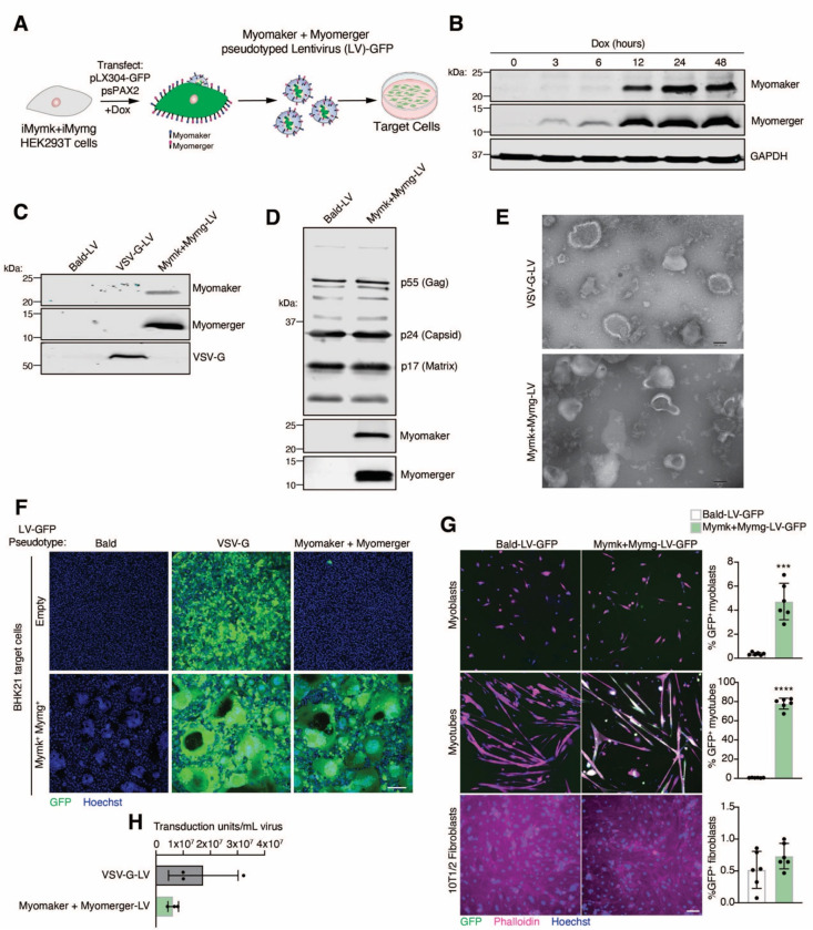
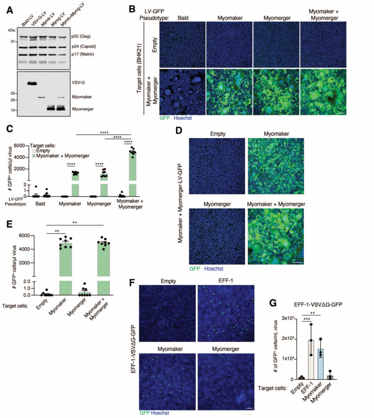
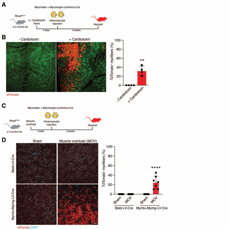
Entry of enveloped viruses into cells is mediated by fusogenic proteins that form a complex between membranes to drive rearrangements needed for fusion. Skeletal muscle development also requires membrane fusion events between progenitor cells to form multinucleated myofibers. Myomaker and Myomerger are muscle-specific cell fusogens, but do not structurally or functionally resemble classical viral fusogens. We asked if the muscle fusogens could functionally substitute for viral fusogens, despite their structural distinctiveness, and fuse viruses to cells. We report that engineering of Myomaker and Myomerger on the membrane of enveloped viruses leads to specific transduction of skeletal muscle. We also demonstrate that locally and systemically injected virions pseudotyped with the muscle fusogens can deliver micro-Dystrophin (μDys) to skeletal muscle of a mouse model of Duchenne muscular dystrophy. Through harnessing the intrinsic properties of myogenic membranes, we establish a platform for delivery of therapeutic material to skeletal muscle.


Conflict of interest statement

Declaration of interests

The authors declare competing financial interests: S.M.H. and D.P.M. have filed patent applications on this work through Cincinnati Children’s Hospital Medical Center.

Figures

Figure 1
Figure 1. Engineering and characterization of lentiviruses pseudotyped with Myomaker and Myomerger.
(A) Schematic showing production of lentiviruses (LV) from Myomaker- and Myomerger-expressing HEK293T cells. (B) Western blot for Myomaker, Myomerger, and GAPDH on HEK293T cell lysates after various times of doxycycline treatment. (C) Western blot on concentrated viral particles for Myomaker, Myomaker, and VSV-G. (D) Western blot on Bald-LVs and Mymk+Mymg-LVs for viral components and the muscle fusogens. (E) Representative images from transmission electron microscopy on VSV-G-LVs and Mymk+Mymg-LVs. Scale bar, 100 nm. (F) Representative images of empty and Mymk+Mymg target cells after application of Bald GFP-encoding LVs or LVs pseudotyped with VSV-G or Myomaker and Myomerger. Nuclei were stained with Hoechst. Scale bar, 100 μm. (G) Quantification of titers for VSV-G-LVs and Mymk+Mymk-LVs prior to concentration of supernatants. Each sample is an average of 3–4 replicates from independent viral preparations. Titers were determined on Mymk+Mymg BHK21 cells. (H) Representative images from cultures of differentiated myotubes, proliferating myoblasts, and fibroblasts that were treated with Bald-LV-GFP or Mymk+Mymg-LV-GFP. Cells were stained with Phalloidin and Hoechst. Scale bar, 100 μm. GFP+ cells were quantified for each cell type and histograms are shown on the right. Data are presented as mean ± standard deviation. Statistical test used was an unpaired t-test with Welch’s correction; ***p<0.001, ****p<0.0001.
Figure 2
Figure 2. The contribution of Myomaker and Myomerger on viral and cell membranes for pseudotyping.
(A) Western blot analysis of viral lysates for the indicated proteins. (B) Representative images for GFP+ cells, as an indication of fusion between the indicated lentiviral (LV) pseudotypes and either empty or Myomaker and Myomerger BHK21 target cells. Scale bar, 100 μm. (C) Quantification of viral infection from (B) expressed as # of GFP+ cells/μl of virus and assayed from viral supernatants. Pseudotypes with Myomaker alone or Myomerger alone exhibit similar transduction, while an increase is observed with pseudotypes with Myomaker and Myomerger together. (D) Lentiviruses pseudotyped with Myomaker and Myomerger were placed on BHK21 target cells expressing one or both fusogens. Representative images for GFP+ cells are shown. Scale bar, 100 μm. (E) Quantification of GFP+ cells from (D) as a metric for viral transduction. (F) VSVΔG-GFP pseudotyped with EFF-1 was applied to the indicated BHK21 target cells. EFF-1-VSVΔG-GFP exhibited similar transduction of target cells expressing EFF-1 or Myomaker. Scale bar, 100 μm. (G) Quantification of viral infection from (F) from viral supernatants (prior to concentration). Data are presented as mean ± standard deviation. Statistical tests used were (C) Two-way ANOVA with Tukey’s correction for multiple comparisons; (E) One-way ANOVA (Kruskal-Wallis test) with Dunn’s correction for multiple comparisons; (G) Repeated measures one-way ANOVA with Tukey’s correction for multiple comparisons; **p<0.01, ***p<0.001, ****p<0.0001.
Figure 3
Figure 3. Mymk+Mymg-LVs transduce activated skeletal muscle in vivo.
(A) Lentiviruses encoding for Cre recombinase and pseudotyped with Myomaker and Myomerger (Mymk+Mymg-LV-Cre) were produced and delivered to the tibialis anterior muscles of RosatdTom mice through direct intramuscular injection. Some tibialis anterior muscles were injured with cardiotoxin prior to receiving lentivirus. (B) Representative images are shown displaying tdTomato+ myofibers after injury and regeneration. Green is muscle autofluorescence. The percentage of tdTomato+ myofibers is shown (right panel). Scale bar, 100 μm. (C) Similar setup as in (A) except muscle overload, which causes hypertrophy, was performed and lentivirus was injected into the plantaris. (D) Representative images are shown for sham and overloaded muscles treated intramuscularly with Bald-LV-Cre or Mymk+Mymg-LV-Cre. Scale bar, 50 μm. Quantification of the percentage of tdTomato+ myofibers (right panel). Data are presented as mean ± standard deviation and combined from at least two independent lentiviral preparations. Statistical tests used were (B) unpaired t-test with Welch’s correction; (D) Two-way ANOVA with Tukey’s correction for multiple comparisons; **p< 0.01, ****p< 0.0001.
Figure 4
Figure 4. Myofibers and myogenic progenitors in dystrophic mice are transduced by lentiviruses pseudotyped with Myomaker and Myomerger.
(A) Mdx4cv; RosatdTom mice were used as recipients for Bald-LV-Cre or Mymk+Mymg-LV-Cre. Tibialis anterior muscles receiving lentiviruses through intramuscular injection were uninjured or previously injured with cardiotoxin. (B) Representative images showing tdTomato+ myofibers with and without cardiotoxin injury. Green is muscle autofluorescence. Scale bar, 100 μm. Quantification of the percentage of tdTomato+ myofibers (right panel). (C) Top panel displays experimental design for analysis of muscle progenitors. Middle panels show representative FACS plots and quantification for a7-Integrin+ myogenic cells (y-axis) and tdTomato+ cells (x-axis) from muscle that received Bald-LV-Cre or Mymk+Mymg-LV-Cre. Bottom panels show representative FACS plots and quantification for tdTomato+ non-myogenic interstitial cells. (D) Top panel is the experimental design using lentivirus encoding for luciferase that was pseudotyped with Bald, VSV-G, or Myomaker and Myomerger. After cardiotoxin injury, lentiviruses were injected intramuscularly and bioluminesence measured through IVIS imaging multiple time points after viral delivery. Representative pseudocolored images show a progressive increase in bioluminescence in the muscles treated with Myomaker and Myomerger-pseudotyped virus. Bottom panel is quantification of bioluminescence for muscles transduced with lentivirus containing Myomaker and Myomerger. Data are presented as mean ± standard deviation and combined from at least two independent lentiviral preparations. Statistical tests used were (B) Unpaired t-test with Welch’s correction; (C) unpaired t-test with Welch’s correction; (D) Friedman test with Dunn’s correction for multiple comparisons; **p<0.01, ***p<0.001, ***p< 0.0001.
Figure 5
Figure 5. Tropism of Myomaker+Myomerger lentiviruses is specific for skeletal muscle after systemic delivery.
(A) Schematic of experimental plan to assess transduction of lentiviruses delivered to mdx4cv; RosatdTom mice through three retro-orbital injections two weeks apart. (B) Representative images showing tdTomato+ myofibers from various skeletal muscle tissues after retro-orbital delivery of Cre-encoding lentiviruses pseudotyped with Bald or Myomaker and Myomerger. Scale bar, 100 μm. (C) Quantification of the percentage of tdTomato+ myofibers. (D) Representative images analyzing tdTomato+ cells from non-skeletal muscle tissues. No positive cells were detected. Quantification is shown on the right. Scale bar, 100 μm. (E) Experimental plan for systemic delivery of lentiviruses after muscle overload. (F) Representative images showing tdTomato+ myofibers in the plantaris. The percentage of tdTomato+ myofibers is also shown (right panel). Scale bar, 50 μm. Data are presented as mean ± standard deviation and combined from at least two independent lentiviral preparations. Statistical tests used were (C) Unpaired t-tests with Welch’s correction; (F) Two-way ANOVA with Tukey’s correction for multiple comparisons; *p<0.05, ****p<0.0001.
Figure 6
Figure 6. Lentiviruses pseudotyped with the muscle fusogens deliver therapeutic material to skeletal muscle.
(A) Schematic for experimental plan to assess transduction of Myomaker and Myomerger-pseudotyped lentiviruses packaged with μDystrophin (μDys) after intramuscular injection. (B) Immunostaining for dystrophin from tibialis anterior muscles from wild-type or mdx4cv mice that were injected with Bald- or Myomaker and Myomerger-lentiviruses encoding μDys. Scale bar, 100 μm. (C) Quantification of the percentage of μDys+ myofibers from (B). (D) Design to assess systemic delivery of lentiviruses in mdx4cv mice. (E) Immunostaining for dystrophin in mdx4cv diaphragms treated with Bald-LV-μDys or Mymk+Mymg-LV-μDys. Scale bar, 50 μm. Quantification of μDys+ myofibers is shown on the right. (F) Quantification of centrally nucleated myofibers in the diaphragms from wild-type mice and mice treated with Bald-LV-μDys or Mymk+Mymg-LV-μDys. (G) Masson’s trichrome staining of diaphragms treated systemically with the indicated lentivirus. Scale bar, 100 μm. Quantification of blue trichrome area is shown on the right. Data are presented as mean ± standard deviation and combined from at least two lentiviral preparations. Statistical tests used were (C), (E-F) One-way ANOVA with Tukey’s correction for multiple comparisons; ***p<0.001, ****p<0.0001.

References

    1. Avinoam O., Fridman K., Valansi C., Abutbul I., Zeev-Ben-Mordehai T., Maurer U.E., Sapir A., Danino D., Grunewald K., White J.M., et al. (2011). Conserved eukaryotic fusogens can fuse viral envelopes to cells. Science 332, 589–592. - PMC - PubMed
    1. Bi P., Ramirez-Martinez A., Li H., Cannavino J., McAnally J.R., Shelton J.M., Sanchez-Ortiz E., Bassel-Duby R., and Olson E.N. (2017). Control of muscle formation by the fusogenic micropeptide myomixer. Science 356, 323–327. - PMC - PubMed
    1. Brukman N.G., Uygur B., Podbilewicz B., and Chernomordik L.V. (2019). How cells fuse. J Cell Biol 218, 1436–1451. - PMC - PubMed
    1. Buchlis G., Podsakoff G.M., Radu A., Hawk S.M., Flake A.W., Mingozzi F., and High K.A. (2012). Factor IX expression in skeletal muscle of a severe hemophilia B patient 10 years after AAV-mediated gene transfer. Blood 119, 3038–3041. - PMC - PubMed
    1. Bulcha J.T., Wang Y., Ma H., Tai P.W.L., and Gao G. (2021). Viral vector platforms within the gene therapy landscape. Signal Transduct Target Ther 6, 53. - PMC - PubMed

Publication types