[Preprint]. 2023 Aug 28:2023.03.20.23287465.
doi: 10.1101/2023.03.20.23287465.

BRCA1 secondary splice-site mutations drive exon-skipping and PARP inhibitor resistance

Affiliations

BRCA1 secondary splice-site mutations drive exon-skipping and PARP inhibitor resistance

Ksenija Nesic et al. medRxiv. .

Update in

  • BRCA1 secondary splice-site mutations drive exon-skipping and PARP inhibitor resistance.
    Nesic K, Krais JJ, Wang Y, Vandenberg CJ, Patel P, Cai KQ, Kwan T, Lieschke E, Ho GY, Barker HE, Bedo J, Casadei S, Farrell A, Radke M, Shield-Artin K, Penington JS, Geissler F, Kyran E, Betsch R, Xu L, Zhang F, Dobrovic A, Olesen I, Kristeleit R, Oza A, McNeish I, Ratnayake G, Traficante N; Australian Ovarian Cancer Study; DeFazio A, Bowtell DDL, Harding TC, Lin K, Swisher EM, Kondrashova O, Scott CL, Johnson N, Wakefield MJ. Nesic K, et al. Mol Cancer. 2024 Aug 5;23(1):158. doi: 10.1186/s12943-024-02048-1. Mol Cancer. 2024. PMID: 39103848 Free PMC article.

Abstract

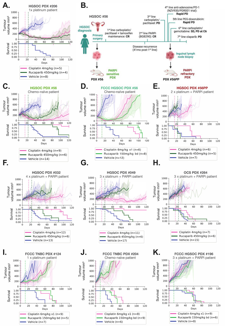
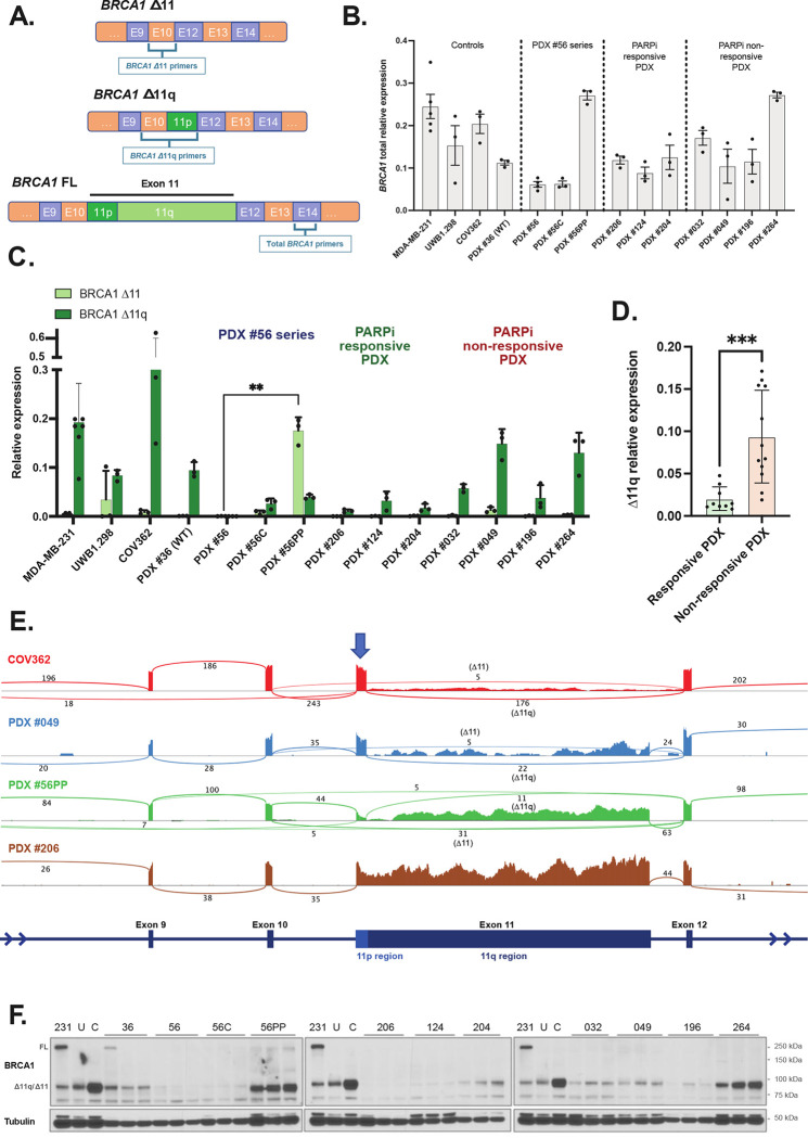
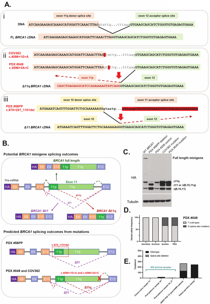
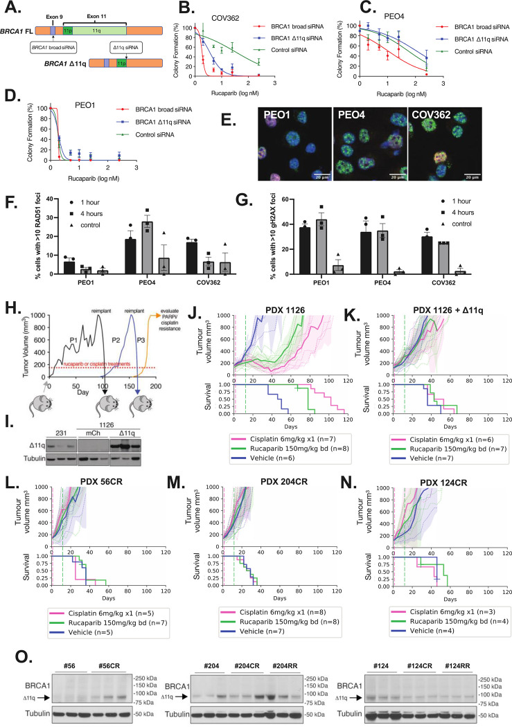
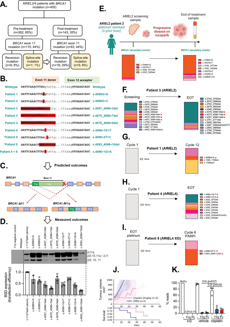
BRCA1 splice isoforms Δ11 and Δ11q can contribute to PARP inhibitor (PARPi) resistance by splicing-out the mutation-containing exon, producing truncated, partially-functional proteins. However, the clinical impact and underlying drivers of BRCA1 exon skipping remain undetermined. We analyzed nine ovarian and breast cancer patient derived xenografts (PDX) with BRCA1 exon 11 frameshift mutations for exon skipping and therapy response, including a matched PDX pair derived from a patient pre- and post-chemotherapy/PARPi. BRCA1 exon 11 skipping was elevated in PARPi resistant PDX tumors. Two independent PDX models acquired secondary BRCA1 splice site mutations (SSMs), predicted in silico to drive exon skipping. Predictions were confirmed using qRT-PCR, RNA sequencing, western blots and BRCA1 minigene modelling. SSMs were also enriched in post-PARPi ovarian cancer patient cohorts from the ARIEL2 and ARIEL4 clinical trials. We demonstrate that SSMs drive BRCA1 exon 11 skipping and PARPi resistance, and should be clinically monitored, along with frame-restoring secondary mutations.


Figures

Figure 1.
Figure 1.. PDX models with BRCA1 exon 11 mutations from PARPi-treated patients are PARPi-resistant.
HGSOC, OCS and TNBC PDX models with BRCA1 exon 11 mutations were treated with cisplatin or the PARPi rucaparib as indicated by hashed vertical lines, and tumor volume assessed twice weekly. Mean tumor volume (mm3) ± 95% CI (hashed lines are individual mice) and corresponding Kaplan–Meier survival analysis. Censored events are represented by crosses on Kaplan–Meier plot; n = individual mice. Detailed patient clinical data can be found in Supplementary table 1. Details of time to harvest, progression and treatment P values for each PDX can be found in Supplementary Table 2. (A) HGSOC PDX #206 had a complete response to both cisplatin and rucaparib (B) Patient #56 timeline, showing generation of the matched HGSOC PDX #56 (chemo-naïve) and #56PP (post-chemotherapy/PARPi patient). Created with BioRender.com. CR = Complete response; PD = Progressive disease; SD = Stable disease; C6 = cycle 6. (C) In vivo treatment data for HGSOC PDX #56 (*previously published (7)). (D) The FCCC derivative of PDX #56 was classified as sensitive to cisplatin and rucaparib, correlating with responses observed for the original lineage treated at WEHI (Figure 1B and Table S2). (E) HGSOC PDX #56PP was derived from patient #56 following multiple lines of therapy, including PARP inhibitor, and was refractory to both cisplatin and rucaparib. (F) HGSOC PDX #032 had progressive disease on rucaparib and stable disease on cisplatin. (G) HGSOC PDX #049 had a partial response to cisplatin and stable disease following rucaparib. (H) OCS PDX #264 had progressive disease on cisplatin and rucaparib. (I) TNBC PDX #124 was classified as sensitive to cisplatin and rucaparib, (J) as was TNBC PDX #204. (K) HGSOC PDX #196 was resistant to both cisplatin and rucaparib.
Figure 2.
Figure 2.. High BRCA1 Δ11q/Δ11 isoform expression observed in PARPi-resistant PDX models.
(A) Schematic for design of quantitative two-step PCR (qRT-PCR) assays. (B) Relative total BRCA1 expression for each PDX (mean ± SD). Δ11q-high cell line MDA-MB-231 (11) was included as a technical control on each qRT-PCR plate, while UWB1.289 and COV362 were included as additional cell line controls. PDX #36 was included as a Δ11q-high PDX control. (C) Relative Δ11 and Δ11q expression for each PDX (mean ± SD). PDX #56PP had the highest Δ11 expression relative to other PDX (P=0.0079 compared to matched PDX #56), while PDX #049 and #264 had the highest Δ11q levels relative to other PDX (classifications in Supp. Tables 3 and 4). (D) Δ11q levels in PARPi responsive PDX models (<2 prior lines of platinum in patient) were lower than levels in non-responsive PDX models (≥2 prior lines of platinum + PARPi) (P=0.0007). (E) RNA sequencing results for a subset of models with either high Δ11q (cell line COV362, PDX #049), high Δ11 (PDX #56PP) or low Δ11/Δ11q (#206) and presented as sashimi plots. BRCA1 exon regions are displayed at bottom of plot, and sequencing read coverage across each exon is represented as a histogram above. The lines connecting exons represent splicing events detected, and the numbers indicate the number of reads assigned to a given event. The blue arrow indicates the exon 11p region, which is retained in the Δ11q isoform. (F) Lysates from nuclear extracts from 3 independent tumors were probed for BRCA1 expression by immunoblotting. Bands at the anticipated sizes for full length (FL) BRCA1 and the Δ11/Δ11q isoforms are marked. Tubulin immunoblotting is included as a loading control. Gels were run simultaneously with cell line lysates included as controls for each gel. MDA-MB-231 (231) cells are a BRCA1 wild-type control with full-length (FL) and Δ11q expression, UWB1.289 (U) and COV362 (C) cells with exon 11 variants and Δ11/Δ11q expression. Statistical comparisons were made using an unpaired t-test with Welch’s correction.
Figure 3.
Figure 3.. Secondary splice site mutations drive alternative BRCA1 isoform expression.
(A) Schematic in section i) shows the BRCA1 exon 11q donor splice site and its interaction with the exon 12 acceptor splice site under wildtype conditions with no exon skipping. When the donor site is disrupted by a mutation (section ii), like in PDX #049 and COV362, the exon 11p donor site can be used preferentially, thus leading to 11q region skipping. In PDX #56PP the exon 11 acceptor site is disrupted by a large deletion, thus the next acceptor site for exon 12 may be used instead, leading to full exon 11 skipping (section iii). (B) This schematic shows the BRCA1 mini-gene design, and splicing outcomes predicted for each secondary splice site mutation found in PDX and cell line models. (C) Splicing predictions for each secondary splice-site mutation modelled by the mini-gene were confirmed by immunoblotting for the HA tag. The PDX #56PP deletion was confirmed to drive high Δ11 and potentially also isoform Δ(9,10,11q) expression. COV362 and PDX #049 secondary splice site mutations were confirmed to drive high Δ11q relative to the wild-type (WT) BRCA1 control and the primary deleterious BRCA1 mutation found in PDX #206. (D) Archival material (pre-PARPi) for patient #049 was screened for the secondary splice site mutation found in PDX #049. While the ascites from which the PDX was derived harboured the splice site mutation, the archival sample did not. (E) Archival material from the original debulking surgery (1C), or in a sample collected after 1st line platinum therapy (3A and a second DNA extraction 3A_21), for PDX #56PP was tested for the secondary splice site deletion found in the PDX using highly sensitive droplet digital PCR. While no deletion was detected in archival samples, poor droplet amplification in these samples limits interpretation. T2 = transplant/passage 2 PDX aliquot.
Figure 4.
Figure 4.. BRCA1 Δ11q isoform expression drives PARPi resistance in cell line and PDX
(A-D) COV362 was sensitised to PARPi rucaparib following siRNA knockdown of either Δ11q specifically, or broad knockdown BRCA1, with schematic of siRNA design shown in (A). Mean ± SEM are shown for three independent colony formation experiments. Knockdown was confirmed by qRT-PCR (Supp Figure S6). Controls are PEO1 (HR deficient) and PEO4 (HR proficient). (E-F) COV362 demonstrated some capacity to form RAD51 foci relative to control 1 hour after DNA damage (10 Gy irradiation; not significant; P value = 0.153). (G) DNA damage following irradiation was confirmed by gH2AX foci (mean ± SEM percent of geminin positive cells with ≥10 RAD51 or gH2AX foci). (H) PDX #1126 (BRCA1 exon 13 mutation) tumors expressing either mCherry or BRCA1-Δ11q were treated with cisplatin or rucaparib. Treated tumors were transplanted into recipient mice for further treatment (Supp Figure S10B–C). (I) BRCA1-Δ11q protein expression was assessed in PDX #1126 infected with BRCA1-Δ11q or mCherry encoded lentivirus following in vivo selection with cisplatin. MDA-MB-231 (231) lysate was included as a control. (J) Cisplatin, rucaparib and vehicle responses were assessed in PDX #1126. Treatment end = vertical hashed lines. Mean tumor volume (mm3) ± 95% CI (hashed lines are individual mice) and corresponding Kaplan–Meier survival analysis. Censored events = crosses on Kaplan–Meier plot; n = individual mice. (K) Cisplatin and rucaparib responses for PDX #1126 tumors with ectopic BRCA1-Δ11q expression following in vivo selection for cisplatin resistance. (L) PDX #56 was subjected to in vivo selection for cisplatin resistance as described in (H). Cisplatin and rucaparib responses following 3 passages of cisplatin re-treatment of PDX #56 (PDX #56CR). (M) Cisplatin and rucaparib resistant PDX #204 (PDX #204CR) were obtained following 3 passages of cisplatin re-treatments of PDX #204. Resistance was similarly obtained using rucaparib re-treatments (Supp Figure S10E). (N) Cisplatin resistant PDX #124 (PDX #124CR) was obtained following 4 passages of cisplatin re-treatment. Resistance via rucaparib re-treatments was also achieved (Supp Figure S10F). (O) Three independent tumors per PDX lineage were probed for BRCA1 expression by immunoblotting. Bands at the anticipated sizes for full length (FL) BRCA1 and the Δ11/Δ11q isoforms are marked. Tubulin immunoblotting is included as a loading control. Corresponding mRNA analysis is presented in Supp Figure S10E.
Figure 5.
Figure 5.. BRCA1 secondary splice-site mutations are enriched in ARIEL2/4 clinical trial patient samples following PARPi treatment.
(A) BRCA1 secondary splice-site mutations increased form 1% (pre-PARPi) to 8% (post-PARPi) in patient tumor/plasma samples from the ARIEL2 and ARIEL4 clinical trials. (B) The BRCA1 (NM_007294.4) exon 11 donor splice-site mutations identified in these patients and the DNA sequence context are presented. *Patient 5 is an ARIEL4 chemotherapy to PARPi cross-over (XO) arm patient, not included in part A (n=5). (C-D) The predicted outcomes based on these disruptions of the exon 11 donor splice site (detailed in Supp Table 9) were confirmed for most mutations (D) using the previously described BRCA1 minigene system (11). Mutations driving lower levels of Δ11/Δ11q also had a reduced minigene transfection efficiencies relative to other samples. (E) Summary of BRCA1 secondary events detected in Patient 2 (ARIEL2) before and after PARPi therapy, and their relative proportions in each sample (by colour). (F) A number of BRCA1 secondary events were also detected in the screening biopsy for platinum and rucaparib resistant Patient 3 (ARIEL2) prior to PARPi, and the number increased at end of rucaparib treatment (EOT). (G) In contrast, platinum resistant (4 prior lines of platinum) Patient 1 (ARIEL2) had no secondary BRCA1 events detected at first cycle of rucaparib, and had stable disease (SD) on treatment. Three secondary events were detected at cycle 12 of treatment, including a splice-site mutation c.4096G>A. (H) Patient 4 (ARIEL4) was partially platinum sensitive (2 prior lines) with no secondary BRCA1 events detected at cycle 1 of rucaparib. The EOT plasma sample was positive for multiple reversion events and two splice site mutations (4096+1G>T and 4096+1G>A) confirmed by minigene to alter splicing (D). (I) Patient 5 (ARIEL4) was platinum resistant and was enrolled in the chemotherapy arm of ARIEL4. They then crossed over to receive rucaparib treatment where they had stable disease. There were no secondary events detected prior to starting rucaparib, but at cycle 6 there were two BRCA1 splice-site mutations detected (c.4096+2T>C and c.4093_4096+10del), without other reversion events. c.4096+2T>C was found to drive alternative BRCA1 splicing (D). Red stars (*) indicate SSM’s detected in patient examples (E-I). (J) SUM149 cells expressing BRCA1-Δ11q (11q) or full length BRCA1 (FL) were mixed equally (0.1%) with a BRCA1-null (−) SUM149 derivative cell line (99.8%) (11). These cell mixtures were injected into mice and treated with vehicle (DPBS) or 6mg/kg cisplatin. Mean tumor volume (mm3) ± 95% CI (hashed lines are individual mice) and corresponding Kaplan– Meier survival analysis. Censored events = crosses on Kaplan–Meier plot; n = individual mice. (K) Targeted sequencing was used to measure the representation of each SUM149 derivative in tumors from (J) and from the cell mixture prior to implantation (t= 0). 11q representation increased 9.2-fold (P = 0.07) while FL representation increased 5.2-fold (P= 0.03) in cisplatin treated tumors compared to vehicle treated tumors. Each datapoint indicates read percentage for independent tumors and P value shown from unpaired t-tests for the indicated comparisons.

References

    1. Moore K, Colombo N, Scambia G, Kim BG, Oaknin A, Friedlander M, et al. Maintenance Olaparib in Patients with Newly Diagnosed Advanced Ovarian Cancer. N Engl J Med 2018;379(26):2495–505 doi 10.1056/NEJMoa1810858. - DOI - PubMed
    1. González-Martín A, Pothuri B, Vergote I, DePont Christensen R, Graybill W, Mirza MR, et al. Niraparib in Patients with Newly Diagnosed Advanced Ovarian Cancer. New England Journal of Medicine 2019;381(25):2391–402 doi 10.1056/NEJMoa1910962. - DOI - PubMed
    1. Ray-Coquard I, Pautier P, Pignata S, Pérol D, González-Martín A, Berger R, et al. Olaparib plus Bevacizumab as First-Line Maintenance in Ovarian Cancer. New England Journal of Medicine 2019;381(25):2416–28. - PubMed
    1. Sakai W, Swisher EM, Karlan BY, Agarwal MK, Higgins J, Friedman C, et al. Secondary mutations as a mechanism of cisplatin resistance in BRCA2-mutated cancers. Nature 2008;451(7182):1116–20 doi 10.1038/nature06633. - DOI - PMC - PubMed
    1. Edwards SL, Brough R, Lord CJ, Natrajan R, Vatcheva R, Levine DA, et al. Resistance to therapy caused by intragenic deletion in BRCA2. Nature 2008;451(7182):1111–5 doi 10.1038/nature06548. - DOI - PubMed

Publication types