[Preprint]. 2023 Mar 21:rs.3.rs-1812836.
doi: 10.21203/rs.3.rs-1812836/v1.

Type I interferon-dependent IFIT3 signaling is critical for viral clearance in airway neutrophils

Affiliations

Type I interferon-dependent IFIT3 signaling is critical for viral clearance in airway neutrophils

Camilla Margaroli et al. Res Sq. .

Update in

Abstract

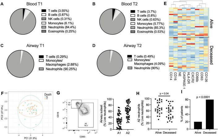
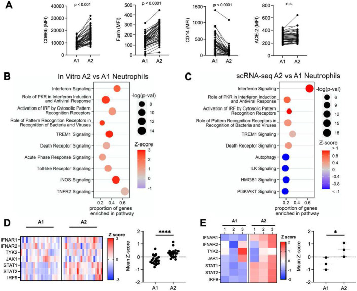
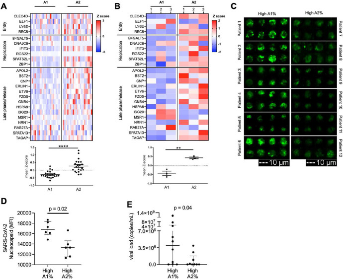
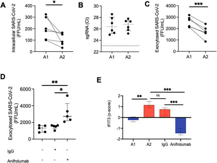
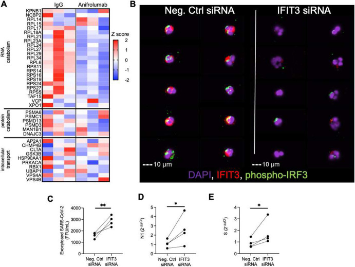
Neutrophilic inflammation characterizes several respiratory viral infections including COVID-19-related ARDS, although its contribution to disease pathogenesis remains poorly understood. Here, we identified two neutrophil subpopulations (A1 and A2) in the airway compartment of 52 severe COVID-19 subjects, where loss of the A2 subset correlated with increased viral burden and reduced 30-days survival. A2 neutrophils showcased a discrete antiviral response with an increased interferon signature. Blockade of type I interferon attenuated viral clearance in A2 neutrophils and downregulated IFIT3 and key catabolic genes, demonstrating direct antiviral neutrophil function. Knockdown of IFIT3 in A2 neutrophils led to loss of IRF3 phosphorylation with consequent reduced viral catabolism, providing the first discrete mechanism of type I interferon signaling in neutrophils. The identification of this novel neutrophil phenotype and its association with severe COVID-19 outcomes emphasizes its likely importance in other respiratory viral infections and potential for new therapeutic approaches in viral illness.


Conflict of interest statement

Conflict of interest: The authors have declared that they have no conflict of interest exists.

Declaration of Interests: The authors declare no competing interests.

Figures

Figure 1.
Figure 1.. Airway neutrophil subsets associate with survival.
Blood and airway immune cell frequencies (live and CD45+) and profiles were determined by flow cytometry. COVID-19 patients displayed blood neutrophilia (A) upon ICU admission (T1, n = 52) (Normal neutrophil frequencies: 40-65%). These profiles were maintained at time point 2 (T2, n = 28) (B). Airway immune cell frequencies in mBAL displayed marked neutrophil infiltration, which was maintained through both time points (C-D). (E) No significant difference was observed between surviving and deceased patient for individual surface markers, or as a combined profile by principal component analysis (F). (G) Presence of specific neutrophil subsets, including airway neutrophils profiles matching the A1 and A2 populations. (H) A2 neutrophil frequency at time of admission discriminated 30-day mortality (Alive =28 , Deceased =24). (I) Low frequencies of the A2 population correlated with mortality (Alive =3 , Deceased =10). Results are shown as median and interquartile range in G and H. Statistical analysis was performed using the unpaired t-test upon normality testing and the Fisher’s exact test for unpaired analysis.
Figure 2.
Figure 2.. A2 neutrophils show antiviral gene signatures.
(A) Airway neutrophils were profiled by flow cytometry at time point 1 (n = 52). A1 and A2 neutrophils expression of surface CD66b, CD14, furin and ACE-2 (MFI: median fluorescence intensity). (B) Pathway analysis for genes enriched in A2 neutrophils generated in vitro (n=3 donors). (C) Pathway analysis for genes enriched in A2 vs A1 BAL neutrophils from scRNA-seq (n=21 patients). (D) Type I interferon pathway gene expression for A2 vs A1 BAL neutrophils from scRNA-seq with mean z-score (n=21 patients). (E) Type I interferon pathway gene expression for A2 vs A1 neutrophils generated ex vivo mean z-score (n=3 per group). Data are shown as median and interquartile range. Statistical analysis was performed using Wilcoxon matched-pair signed rank’s test for paired analysis, Wilcoxon rank-sum test for unpaired analysis. * p<0.05; **** p<0.0001.
Figure 3.
Figure 3.. A2 neutrophils show differential anti-SARS-CoV-2 responses.
(A) Expression analysis of genes implicated in SARS-CoV-2 intracellular antiviral response with mean z-score. Data were obtained from sc-RNA-seq and each column represents a patient (n=21 per group). (B) A1 and A2 neutrophils generated using an in vitro transmigration model showed differential gene expression for SARS-CoV-2 intracellular antiviral response. (C) Airway neutrophils from a subset of patients with high A1 or high A2 frequencies (n=6 per group) were stained for SARS-CoV-2 nucleocapsid (green) and acquired by image cytometry (see Fig. S3). (D) Patients with high A1% showed increased presence of intracellular SARS-CoV-2 in airway neutrophils. (E) Patients with high A1% showed increased presence of extracellular SARS-CoV-2 in the mBAL supernatant (n=19 patients). Results are shown as median and interquartile range. Statistical analysis was performed using Wilcoxon matched-pair signed rank’s test for paired analysis or the Wilcoxon rank-sum test for unpaired analysis.
Fig. 4.
Fig. 4.. A2 neutrophils contribute to clearance of SARS-CoV-2.
(A) A1 and A2 neutrophils incubated with SARS-CoV-2 (MOI = 1), with A2 neutrophils had reduced intracellular virions of infectious SARS-CoV-2 (n=6 neutrophil donors). (B) Subgenomic RNA (sgRNA) detection by RT-PCR of SARS-CoV-2 in A1 and A2 neutrophils (dotted line represents positive control, n = 6 neutrophil donors). (C) A1 and A2 neutrophils show differential exocytosis of infectious SARS-CoV-2 (n=6 neutrophil donors). (D) Type I interferon blockade with Anifrolumab increased exocytosis of infectious SARS-CoV-2 in A2 neutrophils (n=5 neutrophil donors) compared to IgG control or media alone. (E) IFIT3 expression by RNASeq. (FFU = foci forming units). Data are shown as median and interquartile range. Statistical analysis was performed using Wilcoxon matched-pair signed rank’s test or ANOVA with Tukey’s test for multiple comparisons. * p<0.05; ** p<0.01; *** p<0.001; n.s.: not significant.
Fig. 5
Fig. 5. IFIT3 signaling modulates viral clearance in A2 neutrophils
(A) Expression of genes in the macromolecule catabolic processes (GO: 0009057). (B) Image cytometry analysis of airway neutrophils for IFIT3 (red) and phospho-IRF3 (green) expression. (DAPI, purple). (C) IFIT3 knockdown increased exocytosis of infectious SARS-CoV-2 in A2 neutrophils (n=4 neutrophil donors). (D-E) IFIT3 knockdown modulates viral RNA catabolism (N1 and Spike protein RNA) in A2 neutrophils (n=4 neutrophil donors). (FFU = foci forming units). Data are shown as median and interquartile range. Statistical analysis was performed using Wilcoxon matched-pair signed rank’s test, * p<0.05; ** p<0.01.

References

    1. Collaborators, G.B.D.L.R.I. Estimates of the global, regional, and national morbidity, mortality, and aetiologies of lower respiratory infections in 195 countries, 1990-2016: a systematic analysis for the Global Burden of Disease Study 2016. Lancet Infect Dis 18, 1191–1210 (2018). - PMC - PubMed
    1. Hariri L. & Hardin C.C. Covid-19, Angiogenesis, and ARDS Endotypes. N Engl J Med 383, 182–183 (2020). - PubMed
    1. Johansson C. & Kirsebom F.C.M. Neutrophils in respiratory viral infections. Mucosal Immunol 14, 815–827 (2021). - PMC - PubMed
    1. Liu J., et al. Neutrophil-to-lymphocyte ratio predicts critical illness patients with 2019 coronavirus disease in the early stage. J Transl Med 18, 206 (2020). - PMC - PubMed
    1. Mathew D., et al. Deep immune profiling of COVID-19 patients reveals distinct immunotypes with therapeutic implications. Science (2020). - PMC - PubMed

Publication types

LinkOut - more resources