[Preprint]. 2023 Mar 22:2023.03.20.530986.
doi: 10.1101/2023.03.20.530986.

Structural basis for functional properties of cytochrome c oxidase

Affiliations

Structural basis for functional properties of cytochrome c oxidase

Izumi Ishigami et al. bioRxiv. .

Abstract

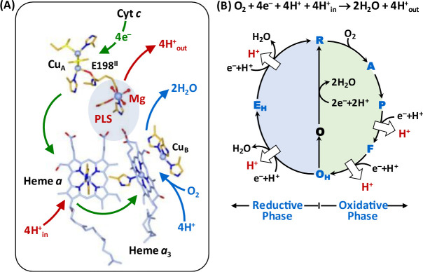
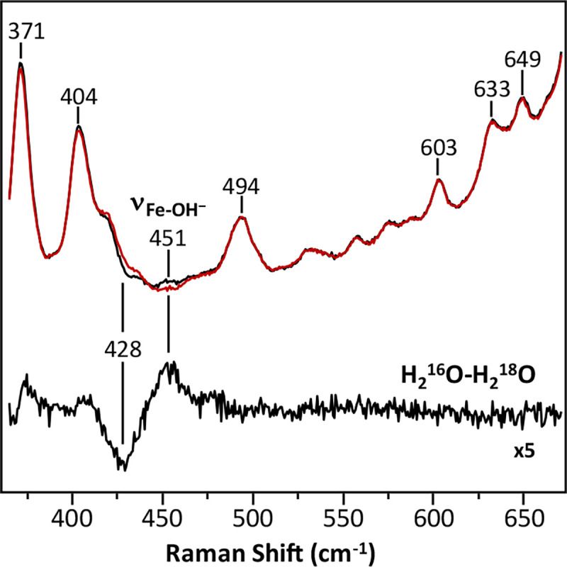
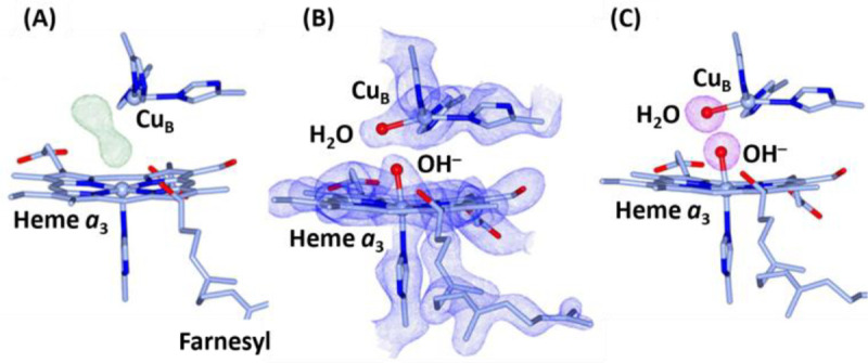
Cytochrome c oxidase (CcO) is an essential enzyme in mitochondrial and bacterial respiration. It catalyzes the four-electron reduction of molecular oxygen to water and harnesses the chemical energy to translocate four protons across biological membranes, thereby establishing the proton gradient required for ATP synthesis1. The full turnover of the CcO reaction involves an oxidative phase, in which the reduced enzyme (R) is oxidized by molecular oxygen to the metastable oxidized OH state, and a reductive phase, in which OH is reduced back to the R state. During each of the two phases, two protons are translocated across the membranes2. However, if OH is allowed to relax to the resting oxidized state (O), a redox equivalent to OH, its subsequent reduction to R is incapable of driving proton translocation2,3. How the O state structurally differs from OH remains an enigma in modern bioenergetics. Here, with resonance Raman spectroscopy and serial femtosecond X-ray crystallography (SFX)4, we show that the heme a3 iron and CuB in the active site of the O state, like those in the OH state5,6, are coordinated by a hydroxide ion and a water molecule, respectively. However, Y244, a residue covalently linked to one of the three CuB ligands and critical for the oxygen reduction chemistry, is in the neutral protonated form, which distinguishes O from OH, where Y244 is in the deprotonated tyrosinate form. These structural characteristics of O provide new insights into the proton translocation mechanism of CcO.


Figures

Fig. 1.
Fig. 1.. Oxygen reduction reaction catalyzed by bCcO.
(A) Schematic illustration of the four redox active metal centers in bCcO and the electron and proton transfers associated with the O2 reduction reaction. The entry of O2 and four substrate protons into the BNC, as well as the release of the product water molecules out of it, are indicated by the blue arrows. The associated entry of four electrons into the BNC and the translocation of four pumped protons across the membrane are indicated by the green and red arrows, respectively. The putative PLS between heme a3 and the Mg center is highlighted by the light blue background. (B) The overall O2 reduction reaction and the associated mechanism. The P intermediate is a general term for the PM and PR intermediates. The entry of the electrons and substrate protons into the BNC and the release of the product water molecules are indicated in each step of the reaction as described in the main text. The coupled proton translocation reactions are indicated by the white arrows. If the OH intermediate produced at the end of the oxidative phase is allowed to relax to the resting O state, its reduction to R does not support proton translocation.
Fig. 2.
Fig. 2.. Resonance Raman spectrum of the O state of bCcO in H216O (black) versus H218O (red).
The H216O-H218O difference spectrum (expanded by 5 fold) is shown at the bottom. The oxygen sensitive mode in the H216O sample centered at 451 cm−1 that shifted to 428 cm−1 in the H218O sample is assigned to the νFe-OH mode.
Fig. 3.
Fig. 3.. Electron density maps of the BNC in the O state of bCcO.
(A) The FO-FC electron density map (contoured at 7.0 σ) showing clear 2-lobe electron density associated with the BNC ligands. (B) The 2FO-FC electron density map (contoured at 2.5 σ) obtained with the electron density modeled with a hydroxide ion coordinated to the heme a3 iron and a water molecule coordinated to CuB. (C) The Polder map (contoured at 7.0 σ) associated with the BNC ligands.
Fig. 4.
Fig. 4.. Protonation state of Y244 in the PR (A) and O (B) states.
The post translationally modified Y244 forms a H-bonding network with the OH group of the farnesyl side chain of heme a3, a water molecule (W1) and T316. In the PR state, an additional water, W2, is recruited into the BNC to stabilize the tyrosinate configuration of Y244. This water is absent in the O state reported here, as evident in the 2FO-FC electron density map (contoured at 1.0 σ) shown in the lower inset, signifying that Y244 is in the neutral protonated state. For clarity the BNC ligands are not shown. The oxygen atom and hydrogen atoms are shown as red and white spheres.
Fig. 5.
Fig. 5.. Hypothesized ligand transformation in the BNC during the CcO reaction cycle.
The oxidative and reductive phase of the reaction cycle are highlighted with green and blue backgrounds, respectively. The white arrows indicate the proton translocation reactions associated with the reaction cycle. The lifetimes associated with the PRF, FOH and OHO transitions are indicated in red.
Fig. 6.
Fig. 6.. Functional role of Y244 during the CcO reaction cycle.
(A) During the active turnover, Y244 cycles between the neutral form (YH), tyrosyl radical form (Y) and deprotonated tyrosinate form (Y) as depicted by the blue cycle. In all the intermediate states active in proton translocation, Y244 is in the Y form (in red), which ensures the tight coupling of electron transfer (green arrows) and substrate proton transfer (blue arrows), via the D and K-channel into the BNC, to drive the proton translocation (red arrows) via the D or H channel from the N to P-side of the membrane. (B) The protonation of the tyrosinate to the YH form (red) in the O state disables the proton translocation upon its reduction to R.

References

    1. Wikstrom M., Krab K. & Sharma V. Oxygen Activation and Energy Conservation by Cytochrome c Oxidase. Chemical reviews 118, 2469–2490, doi:10.1021/acs.chemrev.7b00664 (2018). - DOI - PMC - PubMed
    1. Verkhovsky M. I., Jasaitis A., Verkhovskaya M. L., Morgan J. E. & Wikstrom M. Proton translocation by cytochrome c oxidase. Nature 400, 480–483, doi:10.1038/22813 (1999). - DOI - PubMed
    1. Bloch D. et al. The catalytic cycle of cytochrome c oxidase is not the sum of its two halves. Proceedings of the National Academy of Sciences of the United States of America 101, 529–533, doi:10.1073/pnas.0306036101 (2004). - DOI - PMC - PubMed
    1. Boutet S. et al. High-resolution protein structure determination by serial femtosecond crystallography. Science 337, 362–364, doi:10.1126/science.1217737 (2012). - DOI - PMC - PubMed
    1. Han S., Ching Y. C. & Rousseau D. L. Ferryl and hydroxy intermediates in the reaction of oxygen with reduced cytochrome c oxidase. Nature 348, 89–90, doi:10.1038/348089a0 (1990). - DOI - PubMed

Publication types