[Preprint]. 2023 Mar 24:2023.03.22.533831.
doi: 10.1101/2023.03.22.533831.

Genipin Crosslinks the Extracellular Matrix to Rescue Developmental and Degenerative Defects, and Accelerates Regeneration of Peripheral Neurons

Affiliations

Genipin Crosslinks the Extracellular Matrix to Rescue Developmental and Degenerative Defects, and Accelerates Regeneration of Peripheral Neurons

Kenyi Saito-Diaz et al. bioRxiv. .

Update in

Abstract

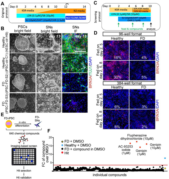
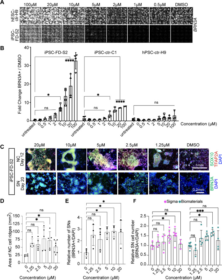
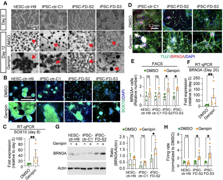
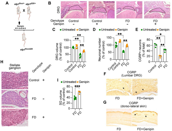
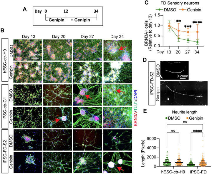
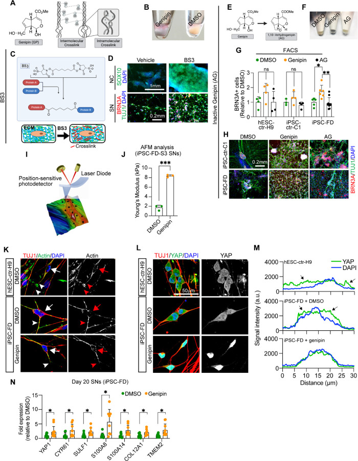
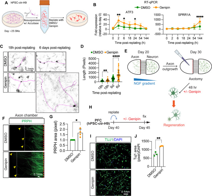
The peripheral nervous system (PNS) is essential for proper body function. A high percentage of the population suffer nerve degeneration or peripheral damage. For example, over 40% of patients with diabetes or undergoing chemotherapy develop peripheral neuropathies. Despite this, there are major gaps in the knowledge of human PNS development and therefore, there are no available treatments. Familial Dysautonomia (FD) is a devastating disorder that specifically affects the PNS making it an ideal model to study PNS dysfunction. FD is caused by a homozygous point mutation in ELP1 leading to developmental and degenerative defects in the sensory and autonomic lineages. We previously employed human pluripotent stem cells (hPSCs) to show that peripheral sensory neurons (SNs) are not generated efficiently and degenerate over time in FD. Here, we conducted a chemical screen to identify compounds able to rescue this SN differentiation inefficiency. We identified that genipin, a compound prescribed in Traditional Chinese Medicine for neurodegenerative disorders, restores neural crest and SN development in FD, both in the hPSC model and in a FD mouse model. Additionally, genipin prevented FD neuronal degeneration, suggesting that it could be offered to patients suffering from PNS neurodegenerative disorders. We found that genipin crosslinks the extracellular matrix, increases the stiffness of the ECM, reorganizes the actin cytoskeleton, and promotes transcription of YAP-dependent genes. Finally, we show that genipin enhances axon regeneration in an in vitro axotomy model in healthy sensory and sympathetic neurons (part of the PNS) and in prefrontal cortical neurons (part of the central nervous system, CNS). Our results suggest genipin can be used as a promising drug candidate for treatment of neurodevelopmental and neurodegenerative diseases, and as a enhancer of neuronal regeneration.


Conflict of interest statement

Competing interests: L.S. is a scientific co-founder and consultant and has received research support from BlueRock Therapeutics.

Figures

Figure 1.
Figure 1.. Chemical screen on FD sensory neurons.
A) Differentiation protocol adapted from Chambers et al., 2012. B) Bright filed imaging shows normal morphology of undifferentiated hPSCs in all lines (left column). SN differentiation is impaired in both FD lines, but normal in healthy control lines (right two columns). C) Differentiation protocol adapted to high-throughput screening conditions. D) The SN differentiation protocol is most efficient in 96 wells when the cells are fed three times. E) Cartoon of the screen set up. F) 640 compounds from the LOPAC chemical library (the first half) were screened. Controls included DMSO only wells, healthy hPSC-ctr-H9 with DMSO (blue dot = positive control), iPSC-FD-S2 iPSCs with DMSO (yellow dot=negative control). Hit compounds were called when the fold change (FC) over the average of all DMSO wells was above the average of all compounds plus 3 SDs (here 3.8, red dots). Every compound was screened at 1 mM and 10 mM. 16 images were taken for each well, all wells were imaged for the ratio of BRN3A+/DAPI+ staining.
Figure 2.
Figure 2.. Validation of the hit compound genipin.
A) Titration of genipin during the SN differentiation in healthy hPSC-ctr-H9 and iPSC-FD-S2 iPSCs. Differentiation protocol depicted in Fig. 1a was used. B) Quantification of genipin titration based on IF of BRN3A+ SNs. n=3–4 biological replicates. C) Titration of genipin in iPSC-FD-S2 cells during differentiation into SN-biased neural crest cells (top row) and SNs (bottom row). Cells were treated with indicated concentrations of genipin starting on day 2. Cells were then fixed on the indicated days and stained for SOX10 and TFAP2A (top) or BRN3A (bottom) and DAPI. D and E) Quantification of size of NC ridges and number of SNs upon genipin treatment. D) Area of ridges in c marked by DAPI staining (n=4–7 biological replicates) and E) number of BRN3A+ SNs in c were quantified (n=3–5 biological replicates). F) Genipin commercially obtained from both Sigma and Biomaterials rescues SN differentiation in iPSC-FD-S2. Cells were differentiated in the presence of genipin from either source starting on day 2. Cells were fixed on day 20 and stained for BRN3A and DAPI. n=3–7 biological replicates. All graphs show mean ± s.d. For B, D, E, and F, one-way ANOVA, *p<0.05, **p<0.005.
Figure 3.
Figure 3.. Genipin rescues neural crest and sensory neuron-related phenotypes in FD.
A) hPSC lines look normal at the pluripotent stage (day 0), but differentiation into NC cells is diminished in the FD lines (DMSO, dark ridges/red arrow indicate NC cells); this is rescued by genipin (10 µM) treatment. B and C) SOX10 expression is restored in NC cells upon genipin treatment. Cells were differentiated in the presence of genipin (10 µM) and fixed on day 12. B) Cells were stained for SOX10 and DAPI and analyzed by IF. n=5 biological replicates. C) Genipin increases SOX10 expression. RNA isolated from FD cells differentiated in the presence of genipin (10 µM) was analyzed by RT-qPCR. n=6 biological replicates. D-F) Genipin restores SN differentiation. iPSC-FD-S2 and iPSC-FD-S3 cells were treated with genipin (10 µM) and differentiated into SNs. RNA was isolated on day 20 or cells were fixed and stained using the indicated antibodies and analyzed by D) IF (n=5 biological replicates), E) intracellular flow cytometry (n=3–4 biological replicates), and F) RT-qPCR (n=6 biological replicates). G) Western blot analysis confirms the increase in SN production upon genipin treatment. Cells differentiated with genipin (10 µM) were lysed on day 20 and immunoblotted with the indicated antibodies (left) and quantified (right). n=3–4 biological replicates. H) Genipin increases firing rate of FD SNs. Cells were differentiated in the presence of genipin (10 µM) and firing rate was analyzed by MEA. n=4–6 biological replicates. In C, E, F, G, H, Two-tailed Student’s t-test. ns, non-significant, *p<0.05, **p<0.005. All graphs show mean ± s.d. Data from iPSC-FD-S2 and iPSC-FD-S3 are pooled as FD in C and F.
Figure 4.
Figure 4.
Genipin rescues FD peripheral deficits in vivo. A) Breeding and treatment schematic. B) Representative H&E-stained transverse sections through lumbar (L1) dorsal root ganglia (DRG) of untreated control, genipin-treated control, untreated FD, and genipin-treated FD E18.5 embryos, at their largest dimensions. C) Volumes of lumbar (L1) DRGs of untreated (n=6) and genipin-treated (n=5) controls, and untreated (n=6) and genipin-treated (n=6) FD E18.5 embryos, displayed as percentage of control. D) Neuronal counts of lumbar (L1) DRGs of untreated (n=6) and genipin-treated (n=5) controls, and untreated (n=6) and genipin-treated (n=6) FD E18.5 embryos, displayed as percentage of control. E) Percentage of CGRP-positive neurons in lumbar (L1) DRGs of control untreated (n=3), FD untreated (n=3), and genipin-treated FD (n=3). F) Representative images of transverse sections through lumbar (L1) DRGs of untreated FD and genipin-treated FD E18.5 embryos immunostained with CGRP. G) Representative images of transverse sections through dorso-lateral skin of untreated FD and genipin-treated FD E18.5 embryos immunostained with CGRP. Arrows show positive signal. H) Representative H&E-stained sections of stellate ganglia (SG) of genipin-treated control, untreated FD, and genipin-treated FD E18.5 embryos. I) Volumes of SG of untreated (n=6) and genipin-treated (n=3) FD E18.5 embryos, plotted as percentage of control. All graphs show mean ± s.d.. For C, D, E, one-way ANOVA followed by Tuckey HSD post hoc test. For I, two-tailed t-test. *p<0.05, **p<0.01, ***p<0.001
Figure 5.
Figure 5.. Genipin rescues sensory neuron degeneration in FD.
A) Schematic of survival assay. B) Healthy or FD cells were treated with genipin from day 12 on (when neurons are born) and monitored for survival for 21 days. Cells were fixed on the indicated days and stained for BRN3A, TUJ1 and DAPI. Arrows indicate healthy, ganglia-like SN clusters. C) Image quantification of B. n=8, Two-way ANOVA followed by Šídák multiple comparisons. **p<0.005, ***p<0.001, ****p<0.0001. D,E, Genipin increases neurite length and number in FD cells. D) Representative images of neurite length. E) Measurement of neurite length (number of pixels) of D. n=6–8 biological replicates, one-way ANOVA followed by Tukey’s multiple comparisons. ns, non-significant, ****p<0.0001. All graphs show mean ± s.d. Data from iPSC-FD-S2 and iPSC-FD-S3 are pooled as FD in C, and E.
Figure 6.
Figure 6.. Genipin rescues FD phenotypes via crosslinking of extracellular matrix proteins and activates.
A) Genipin chemical structure and schematic of intermolecular and intramolecular crosslinking. B) Treatment with genipin turns cells blue. C) Schematic of BS3 crosslinking action and its extracellular location. D) BS3 rescues the NC and SN differentiation defect in FD. FD cells were differentiated in the presence of DMP and fixed on day 12 (NC) and day 20 (SN). Following staining using the indicated antibodies. n=3–5 biological replicates. E-H) 1,10-anhydrogenipin (AG) does not rescue the SN defect in FD. E) AG chemical structure. F) AG does not turn cells blue. G, H) AG does not promote FD SN differentiation. Cells were differentiated into SNs in the presence of genipin or AG. SNs were fixed on day 20 and stained for BRN3A for intracellular flow cytometry analysis (G, n=6–8 biological replicates), or stained for BRN3A, TUJ1, and DAPI for IF (H, n=6–8 biological replicates). I, J) Genipin-mediated crosslinking increases ECM stiffness. I) AFM Experiment schematics. J) Genipin increases the Young’s modulus of SNs. iPSC-FD-S3 SNs were fixed on day 25 and analyzed by AFM (n=3 biological replicates). K-M) Genipin reorganizes the actin cytoskeleton and induces transcription of YAP-dependent genes. K) Genipin partially rescues the differences of actin expression pattern in healthy and FD SNs. Day 20 SNs were fixed and stained for the indicated antibodies. Images were obtained by confocal microscopy. Actin signal in the cell body (arrows) or the axons (arrowheads) are highlighted. L, M) YAP localization changes in the presence of genipin. Day 20 SNs were fixed and stained for the indicated antibodies. M) YAP and DAPI signal intensity from images on L) was measured and plotted n=6–7 biological replicates). Arrows indicate YAP signal outside of the nucleus (stained with DAPI). N) Expression of YAP-dependent genes. RNA from FD SNs treated with DMSO or Genipin was extracted on day 20 and gene expression was analyzed by RT-qPCR (n=7–9 biological replicates). All graphs show mean ± s.d.. For G, one-way ANOVA followed by Tukey’s multiple comparisons. For J, two-tailed t test with Welch’s correction. For N, two-tailed t test. ns, non-significant, *p<0.05, **p<0.005, ***p<0.001. In G, M, and L data from iPSC-FD-S2 and iPSC-FD-S3 are pooled as FD.
Figure 7.
Figure 7.
Genipin accelerates axon regeneration after injury in different neuronal types. A-D) Genipin enhances rengeneration of healthy SNs. A) Experiment schematics. B) Genipin increases expresison of injury-related genes. RNA from hPSC-ctr-H9 SNs was isolated at indicated times and gene expression was measured by RT-qPCR (n=4–7 biological replicates). C,D) Genipin increases axon length after injury. Day 25 SNs from hPSC-ctr-H9 cells were fixed at indicated times after replating in the presence of DMSO or Genipin, and stained for TUJ1. Axons were traced (magenta) and measurement were plotted in D (n>20 cells from 5 biological replicates). E-G) Genipin increases length and complexity of axons from sympathetic neurons. E) Experiment schematics. For details, see Methods. F,G) Axons from sympathetic neurons were removed. After 48 h treatment with Genipin, neurons were fixed and stained for PRPH. Axons were measured (arrowheads indicate length of axons) and the pixels with high PRPH signal were measuered and graphed in J (n=4 biological replicates). H-J) Genipin promotes axon regeneration in prefrontal cortical (PFC) neurons. H) Experiment schematics. For details, see Methods. I,J) hPSC-ctr-H9 PFC neurons reaplted and incuabted with Genipin for 5 days. Neurons were then fixed and stained for TUJ1 as shown in I. Measurement of the pixels with high TUJ1 signal were normalized to DAPI and plotted in J (n=3 biological replicates). All graphs show mean ± s.d. For B, two-way ANOVA followed by Šídák multiple comparisons. For D, G, and H, two-tailed t test. *p<0.05, **p<0.005, ***p<0.001, ****p<0.0001.

References

    1. Meng J., Zhang Q., Yang C., Xiao L., Xue Z., and Zhu J. (2019). Duloxetine, a Balanced Serotonin-Norepinephrine Reuptake Inhibitor, Improves Painful Chemotherapy-Induced Peripheral Neuropathy by Inhibiting Activation of p38 MAPK and NF-κB. Front Pharmacol 10, 365. 10.3389/fphar.2019.00365. - DOI - PMC - PubMed
    1. Norcliffe-Kaufmann L., Slaugenhaupt S.A., and Kaufmann H. (2017). Familial dysautonomia: History, genotype, phenotype and translational research. Prog Neurobiol 152, 131–148. 10.1016/j.pneurobio.2016.06.003. - DOI - PubMed
    1. O’Connor A.B. (2009). Neuropathic pain: quality-of-life impact, costs and cost effectiveness of therapy. Pharmacoeconomics 27, 95–112. 10.2165/00019053-200927020-00002. - DOI - PubMed
    1. Benam K.H., Dauth S., Hassell B., Herland A., Jain A., Jang K.-J., Karalis K., Kim H.J., MacQueen L., Mahmoodian R., et al. (2015). Engineered in vitro disease models. Annu Rev Pathol 10, 195–262. 10.1146/annurev-pathol-012414-040418. - DOI - PubMed
    1. Blumenfeld A., Slaugenhaupt S.A., Liebert C.B., Temper V., Maayan C., Gill S., Lucente D.E., Idelson M., MacCormack K., Monahan M.A., et al. (1999). Precise genetic mapping and haplotype analysis of the familial dysautonomia gene on human chromosome 9q31. Am J Hum Genet 64, 1110–1118. 10.1086/302339. - DOI - PMC - PubMed

Publication types