[Preprint]. 2023 Mar 16:rs.3.rs-2675436.
doi: 10.21203/rs.3.rs-2675436/v1.

The Genetic and Epigenetic Features of Bilateral Wilms Tumor Predisposition: A Report from the Children's Oncology Group AREN18B5-Q Study

Affiliations

The Genetic and Epigenetic Features of Bilateral Wilms Tumor Predisposition: A Report from the Children's Oncology Group AREN18B5-Q Study

Andrew J Murphy et al. Res Sq. .

Update in

Abstract

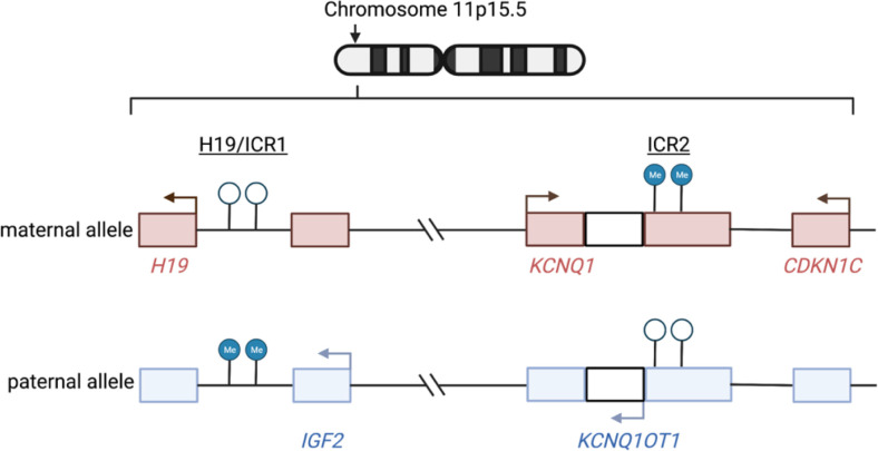
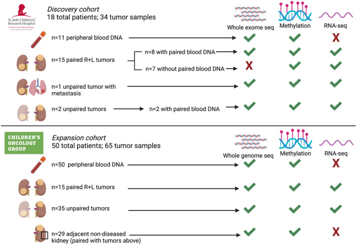
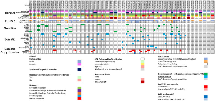
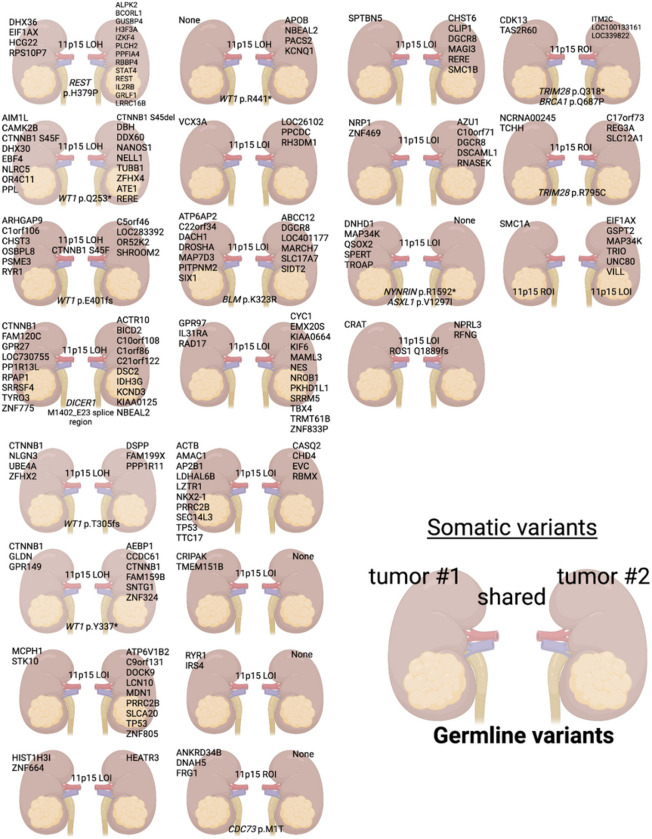
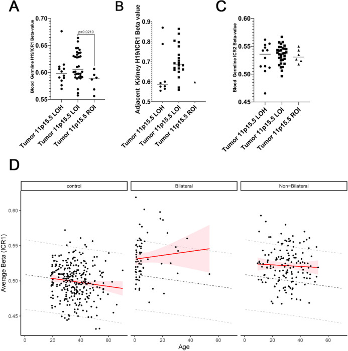
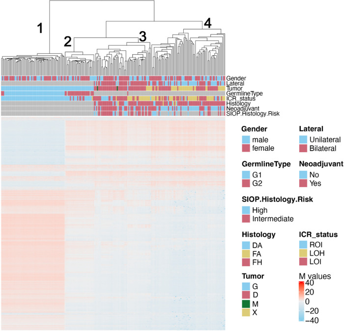
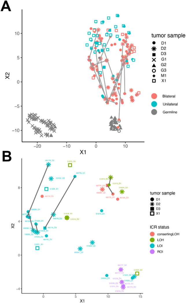
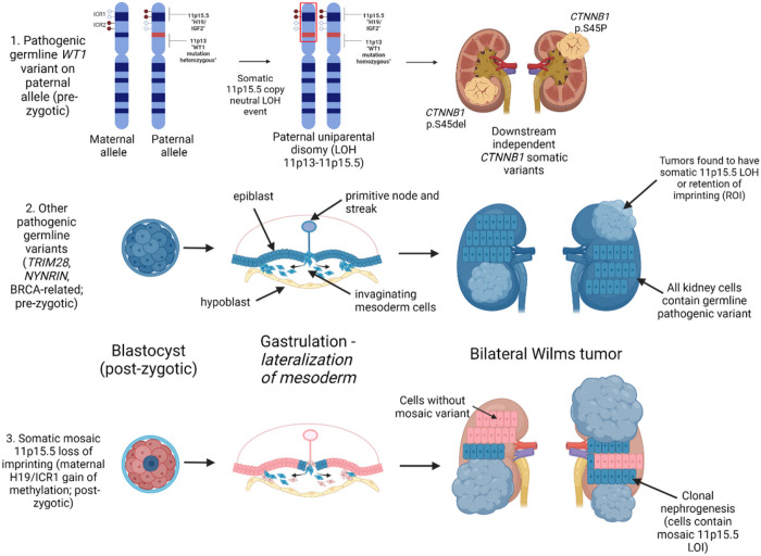
This study comprehensively evaluated the landscape of genetic and epigenetic events that predispose to synchronous bilateral Wilms tumor (BWT). We performed whole exome or whole genome sequencing, total-strand RNA-seq, and DNA methylation analysis using germline and/or tumor samples from 68 patients with BWT from St. Jude Children's Research Hospital and the Children's Oncology Group. We found that 25/61 (41%) of patients evaluated harbored pathogenic or likely pathogenic germline variants, with WT1 (14.8%), NYNRIN (6.6%), TRIM28 (5%) and the BRCA-related genes (5%) BRCA1, BRCA2, and PALB2 being most common. Germline WT1 variants were strongly associated with somatic paternal uniparental disomy encompassing the 11p15.5 and 11p13/WT1 loci and subsequent acquired pathogenic CTNNB1 variants. Somatic coding variants or genome-wide copy number alterations were almost never shared between paired synchronous BWT, suggesting that the acquisition of independent somatic variants leads to tumor formation in the context of germline or early embryonic, post-zygotic initiating events. In contrast, 11p15.5 status (loss of heterozygosity, loss or retention of imprinting) was shared among paired synchronous BWT in all but one case. The predominant molecular events for BWT predisposition include pathogenic germline variants or post-zygotic epigenetic hypermethylation at the 11p15.5 H19/ICR1 locus (loss of imprinting). This study demonstrates that post-zygotic somatic mosaicism for 11p15.5 hypermethylation/loss of imprinting is the single most common initiating molecular event predisposing to BWT. Evidence of somatic mosaicism for 11p15.5 loss of imprinting was detected in leukocytes of a cohort of BWT patients and long-term survivors, but not in unilateral Wilms tumor patients and long-term survivors or controls, further supporting the hypothesis that post-zygotic 11p15.5 alterations occurred in the mesoderm of patients who go on to develop BWT. Due to the preponderance of BWT patients with demonstrable germline or early embryonic tumor predisposition, BWT exhibits a unique biology when compared to unilateral Wilms tumor and therefore warrants continued refinement of its own treatment-relevant biomarkers which in turn may inform directed treatment strategies in the future.

Keywords: 11p15.5; Bilateral Wilms tumor; WT1; Wilms tumor predisposition.


Conflict of interest statement

Conflict of Interest Statement: The authors have no conflicts of interest to declare.

Figures

Figure 1
Figure 1. Mechanisms of dysregulated imprinting at chromosome 11p15.5.
Chromosome 11p15.5 houses a cluster of imprinted genes (including IGF2, KCNQ1, CDKN1C) and noncoding RNAs (including H19, KCNQ1OT1) that are either expressed only from the maternal or paternal allele. The expression of these genes is regulated by differential methylation at two imprinting control regions (ICR) – 11p15.5 H19/ICR1 and KCNQ1OT1/ICR2. In normal physiology, H19/ICR1 is methylated on the paternal allele only and IGF2is expressed from this allele. In contrast, ICR2, located at the promoter region of KCNQ1OT1 is methylated on the maternal allele only, leading to expression of KCNQ1 and CDKN1C from the maternal allele. The normal physiologic imprinting status at 11p15.5 is referred to as 11p15.5 retention of imprinting (ROI) throughout this manuscript. Two predominant mechanisms occur that disrupt imprinting and result in biallelic expression of IGF2: 1) 11p15.5 H19/ICR1 loss of imprinting (LOI) refers to site-specific epigenetic gain of methylation at H19/ICR1 and 2) 11p15.5 copy neutral loss of heterozygosity (cn-LOH) refers to genetic deletion of the maternal allele and duplication of the paternal allele (paternal uniparental disomy). With 11p15.5 LOI, there is hypermethylation of H19/ICR1 and normal methylation of KCNQ1OT1/ICR2. With 11p15.5 LOH, there is hypermethylation of H19/ICR1 and hypomethylation of KCNQ1OT1/ICR2. Arrows indicate active transcription reflective of the normal physiologic imprinting pattern. Graphic made with biorender.com
Figure 2
Figure 2. Specimens and molecular assays used in the current study.
Whole exome or whole genome sequencing germline variant calls were made using DNA obtained from 61 patients with available DNA from peripheral blood (n=11 SJCRH and n=50 COG). Paired tumor sets are samples from both right and left tumors in a patient with synchronous BWT (n=30). Unpaired tumors are samples from either the right or left tumor in a patient with synchronous BWT, but for whom one side was not available for analysis (n=37). Adjacent non-diseased kidney was confirmed by a pathologist and came from patients with tumors in the study. R – right L – left.
Figure 3
Figure 3. Spectrum of clinical, genetic, and epigenetic features associated with bilateral Wilms tumor.
Paired synchronous BWT (n=24) are outlined in paired boxes on the left side of the graphic. Additional paired synchronous BWT (n=7) that did not have a germline peripheral blood sample available are shown in paired boxes on the right side of the graphic. These 7 paired synchronous BWT specimens were analyzed for methylation and RNA-seq only. Gray boxes indicate when a given finding could not be assessed due to sample availability. Unpaired specimens from BWT patients are outlined in the adjacent boxes without spacing in the center of the graphic.
Figure 4
Figure 4
Somatic variants are almost never shared in paired synchronous bilateral Wilms tumor, while chromosome 11p15.5 status is shared in nearly every case. The top 4 rows contain 15 sets of paired synchronous BWT samples obtained from the Children’s Oncology Group and the bottom 4 rows are 8 sets from St. Jude Children’s Research Hospital. Unique/independent somatic variants are indicated over each tumor/kidney. Any shared somatic variants or 11p15.5 status (LOI, LOH, ROI) are indicated between the two kidneys. Any germline variants in pediatric cancer or WT predisposition genes are shown at the bottom of the pair. Graphic made with biorender.com.
Figure 5
Figure 5
(A) Patients with BWT containing 11p15.5 LOI were found to have a statistically significant increase in 11p15.5 H19/ICR1 methylation detected in the peripheral blood when compared to patients with tumors having 11p15 retention of imprinting (p=0.0210). (B) 11p15.5 LOI was often detectable above the threshold value (b >=0.7) for loss of imprinting in adjacent non-diseased kidney tissues. (C) No differences were detected among patient groups at 11p15.5 KCNQ1OT1/ICR2. Of note, the single sample outlier with tumor 11p15.5 LOH, hypermethylation at H19/ICR1, and hypomethylation at KCNQ1OT1/ICR2detected in the peripheral blood was confirmed to have mosaicism for 11p15 LOH detected in peripheral blood by whole genome sequencing. (D) An increase in H19/ICR1 methylation detected in peripheral blood was noted between BWT patients, BWT long-term survivors, and healthy community controls. 11p15.5 H19/ICR1 methylation increases with age in BWT patients and long-term survivors but decreases with age in healthy community controls and in unilateral WT long-term survivors. Dashed lines demonstrate the predicted change in methylation by age derived from the control group. Shaded red areas indicate the 95% predication interval.
Figure 6
Figure 6
Unsupervised hierarchical clustering of methylation M-values from the top 10,000 most variable probes in the 850K EPIC Methylation Beadchip array demonstrates distinct clustering of DNA samples derived from peripheral blood (cluster 1), adjacent non-diseased kidney (cluster 2), a cluster of predominantly BWT (cluster 3), and a cluster of predominantly unilateral WT (cluster 4). Within the BWT cluster (cluster 3), samples with 11p15.5 LOH, 11p15.5 LOI, and 11p15.5 ROI cluster together. Notably, the BWT cluster (cluster 3) joins with adjacent non-diseased kidney (cluster 2) more closely than the unilateral WT cluster. Germline type: G1 – blood, G2 – kidney. Histology – DA – diffuse anaplasia, FA – focal anaplasia, FH – favorable histology. Tumor type: G – germline sample, D – primary tumor sample, M – metastatic sample, X – patient-derived xenograft.
Figure 7
Figure 7. TSNE clustering analysis of 850K EPIC Methylation Beadchip array.
(A) Predominant clusters of unilateral (teal) and bilateral (salmon) WT. BWT cluster closer to adjacent non-diseased kidney (gray triangles) than unilateral WT. Gray lines connect synchronous BWT. However, large differences in synchronous BWT correlated with differential tumor purity in each DNA sample. (B) Clustering of BWT with a tumor purity filter applied (excluding specimens with tumor purity < 80%) shows near adjacent clustering of paired synchronous BWT specimens. Within BWT, samples cluster according to 11p15.5 status: 11p15.5 LOI (light blue), 11p15.5 LOH (red and green), and 11p15.5 ROI (purple). The greatest difference between paired synchronous BWT samples is seen in samples with 11p15.5 LOI (samples connected by dashed lines).
Figure 8
Figure 8. Summary of Proposed Mechanisms for Bilateral Wilms tumor development.
The top panel depicts the molecular sequence leading to BWT in patients with pathogenic heterozygous WT1 germline variants. Here, somatic 11p15.5 copy neutral loss of heterozygosity causes biallelic inactivation of WT1 and biallelic expression of IGF2, a sequence which is often followed by downstream distinct CTNNB1 somatic variants unique to each tumor. The middle panel depicts the general sequence of BWT development due to a pre-zygotic pathogenic germline variant in which the germline variant is present in all kidney cells. The bottom panel depicts somatic mosaic 11p15.5 loss of imprinting, in which 11p15.5 H19/ICR1 gain of methylation occurs on the maternal allele in a post-zygotic embryonic cell. This event must occur prior to lateralization of cells fated to become mesoderm during embryonic gastrulation. At this time in embryonic development, the cells that give rise to the intermediate mesoderm and therefore the kidneys are anatomically sequestered from one another (right and left). This lateralization results in a somatic mosaic distribution of 11p15.5 LOI throughout the body/mesoderm. Expansion of cellular clones containing the somatic mosaic alteration is termed clonal nephrogenesis and explains the detection of 11p15.5 LOI in adjacent non-diseased kidney tissue. BWT and/or multifocal WT arise from these clonal populations of kidney cells. Graphic made with biorender.com

References

    1. Davidoff A.M. Wilms tumor. Adv Pediatr 59, 247–67 (2012). - PMC - PubMed
    1. Knudson A.G. & Strong L.C. Mutation and cancer: a model for Wilms’ tumor of the kidney. J Natl Cancer Inst 48, 313–24 (1972). - PubMed
    1. Spreafico F. et al. Wilms tumour. Nat Rev Dis Primers 7, 75 (2021). - PubMed
    1. Beckwith J.B., Kiviat N.B. & Bonadio J.F. Nephrogenic rests, nephroblastomatosis, and the pathogenesis of Wilms’ tumor. Pediatr Pathol 10, 1–36 (1990). - PubMed
    1. Chernin G. et al. Genotype/phenotype correlation in nephrotic syndrome caused by WT1 mutations. Clin J Am Soc Nephrol 5, 1655–62 (2010). - PMC - PubMed

Publication types