. 2023 Mar 13:14:1027872.
doi: 10.3389/fpsyg.2023.1027872. eCollection 2023.

What determines the neural response to snakes in the infant brain? A systematic comparison of color and grayscale stimuli

Affiliations

What determines the neural response to snakes in the infant brain? A systematic comparison of color and grayscale stimuli

Julie Bertels et al. Front Psychol. .

Abstract

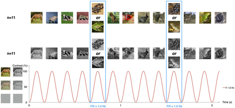
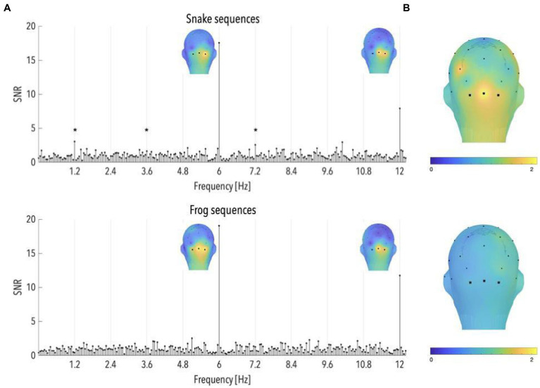
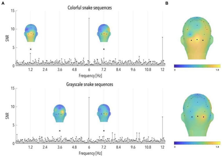
Snakes and primates have coexisted for thousands of years. Given that snakes are the first of the major primate predators, natural selection may have favored primates whose snake detection abilities allowed for better defensive behavior. Aligning with this idea, we recently provided evidence for an inborn mechanism anchored in the human brain that promptly detects snakes, based on their characteristic visual features. What are the critical visual features driving human neural responses to snakes is an unresolved issue. While their prototypical curvilinear coiled shape seems of major importance, it remains possible that the brain responds to a blend of other visual features. Coloration, in particular, might be of major importance, as it has been shown to act as a powerful aposematic signal. Here, we specifically examine whether color impacts snake-specific responses in the naive, immature infant brain. For this purpose, we recorded the brain activity of 6-to 11-month-old infants using electroencephalography (EEG), while they watched sequences of color or grayscale animal pictures flickering at a periodic rate. We showed that glancing at colored and grayscale snakes generated specific neural responses in the occipital region of the brain. Color did not exert a major influence on the infant brain response but strongly increased the attention devoted to the visual streams. Remarkably, age predicted the strength of the snake-specific response. These results highlight that the expression of the brain-anchored reaction to coiled snakes bears on the refinement of the visual system.

Keywords: EEG; color; infancy; snakes; steady-state visual evoked potential.


Conflict of interest statement

The authors declare that the research was conducted in the absence of any commercial or financial relationships that could be construed as a potential conflict of interest.

Figures

Figure 1
Figure 1
Schematic illustration of the experimental paradigm. Animal pictures were presented by sinusoidal contrast modulation at a rate of 6 per second (F = 6 Hz). Snake or frog pictures were presented every fifth stimulus (F = 6/5 = 1.2 Hz), in different trial sequences. Half of the infants were presented with color pictures, the other half with grayscale versions of the same pictures.
Figure 2
Figure 2
(A) Signal-to-noise ratio (SNR) spectra of category-selective and stimulation responses until 12 Hz, in the occipital region (data have been averaged across O1, Oz, and O2), and topographical maps of SNR over posterior scalp regions at stimulation frequencies, for snake and frog sequences (n = 87 and 91, respectively). The topographies at 6 and 12 Hz are shown on their individual maximal color scales. The asterisk indicates a significant discrimination response in the occipital region, of colorful snake pictures. (B) Topographical maps of SNR averaged on the first six harmonics of the category-selective response, for snake and frog sequences (upper and lower part, respectively).
Figure 3
Figure 3
Associations between infants’ age and SNR values averaged on the first six harmonics of the category-selective response, for snake and frog sequences. Red dots refer to infants in the color group, grey dots refer to infants in the grayscale group.
Figure 4
Figure 4
(A) Signal-to-noise ratio (SNR) spectra of category-selective and stimulation responses until 12 Hz, in the occipital region (data have been averaged across O1, Oz, and O2), and topographical maps of SNR over posterior scalp regions at significant category-selective frequencies, for colorful and grayscale snake sequences (n = 55 and 32, respectively). The topographies at the harmonics are shown on their individual maximal color scales. The asterisk indicates a significant discrimination response in the occipital region, of colorful and grayscale snake pictures. (B) Topographical maps of SNR averaged on the first six harmonics of the category-selective response, for colorful and grayscale snake sequences (upper and lower part, respectively).

Similar articles

Cited by

References

    1. Barry-Anwar R., Hadley H., Conte S., Keil A., Scott L. S. (2018). The developmental time course and topographic distribution of individual-level monkey face discrimination in the infant brain. Neuropsychologia 108, 25–31. doi: 10.1016/j.neuropsychologia.2017.11.019, PMID: - DOI - PubMed
    1. Bertels J., Bayard C., Floccia C., Destrebecqz A. (2018). Rapid detection of snakes modulates spatial orienting in infancy. Int. J. Behav. Dev. 42, 381–387. doi: 10.1177/0165025417693955 - DOI
    1. Bertels J., Bourguignon M., de Heering A., Chetail F., De Tiège X., Cleeremans A., et al. . (2020). Snakes elicit specific neural responses in the human infant brain. Sci. Rep. 10:7443. doi: 10.1038/s41598-020-63619-y, PMID: - DOI - PMC - PubMed
    1. Changizi M. A., Zhang Q., Shimojo S. (2006). Bare skin, blood and the evolution of primate colour vision. Biol. Lett. 2, 217–221. doi: 10.1098/rsbl.2006.0440, PMID: - DOI - PMC - PubMed
    1. Courage M. L., Adams R. J. (1990). Visual acuity assessment from birth to three years using the acuity card procedure: cross-sectional and longitudinal samples. Am. J. Optom. Physiol. Optic 67, 713–718. doi: 10.1097/00006324-199009000-00011, PMID: - DOI - PubMed

LinkOut - more resources