. 2022 Dec;11(12):7788-7794.
doi: 10.4103/jfmpc.jfmpc_1136_22. Epub 2023 Jan 17.

Spectrum of magnetic resonance imaging findings in post-COVID-19 patients presenting with rhino-orbito-cerebral mucormycosis in a teaching hospital in Malwa region of Punjab

Affiliations

Spectrum of magnetic resonance imaging findings in post-COVID-19 patients presenting with rhino-orbito-cerebral mucormycosis in a teaching hospital in Malwa region of Punjab

Pushpinder Singh et al. J Family Med Prim Care. 2022 Dec.

Abstract

Background: Rhino-orbito-cerebral-mucormycosis (ROCM) associated with COVID-19 infection was at its peak during and immediately after the second wave of the pandemic in India during 2021. Many of the risk factors were implicated in the development of this deadly fungal infection, when there was a sudden surge of cases, especially who had a history of COVID-19 infection.

Objectives: The objective of the study was to describe the characteristic magnetic resonance imaging (MRI) findings in invasive mucormycosis and to evaluate the extent and severity of invasive mucormycosis.

Materials and methods: A retrospective study was carried out for 4 months which included 60 patients who underwent MRI using a Siemens Avanto 1.5 Tesla scanner. During our study, 68 cases were selected based on clinicoradiological features suspected of ROCM. However, 8 patients were excluded on the basis of exclusion criteria as either there was no definitive evidence of COVID infection or were proven to be negative for mucormycosis on microbiological examination.

Results: On the basis of the spectrum of MRI findings, post-COVID-19 related ROCM was broadly categorized into three stages. Out of total of 60 patients, the disease was localized to the nasal and paranasal sinus region (Stage I) in 7 patients (11.66%), extending to adjacent extrasinus orofacial soft tissue structures (Stage II) in 36 patients (60%), and intracranial extension of disease (Stage III) was seen in 17 patients (28.33%).

Conclusion: In post-COVID-19 patients presenting with clinical features suspected of ROCM, MRI imaging helps in early diagnosis and staging/severity of ROCM, so that timely intervention can be planned to reduce mortality as well as morbidity.

Keywords: Black turbinate sign; COVID-19; ROCM; mucormycosis; orbital cellulitis.


Conflict of interest statement

There are no conflicts of interest.

Figures

Figure 1
Figure 1
Microbiological lab investigation showing. a. Aseptate hyphae with wide angle branching in nasal tissue (KOH wet mount 40×). b. KOH wet mount of debrided tissue from rhinocerebral zygomycosis showing broad aseptate hyphae with sporangiophore with columella and sporangia. c. LPCB mount of mucor. d. Rapidly growing gray colonies of mucor species on SDA media
Figure 2
Figure 2
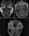
T1 post-contrast MRI sequence in a) Axial b) Coronal planes depicting non-enhancement of left inferior turbinate (long arrow) suggestive of black turbinate sign, whereas contralateral inferior turbinate showing normal enhancement (short arrow)
Figure 3
Figure 3
Involvement of various paranasal sinuses
Figure 4
Figure 4
a) T1 coronal MRI PNS image depicting hypointense material completely filling bilateral ethmoid sinuses (arrow) b) T1 axial post-contrast MRI PNS image depicting patchy to poorly enhancing material in bilateral ethmoid air cells (arrow) c) T1 FS post-contrast MRI PNS axial sections depicting soap bubble appearance in bilateral maxillary sinuses
Figure 5
Figure 5
a) MRI T2 axial image of PNS/face showing heterogeneous material in the bilateral maxillary sinus, with extrasinus extension on the left side in the form of periantral fat stranding and obliteration including premaxillary (long arrow) as well as retroantral fat involvement (short arrow) b) T1 FS post-contrast T1 axial image of PNS/Face showing heterogeneous enhancing material with interspersed non-enhancing areas almost completely filling the bilateral ethmoid sinuses and extending to bilateral orbits including orbital apex, intraconal as well as extraconal compartment (Stage II)
Figure 6
Figure 6
a) T2 and b) T1 FS post-contrast axial images show tenting of eye ball (long arrow) and stretching of the optic nerve (short arrow) along with proptosis suggestive of orbital compartment syndrome
Figure 7
Figure 7
a) Coronal b) Axial sections illustrating non-enhancing necrotic material in the left ethmoid sinus extending to the medial orbital compartment (short arrow) via lamina papyracea erosion and intracranial extension via cribriform plate erosion forming peripheral ring-enhancing area suggestive of abscess in left frontal region (long arrow) in stage III ROCM patient

Similar articles

References

    1. Navarro-Perea C, Canas-Zamarra I, Mencia-Gutierrez E, Revilla-Sanchez E, Lago-Llinas M, Perez-Trigo S, et al. Rhino-orbito-cerebral mucormycosis: Two cases with amaurosis as presentation, medical surgical management and follow-up. Case Rep Ophthalmol Med 2019. 2019:1–7. - PMC - PubMed
    1. Devana SK, Gupta VG, Mavuduru RS, Bora GS, Sharma AP, Parmar KM, et al. Isolated renal mucormycosis in immunocompetent hosts: Clinical spectrum and management approach. Am J Trop Med Hyg. 2019;100:791–7. - PMC - PubMed
    1. Skiada A, Pavleas I, Drogari-Apiranthitou M. Epidemiology and diagnosis of mucormycosis: An update. J Fungi. 2020;6:265. - PMC - PubMed
    1. Prakash H, Chakrabarti A. Epidemiology of Mucormycosis in India. Microorganisms. 2021;9:523. - PMC - PubMed
    1. Bhattacharyya A, Sarma P, Sharma DJ, Das KK, Kaur H, Prajapat M, et al. Rhino-orbital-cerebral-mucormycosis in COVID-19: A systematic review. Indian J Pharmacol. 2021;53:317–27. - PMC - PubMed