Review
. 2022 Dec 1;5(1):10-19.
doi: 10.1016/j.smhs.2022.11.004. eCollection 2023 Mar.

The skeletal muscle fiber periphery: A nexus of mTOR-related anabolism

Affiliations
Review

The skeletal muscle fiber periphery: A nexus of mTOR-related anabolism

Cassidy T Tinline-Goodfellow et al. Sports Med Health Sci. .

Abstract

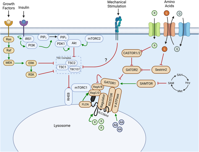
Skeletal muscle anabolism is driven by numerous stimuli such as growth factors, nutrients (i.e., amino acids, glucose), and mechanical stress. These stimuli are integrated by the mechanistic target of rapamycin (mTOR) complex 1 (mTORC1) signal transduction cascade. In recent years, work from our laboratory and elsewhere has sought to unravel the molecular mechanisms underpinning the mTOR-related activation of muscle protein synthesis (MPS), as well as the spatial regulation of these mechanisms within the skeletal muscle cell. These studies have suggested that the skeletal muscle fiber periphery is a region of central importance in anabolism (i.e., growth/MPS). Indeed, the fiber periphery is replete with the substrates, molecular machinery, and translational apparatus necessary to facilitate MPS. This review provides a summary of the mechanisms underpinning the mTOR-associated activation of MPS from cell, rodent, and human studies. It also presents an overview of the spatial regulation of mTORC1 in response to anabolic stimuli and outlines the factors that distinguish the periphery of the cell as a highly notable region of skeletal muscle for the induction of MPS. Future research should seek to further explore the nutrient-induced activation of mTORC1 at the periphery of skeletal muscle fibers.

Keywords: Hypertrophy; Muscle protein synthesis; Periphery; Skeletal muscle; Translation; mTOR.


Conflict of interest statement

The authors declare that they have no known competing financial interests or personal relationships that could have appeared to influence the work reported in this paper.

Figures

Fig. 1
Fig. 1
Canonical understanding of activation of mTORC1 in a variety of cell types. Growth factor and insulin stimulation results in signalling cascades through the Ras/Raf/ERK and the PI3K/Akt pathways, respectively, resulting in the inhibitory phosphorylation of the TSC complex, alleviating inhibition on Rheb. This allows Rheb to allosterically activate mTORC1. Amino acids function to promote mTORC1 recruitment to the lysosome by the Rag GTPases through a variety of upstream regulators, such that it can be activated by lysosomal Rheb. R, Arginine; L, Leucine; Q, Glutamine; AA, Amino acids; SAM, S-adenosylmethionine; Hcy, homocysteine; Met, methionine; SAH, S-adenosylhomocysteine. Images were created using BioRender.com.
Fig. 2
Fig. 2
Overview of the regulation of mTOR action at the skeletal muscle fiber periphery (A) Post-absorptive (i.e., fasted). state. During the post-absorptive period, there are less circulating amino acids, the mTOR-Lysosome complexes are mostly colocalized, but dispersed throughout the cytoplasm, as is VPS34 (B) The cellular state during the post-prandial (i.e., fed). period and following resistance exercise. The influx of amino acids and mechanical stimulation results in the mTOR-lysosome complex moving to the periphery of the cell, where the translational machinery is located. In some instances, certain stimuli (e.g., whole-egg ingestion and leucine ingestion) may stimulate an increase in mTOR-lysosome colocalization. Mechanical stimulation of muscle may also promote phosphorylation of TSC2 and its dissociation from the lysosome, further promoting mTORC1 activation. L, Leucine; Q, Glutamine; P, Phosphorylation modification.

References

    1. Goldberg A.L. Protein synthesis during work-induced growth of skeletal muscle. J Cell Biol. 1968;36(3):653–658. doi: 10.1083/jcb.36.3.653. - DOI - PMC - PubMed
    1. Saxton R.A., Sabatini D.M. mTOR signaling in growth, metabolism, and disease. Cell. 2017;168(6):960–976. doi: 10.1016/j.cell.2017.02.004. - DOI - PMC - PubMed
    1. Hara K., Maruki Y., Long X., et al. Raptor, a binding partner of target of rapamycin (TOR), mediates TOR action. Cell. 2002;110(2):177–189. doi: 10.1016/s0092-8674(02)00833-4. - DOI - PubMed
    1. Kim D.H., Sarbassov D.D., Ali S.M., et al. mTOR interacts with raptor to form a nutrient-sensitive complex that signals to the cell growth machinery. Cell. 2002;110(2):163–175. doi: 10.1016/s0092-8674(02)00808-5. - DOI - PubMed
    1. Kim D.H., Sarbassov D.D., Ali S.M., et al. GbetaL, a positive regulator of the rapamycin-sensitive pathway required for the nutrient-sensitive interaction between raptor and mTOR. Mol Cell. 2003;11(4):895–904. doi: 10.1016/s1097-2765(03)00114-x. - DOI - PubMed

LinkOut - more resources