. 2022 Nov 29;6(1):e1236.
doi: 10.1002/jsp2.1236. eCollection 2023 Mar.

Osteoconductivity and neurotoxicity of silver-containing hydroxyapatite coating cage for spinal interbody fusion in rats

Affiliations

Osteoconductivity and neurotoxicity of silver-containing hydroxyapatite coating cage for spinal interbody fusion in rats

Takema Nakashima et al. JOR Spine. .

Abstract

Background: The use of spinal instrumentation is an established risk factor for postoperative infection. To address this problem, we prepared silver-containing hydroxyapatite coating, consisting of highly osteoconductive hydroxyapatite interfused with silver. The technology has been adopted for total hip arthroplasty. Silver-containing hydroxyapatite coating has been reported to have good biocompatibility and low toxicity. However, no studies about applying this coating in spinal surgery have addressed the osteoconductivity and direct neurotoxicity to the spinal cord of silver-containing hydroxyapatite cages in spinal interbody fusion.

Aim: In this study, we evaluated the osteoconductivity and neurotoxicity of silver-containing hydroxyapatite-coated implants in rats.

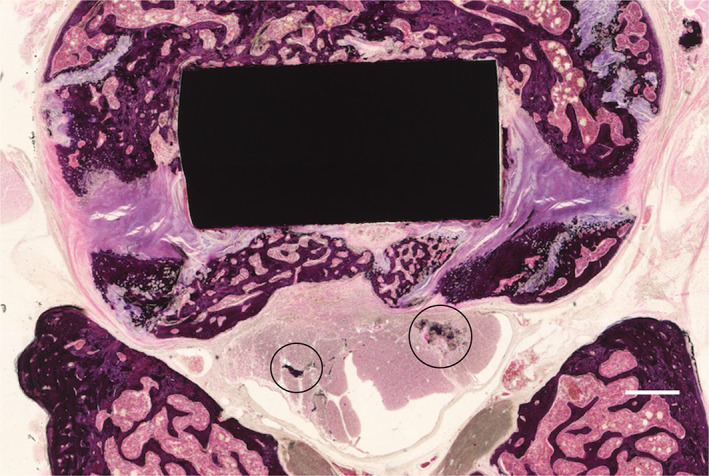
Materials & methods: Titanium (non-coated, hydroxyapatite-coated, and silver-containing hydroxyapatite-coated) interbody cages were inserted into the spine for anterior lumbar fusion. At 8 weeks postoperatively, micro-computed tomography and histology were performed to evaluate the osteoconductivity of the cage. Inclined plane test and toe pinch test were performed postoperatively to assess neurotoxicity.

Results: Micro-computed tomography data indicated no significant difference in bone volume/total volume among the three groups. Histologically, the hydroxyapatite-coated and silver-containing hydroxyapatite-coated groups showed significantly higher bone contact rate than that of the titanium group. In contrast, there was no significant difference in bone formation rate among the three groups. Data of inclined plane and toe pinch test showed no significant loss of motor and sensory function in the three groups. Furthermore, there was no degeneration, necrosis, or accumulation of silver in the spinal cord on histology.

Conclusions: This study suggests that silver-hydroxyapatite-coated interbody cages produce good osteoconductivity and are not associated with direct neurotoxicity.

Keywords: hydroxyapatite; lumbar spinal interbody fusion; neurotoxicity; osteoconductivity; silver.


Conflict of interest statement

The authors declare no conflicts of interest.

Figures

FIGURE 1
FIGURE 1
The surgical procedure of interbody fusion. (A) Median abdominal incision and transperitoneal approach. (B) Ligation of the left iliolumbar vein (white arrow). (C) Anterior access to L3–L4 (white arrow) through the safe interval between the bilateral iliopsoas muscles. (D) Fusion with a titanium piece and a plate with screws.
FIGURE 2
FIGURE 2
(A)) Evaluation of inclined plane test at each observation point. “*” Significant difference at 2 weeks postoperatively between the titanium (Ti) and hydroxyapatite (HA) groups (p = 0.012). “†” Significant difference at 2 weeks postoperatively between the Ti and Ag‐HA groups (p = 0.008). (B) Evaluation of toe pinch test at each observation point.
FIGURE 3
FIGURE 3
Example of microcomputed tomography in the sagittal planes of the interbody fusion at 8 weeks. (A) Titanium (Ti) group, (B) hydroxyapatite (HA) group, (C) silver‐containing hydroxyapatite (Ag‐HA) group. Area within the yellow outlines indicates the region of interest in which bone morphometry on this plane is shown.
FIGURE 4
FIGURE 4
Evaluation of microcomputed tomography assessment of spinal fusion. Bone volume/total volume (BV/TV) at 4 and 8 weeks, postoperatively.
FIGURE 5
FIGURE 5
Photomicrographs taken in the sagittal plane of rat intervertebral disc stained with hematoxylin and eosin (H&E) at 8 weeks postoperatively. (A) titanium (Ti) group, (B) hydroxyapatite (HA) group, (C) silver‐containing hydroxyapatite (Ag‐HA) group (scale bar = 500 μm)
FIGURE 6
FIGURE 6
Photomicrographs taken in the sagittal plane depicting an expanded field of view corresponding to Figure 5. Best, intermediate, and worst images of (A) titanium (Ti) group, (B) hydroxyapatite (HA) group, (C) silver‐containing hydroxyapatite (Ag‐HA) group (scale bar = 500 μm).
FIGURE 7
FIGURE 7
(A) Evaluation of histology assessment of mean affinity index of bone formation. (B) Evaluation of histology assessment of mean affinity index of bone contact. “*” Significant difference between the titanium (Ti) and hydroxyapatite (HA) groups (p = 0.009). “†” Significant difference between the Ti and Ag‐HA groups (p = 0.018)
FIGURE 8
FIGURE 8
Histologic section taken in the axial plane through the silver‐containing hydroxyapatite (Ag‐HA) coated cage. Hematoxylin and eosin (H&E) stain was used (scale bar = 500 μm). Dark stain regions are bone fragments that result from the sectioning process (black circles).

References

    1. Mobbs RJ, Phan K, Malham G, et al. Lumbar interbody fusion: techniques, indications and comparison of interbody fusion options including PLIF, TLIF, MI‐TLIF, OLIF/ATP, LLIF and ALIF. J Spine Surg. 2015;1(1):2‐18. - PMC - PubMed
    1. Martino ADI, Papalia R, Albo E, et al. Infection after spinal surgery and procedures. Eur Rev Med Pharmacol Sci. 2019;23(2):173‐178. - PubMed
    1. McGilvray KC, Easley J, Seim HB, et al. Bony ingrowth potential of 3D‐printed porous titanium alloy: a direct comparison of interbody cage materials in an in vivo ovine lumbar fusion model. Spine J. 2018;18(7):1250‐1260. - PMC - PubMed
    1. Yin D, Liu B, Chang Y, Gu H, Zheng X. Management of late‐onset deep surgical site infection after instrumented spinal surgery. BMC Surg. 2018;18(1):4‐9. - PMC - PubMed
    1. Pappou IP, Papadopoulos EC, Sama AA, Girardi FP, Cammisa FP. Postoperative infections in interbody fusion for degenerative spinal disease. Clin Orthop Relat Res. 2006;444:120‐128. - PubMed