Review
. 2022 Nov 14;6(1):e1231.
doi: 10.1002/jsp2.1231. eCollection 2023 Mar.

Intervertebral disc degeneration and how it leads to low back pain

Affiliations
Review

Intervertebral disc degeneration and how it leads to low back pain

Ashish D Diwan et al. JOR Spine. .

Abstract

The purpose of this review was to evaluate data generated by animal models of intervertebral disc (IVD) degeneration published in the last decade and show how this has made invaluable contributions to the identification of molecular events occurring in and contributing to pain generation. IVD degeneration and associated spinal pain is a complex multifactorial process, its complexity poses difficulties in the selection of the most appropriate therapeutic target to focus on of many potential candidates in the formulation of strategies to alleviate pain perception and to effect disc repair and regeneration and the prevention of associated neuropathic and nociceptive pain. Nerve ingrowth and increased numbers of nociceptors and mechanoreceptors in the degenerate IVD are mechanically stimulated in the biomechanically incompetent abnormally loaded degenerate IVD leading to increased generation of low back pain. Maintenance of a healthy IVD is, thus, an important preventative measure that warrants further investigation to preclude the generation of low back pain. Recent studies with growth and differentiation factor 6 in IVD puncture and multi-level IVD degeneration models and a rat xenograft radiculopathy pain model have shown it has considerable potential in the prevention of further deterioration in degenerate IVDs, has regenerative properties that promote recovery of normal IVD architectural functional organization and inhibits the generation of inflammatory mediators that lead to disc degeneration and the generation of low back pain. Human clinical trials are warranted and eagerly anticipated with this compound to assess its efficacy in the treatment of IVD degeneration and the prevention of the generation of low back pain.

Keywords: GDF6; artificial intelligence; discogenic pain; facial recognition; intervertebral disc degeneration; low back pain; neuropathic pain; nociceptive pain.


Conflict of interest statement

The authors declare no conflicts of interest.

Figures
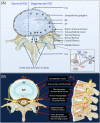
FIGURE 1
FIGURE 1
Schematic showing the innervation of the normal and degenerate intervertebral disc (IVD) (A). The normal IVD is the largest predominately aneural structure in the human body where nerves are confined to the outer annular lamellae. With IVD degeneration and depletion of space‐filling aggrecan from the IVD a significant reduction in internal hydrostatic pressure in the IVD and the production of inflammatory mediators and neurotrophic factors provides conditions that are conducive to the ingrowth of nociceptive nerves (*) from the sinu‐intervertebral nerve into the AF of the IVD and significant increases of peripheral mechanoreceptors in the IVD. Thus, there is an increased perception of pain in the mechanically incompetent degenerate IVD. Peripheral IVD neural structures such as the sympathetic ganglion and dorsal root ganglion and the rami communicantes are highly innervated structures. The DRG communicates with the sensory dorsal horn of the spinal cord. There are also connections to the dural nerve plexus (arrows, *) from the sinu‐vertebral nerve. The facet joint capsule and associated subchondral bone and vertebral bodies adjacent to the IVDs and CEPs are all innervated. The spinal nerve has dorsal and anterior connections to the spinal cord (inset). Neural organization of a lumbar spinal segment (B). AF, annulus fibrosus; ALL, anterior longitudinal ligament; DRG, dorsal root ganglion; NP, nucleus pulposus; PLL, posterior longitudinal ligament; SC, spinal cord. Figure modified from Kallewaard et al. with permission
FIGURE 2
FIGURE 2
Neural growth factors, neurotransmitters, and neural receptors involved in the ingrowth of nerves into the intervertebral disc (IVD) and transmission of sensory signals to the CNS. Composite schematic figure depicting a horizontally bisected IVD (A), sinuvertebral nerve and its branches to the sympathetic and sensory DRG and sensory nerves that communicate with the sensory layers of the dorsal horns of the spinal cord. 1. Nucleus pulposus, 2. Outer AF, 3. Inner AF, 4. Posterior longitudinal ligament, 5. Anterior longitudinal ligament, 6. Sympathetic root ganglion and its pre‐ and post‐ganglionic white and gray ramus communicantes, 7. Postganglionic nerve fibers, 8. DRG and its nerve fibers, pain receptors, neurotrophins and neurotransmitter associated peptides, 9. Sinuvertebral nerve (B) that communicate with the sensory layers (10) (C) of the spinal cord dorsal horn (11) (D). (i) TrkA/TrkB (Tropomyosin receptor kinase A, B; receptor for nerve growth factor [NGF]/brain‐derived neurotrophic factor, BDNF). (ii) Degenerin/epithelial sodium channels (DEG/ENaCs) (ENaCa, b, and c). (iii) Acid‐sensing ion channels (ASIC1, ASIC2, and ASIC3). (iv) TRP family (TRPA1, TRPC1, TRPC6, and TRPV1‐4). (v) Neuron associated neurotransmitter peptides CGRP (calcitonin gene‐related peptide), GFRα1 and GFRα3 (glial cell‐line‐derived neurotrophic receptor subtypes α1 and α3), P2X3 (ATP‐gated ion channel subtype P2X3), SP (substance P), TMP (thiamine monophosphatase), VR1 (vanilloid receptor subtype 1). BDNF, brain derived nerve factor; NGF, nerve growth factor. The horizontally sectioned spinal cord (d) is stained with Nissl stain to show the white and gray matter and DRG. Figure modified from Takahashi et al. with permission

References

    1. Chou R. Low back pain (chronic). BMJ Clin Evid. 2010;2010:1116. - PMC - PubMed
    1. Briggs A, Buchbinder R. Back pain: a National Health Priority Area in Australia? Med J Aust. 2009;190:499‐502. - PubMed
    1. Chou R. In the clinic. Low back pain. Ann Intern Med. 2014;150:ITC6‐1. - PubMed
    1. Vos T, Flaxman AD, Naghavi M, et al. Years lived with disability (YLDs) for 1160 sequelae of 289 diseases and injuries 1990‐2010: a systematic analysis for the Global Burden of Disease Study 2010. Lancet. 2010;380:2163‐2196. - PMC - PubMed
    1. Ehrlich G. Low back pain. Bull World Health Organ. 2003;81:671‐676. - PMC - PubMed