. 2023 May;43(5):e132-e150.
doi: 10.1161/ATVBAHA.123.319120. Epub 2023 Mar 30.

FN (Fibronectin)-Integrin α5 Signaling Promotes Thoracic Aortic Aneurysm in a Mouse Model of Marfan Syndrome

Affiliations

FN (Fibronectin)-Integrin α5 Signaling Promotes Thoracic Aortic Aneurysm in a Mouse Model of Marfan Syndrome

Minghao Chen et al. Arterioscler Thromb Vasc Biol. 2023 May.

Abstract

Background: Marfan syndrome, caused by mutations in the gene for fibrillin-1, leads to thoracic aortic aneurysms (TAAs). Phenotypic modulation of vascular smooth muscle cells (SMCs) and ECM (extracellular matrix) remodeling are characteristic of both nonsyndromic and Marfan aneurysms. The ECM protein FN (fibronectin) is elevated in the tunica media of TAAs and amplifies inflammatory signaling in endothelial and SMCs through its main receptor, integrin α5β1. We investigated the role of integrin α5-specific signals in Marfan mice in which the cytoplasmic domain of integrin α5 was replaced with that of integrin α2 (denoted α5/2 chimera).

Methods: We crossed α5/2 chimeric mice with Fbn1mgR/mgR mice (mgR model of Marfan syndrome) to evaluate the survival rate and pathogenesis of TAAs among wild-type, α5/2, mgR, and α5/2 mgR mice. Further biochemical and microscopic analysis of porcine and mouse aortic SMCs investigated molecular mechanisms by which FN affects SMCs and subsequent development of TAAs.

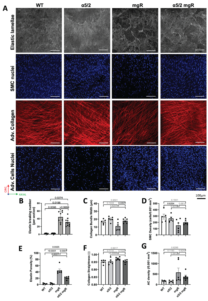
Results: FN was elevated in the thoracic aortas from Marfan patients, in nonsyndromic aneurysms, and in mgR mice. The α5/2 mutation greatly prolonged survival of Marfan mice, with improved elastic fiber integrity, mechanical properties, SMC density, and SMC contractile gene expression. Furthermore, plating of wild-type SMCs on FN decreased contractile gene expression and activated inflammatory pathways whereas α5/2 SMCs were resistant. These effects correlated with increased NF-kB activation in cultured SMCs and mgR aortas, which was alleviated by the α5/2 mutation or NF-kB inhibition.

Conclusions: FN-integrin α5 signaling is a significant driver of TAA in the mgR mouse model. This pathway thus warrants further investigation as a therapeutic target.

Keywords: fibronectin; inflammation; smooth muscle cell; target; thoracic aneurysm.


Conflict of interest statement

DISCLOSURES

JDH is a member of the Scientific Advisory Board of and paid consultant for Heartflow, Inc., an AI-based company focused on non-invasive assessment of coronary artery disease. The other authors declare no conflicts, financial or otherwise.

Figures

Figure 1.
Figure 1.. Fibronectin (FN) and smooth muscle α-actin (SMA) in human and murine ascending thoracic aortas.
(A) Representative image from human ascending aorta tissue from normal donors, non-syndromic aneurysms (NS TAA) and Marfan syndromic aneurysm (MFS TAA) patients stained for FN (cyan) and SMA (red). (B, C) Quantification of immunofluorescence signal intensity of FN and αSMA in A (Con, n=3, NS TAA, n=9, MFS TAA, n=3). (D) Ascending aortas from WT mice (n=6), α5/2 mice (n=7), mgR mice (n=8), and α5/2 mgR double mutant mice (n=10) were stained for FN (red) and SMA (green). Quantification of immunofluorescence intensity of FN and αSMA quantified in (E, F). Data points in B and C reference individual human patients or healthy donors, data points in E and F reference individual mice. Columns indicate mean values with standard errors. Supplemental Fig. S1 shows similar findings for smooth muscle 22α (SM22). Non-parametric one-way ANOVA (Kruskai-Wallis test) for Fig. 1B and 1C, ordinary one-way ANOVA for Fig. 1E and 1F with post-hoc Tukey test.
Figure 2.
Figure 2.. Mouse survival and mechanical parameters in ascending thoracic aortas.
WT, α5/2, mgR and α5/2 mgR mice were examined for survival, expansion of the ascending aorta and critical mechanical characteristics: (A) Male mice survival over time. (B) Body mass and (C) mean blood pressure at 8-9 weeks. (D) Gross appearance and (E) H&E staining of cross section of ascending aortas. (F-I) Bulk geometric and passive biomechanical metrics calculated under ex vivo equivalent systolic conditions (120 mmHg and specimen-specific in vivo axial stretches). (J-M) Variation in outer diameter measured during vasoactive biomechanical testing. For panel A: WT n=10, α5/2 n=10, mgR n=8, α5/2 mgR n=10. For panels B, C, F-M, data points references an individual mice (WT, n=5-6; α5/2, n=5–6; mgR, n=5; α5/2 mgR, n=5). Data points reference individual mice. Columns indicate mean values with standard errors. Kaplan-Meier survival analysis was used for Fig. 2A, ordinary one-way ANOVA for the other panels, with post-hoc Tukey test.
Figure 3.
Figure 3.. Microstructural characterization of ascending aortas under physiological loading.
(A) Representative two-photon images of ascending thoracic aortas acquired ex vivo at under conditions that match diastole in vivo (80 mmHg and specimen-specific in vivo axial stretches) in from WT, α5/2, mgR and α5/2 mgR mice. Top row: elastin; second row: medial SMC nuclei; third row: adventitial collagen; bottom row: adventitial cell nuclei. (B) Number of elastin breaks per histological cross-section fixed at unloaded configuration (ordinary t test comparing mgR and α5/2 mgR showed significance p=0.0340). (C-G) Microstructural metrics quantified from images as in A. Data points reference individual mice. Columns indicate mean values with standard errors. For Fig. 3B: WT, n=6; α5/2, n=7; mgR, n=8; α5/2 mgR, n=10. For Fig. 3C–G: WT, n=5, α5/2, n=5; mgR, n=6; α5/2 mgR, n=5. Non-parametric ANOVA was used for Fig. 3B (Kruskai-Wallis test) , and ordinary one-way ANOVA with post-hoc Tukey test for the other panels.
Figure 4.
Figure 4.. Bulk RNAseq of ascending aortas.
(A) Log10 gene expression values of all samples were subjected to principal component analysis displayed as projection of sample gene expression vectors on the first principal component. (B) Volcano plots and pairwise differentially expressed gene (DEG) number comparisons between WT, α5/2, mgR and α5/2 mgR mice, n=3 for each genotype. Significantly down- and up-regulated genes are visualized as blue or orange points, respectively. Numbers below volcano plots indicate total significantly up- and down- regulated genes. (C-D) Pathway enrichment analysis using Gene Ontology Biological Processes and Fisher’s Exact Test for up- and down-regulated genes between mgR and α5/2 mgR tissue. (E-G) Read count ratio (compare to the mean read count value of WT group) of individual inflammatory, contractile and ECM remodeling genes of interest. Data points reference individual mice. Columns indicate mean values with standard errors in E-G. DEGs indicated E-G in were identified using DESeq2 1.32.0 using an adjusted p-value cutoff of 0.05 and a minimum log2(fold change) of +/− log2(1.3) .
Figure 5.
Figure 5.. Staining for inflammatory markers in murine ascending thoracic aorta.
(A, B) Ascending aorta cross sections from WT, α5/2, mgR and α5/2 mgR mice were stained for the macrophage marker CD68 or the total hematopoietic marker CD45 together with DAPI to mark cell nuclei as indicated. (C, D) Quantification of CD68- and CD45-positive cells area per section area. Data points reference individual mice. Columns indicate mean values with standard errors. For C and D, WT, n=6; α5/2, n=7; mgR, n=8; α5/2 mgR, n=10. Ordinary one-way ANOVA with post-hoc Tukey test for C and D.
Figure 6.
Figure 6.. NF-κB in human and murine ascending thoracic aorta.
(A) Cross sections from human ascending aortas from healthy controls (Con), non-syndromic aneurysms (NS TAA) and Marfan syndromic aneurysms (MFS TAA) stained for total-p65 plus DAPI as indicated. (B) Quantification of p65 nuclear to cytoplasm ratio in human ascending aortas: Con, n=3, NS TAA, n=9, MFS TAA, n=3. (C) Ascending aortic cross sections from WT (n=6), α5/2 (n=7), mgR (n=8) and α5/2 mgR (n=10) mice stained for p65 and DAPI. (D) Quantification of p65 nuclear to cytoplasm ratio in WT, α5/2, mgR and α5/2 mgR in Fig.C. Data points in B reference individual human patients or healthy donors, data points in D reference individual mice. Columns indicate mean values with standard errors. Non-parametric t test for Fig. 6B, ordinary one-way ANOVA for Fig. 6D with post-hoc Tukey test.
Figure 7.
Figure 7.. Direct effects of FN on SMCs.
(A, C) WT or α5/2 mouse aortic SMCs were plated on FN or MG for 24 h then NF-kB activation (p65 phosphorylation) and αSMA, CNN1, SM22 levels assayed by Western blotting. (B, D) Quantification of αSMA, CNN1, SM22 protein levels (normalized by GAPDH) and ratio of phospho-p65 to total p65 signal in Fig. 7A and 7C. Each point in Fig. 7B and 7D corresponds to an individual experiment, n=6 for each group. (E, G) WT or α5/2 mouse aortic SMC on FN and MG were stained for αSMA, CNN1, phospho-myosin light chain (P-MLC) and total-p65. αSMA, CNN1 phospho-MLC median fluorescence intensity and nuclear translocation of p65 quantified in (F, H). Each point in Fig. 7F and H corresponds to an image, n=20 for each group, columns indicate mean values with standard errors. Ordinary t-test for Fig. 7B, D, F and H between FN and MG.

References

    1. Chaudhry SS, Cain SA, Morgan A, Dallas SL, Shuttleworth CA and Kielty CM. Fibrillin-1 regulates the bioavailability of TGFbeta1. J Cell Biol. 2007;176:355–67. - PMC - PubMed
    1. Karimi A and Milewicz DM. Structure of the Elastin-Contractile Units in the Thoracic Aorta and How Genes That Cause Thoracic Aortic Aneurysms and Dissections Disrupt This Structure. Can J Cardiol. 2016;32:26–34. - PMC - PubMed
    1. Humphrey JD, Milewicz DM, Tellides G and Schwartz MA. Cell biology. Dysfunctional mechanosensing in aneurysms. Science. 2014;344:477–9. - PMC - PubMed
    1. Sengle G and Sakai LY. The fibrillin microfibril scaffold: A niche for growth factors and mechanosensation? Matrix Biol. 2015;47:3–12. - PubMed
    1. Creamer TJ, Bramel EE and MacFarlane EG. Insights on the Pathogenesis of Aneurysm through the Study of Hereditary Aortopathies. Genes (Basel). 2021;12. - PMC - PubMed

Publication types