. 2023 Mar 30;13(2):4409-4491.
doi: 10.1002/cphy.c190043.

Aldosterone: Renal Action and Physiological Effects

Affiliations

Aldosterone: Renal Action and Physiological Effects

Jermaine G Johnston et al. Compr Physiol. .

Abstract

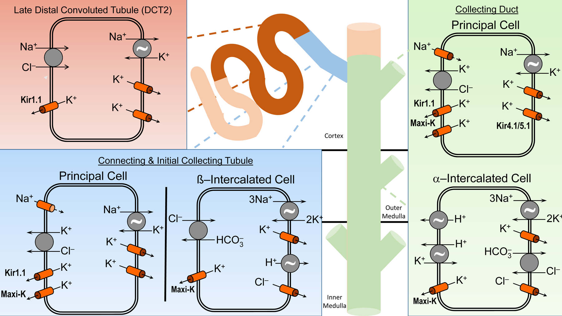
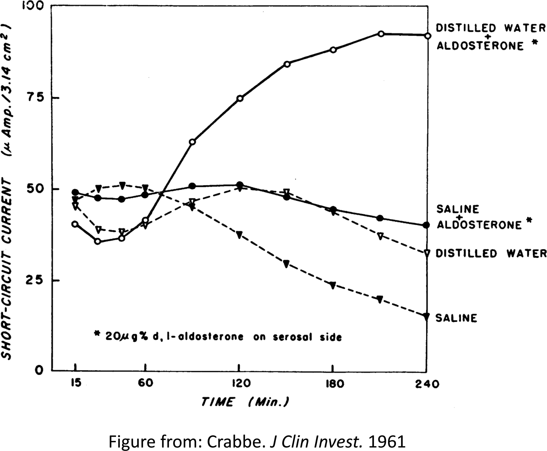
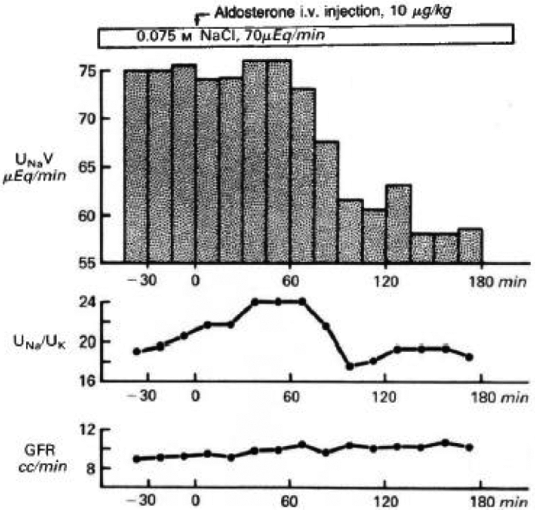
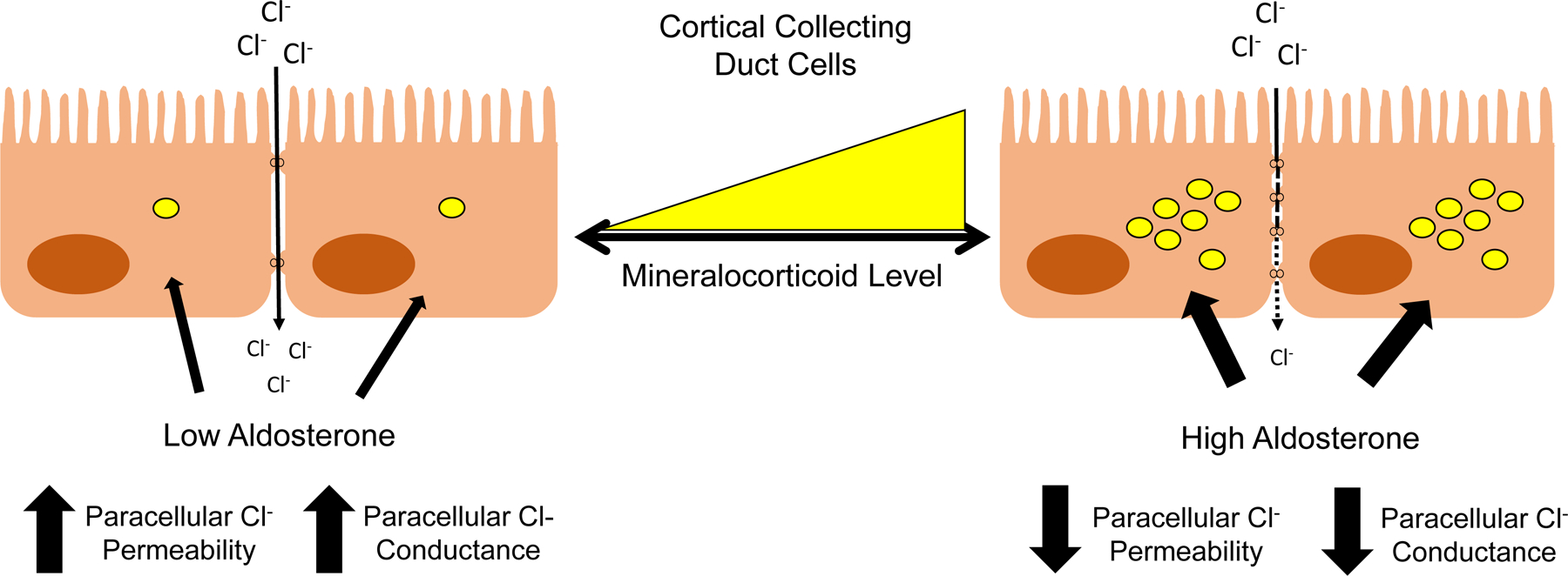
Aldosterone exerts profound effects on renal and cardiovascular physiology. In the kidney, aldosterone acts to preserve electrolyte and acid-base balance in response to changes in dietary sodium (Na+ ) or potassium (K+ ) intake. These physiological actions, principally through activation of mineralocorticoid receptors (MRs), have important effects particularly in patients with renal and cardiovascular disease as demonstrated by multiple clinical trials. Multiple factors, be they genetic, humoral, dietary, or otherwise, can play a role in influencing the rate of aldosterone synthesis and secretion from the adrenal cortex. Normally, aldosterone secretion and action respond to dietary Na+ intake. In the kidney, the distal nephron and collecting duct are the main targets of aldosterone and MR action, which stimulates Na+ absorption in part via the epithelial Na+ channel (ENaC), the principal channel responsible for the fine-tuning of Na+ balance. Our understanding of the regulatory factors that allow aldosterone, via multiple signaling pathways, to function properly clearly implicates this hormone as central to many pathophysiological effects that become dysfunctional in disease states. Numerous pathologies that affect blood pressure (BP), electrolyte balance, and overall cardiovascular health are due to abnormal secretion of aldosterone, mutations in MR, ENaC, or effectors and modulators of their action. Study of the mechanisms of these pathologies has allowed researchers and clinicians to create novel dietary and pharmacological targets to improve human health. This article covers the regulation of aldosterone synthesis and secretion, receptors, effector molecules, and signaling pathways that modulate its action in the kidney. We also consider the role of aldosterone in disease and the benefit of mineralocorticoid antagonists. © 2023 American Physiological Society. Compr Physiol 13:4409-4491, 2023.


Figures

Figure 1:
Figure 1:. Mineralocorticoid and Glucocorticoid synthesis pathways.
Enzymes involved in pathway reactions are in bold
Figure 2:
Figure 2:. The adrenal gland.
The adrenal cortex is made up of three layers: the outermost layer is known as the zona glomerulosa and is the main area for mineralocorticoid production. The middle layer of the cortex is called the zona fasciculata, and mainly synthesizes glucocorticoids. The inner layer of the cortex is known as the zona reticularis, and this section produces androgens. The core of the adrenal gland is known as the adrenal medulla, which is the site of catecholamine production.
Figure 3:
Figure 3:. The renin-angiotensin aldosterone system (RAAS).
In response to a decrease in blood volume, BP decreases, which elicits a response from the kidney to increase BP. This renal response is mediated by the production and secretion of renin by the juxtaglomerular cells in response to decreased arterial pressure sensed by renal baroreceptors and decreased luminal NaCl concentration sensed by the macula densa. The enzyme renin cleaves angiotensinogen, also known as renin substrate to produce angiotensin I, which is further processed to angiotensin II. Angiotensin II increases blood volume by two mechanisms: directly constricting systemic and renal arteries and arterioles, and by stimulating the production of aldosterone from the adrenal gland. The subsequent increase in vascular resistance and the NaCl and water reabsorption restores blood volume and BP toward normal and reduces RAAS activity.
Figure 4.
Figure 4.. Steroid-binding properties of the human mineralocorticoid receptor (MR) expressed in cultured cells.
(A) Scatchard analysis of tritiated aldosterone binding in extracts prepared from pRShMR-transfected COS cells (B and C) Competition of unlabeled steroids for binding with 5 nM [3H] aldosterone in transfected COS cell extracts Abbreviations: Aldo, aldosterone, Doc, deoxycorticosterone; Dex, dexamethasone; Spiro, spironolactone; E2, 17β-estradiol; CS, corticosterone; HC, hydrocortisone; and Prog, progesterone. Figure adapted from Arriza et al (28).
Figure 5:
Figure 5:. Cellular Mechanisms of Na+ and K+ Transport in the Aldosterone-sensitive Distal Nephron (ASDN).
The ASDN, consisting of the late distal convoluted tubule (DCT2), the connecting (CNT) and initial collecting tubule (ICT), and the collecting duct (CD), express the mineralocorticoid receptor (MR) and the high affinity enzyme 11ß-hydroxysteroid dehydrogenase type 2 (11ß-HSD2) which oxidizes cortisol to cortisone and is important in conferring mineralocorticoid specificity to the ASDN. Na+ reabsorption occurs predominantly by the electroneutral NaCl cotransporter in the DCT2, with progressive increasing the proportion of electrogenic Na+ absorption occurring in the CNT, ICT and CD. K+ in the ASDN is secreted by two classes of K+ channels, inwardly-rectifying K+ channels (Kir1.1; also known as the renal outer medullary channel or ROMK) and large conductance Ca2+-activated K+ channels (also known as BK or Maxi-K channels). The apical KCl cotransporter in principal cells is involved in non-conductive K+ secretion. K+ absorption is an active process driven by an apical HKα1 H+K+-ATPase and basolateral K+ channels in intercalated cells and in the principal cells by HKα2 H+K+-ATPase (not shown). The ~ symbol indicates an ATPase.
Figure 6:
Figure 6:. Stimulation by aldosterone of active sodium transport across the toad bladder in vitro.
Experiments performed on paired membranes. The serosal surface of one bladder half was exposed to aldosterone and the corresponding half served as control. Eight toads had been maintained in distilled water, eight in saline, prior to these incubations. Figure adapted from Crabbé et al (146).
Figure 7:
Figure 7:. Effect of aldosterone on urine Na excretion.
Results of a representative clearance experiment. Urine sodium excretion (UNaV), urinary Na:K concentration ratio (Una/UK), and glomerular filtration rate (GFR) are graphed over time before and after intravenous aldosterone (arrow). Adapted from Wingo et al (905).
Figure 8:
Figure 8:. Effect of low versus high mineralocorticoid levels on paracellular chloride (Cl) permeability and conductance.
The low tissue conductance in response to high mineralocorticoid stimulation correlates with a high resistance. At low mineralocorticoid levels, tissue conductance increases to modest levels.
Figure 9:
Figure 9:. The role for the renal aldosterone endothelin feedback system (RAEFS) on mineralocorticoid-stimulated Na+ retention.
Normal mice are shown in black lines and squares. Mice not expressing ET-1 in the collecting duct (CD ET-1 KO) are shown in dashed lines and open circles. Normal mice exhibit transient Na+ retention before escaping from progressive positive Na+ balance. CD ET-1 KO fail to undergo aldosterone escape, with persistent positive daily Na+ balance. Daily Na+ balance over days 1–19. Solid lines are controls, and dotted lines are CD ET-1 KO. Control, n = 9; CD ET-1 KO, n = 6. Data is shown as means + SE. In B, *P < 0.05 within control, day 1 vs. days 2, 3, 6, and 7, Tukey's test; †P < 0.05 within control, day 11 vs. days 9–12 and 14–19, Tukey's test; ‡P < 0.05, significant effect of genotype, repeated-measures ANOVA. Adapted from Lynch et al (506).
Figure 10:
Figure 10:. Diagram of a theoretical renal aldosterone-endothelin feedback system (RAEFS) in the collecting duct.
In response to a low Na+ intake, high aldosterone levels activate mineralocorticoid receptors (MR), but the production of endothelin-1 (ET-1) is attenuated by reduced distal luminal Na+ delivery and flow. Consequently, ET-1 mediated inhibition of ENaC in the collecting duct (CD) is reduced and the CD is poised to enhance Na+ absorption. When aldosterone is inappropriately high for the dietary Na+ content, high luminal Na and flow serve to enhance aldosterone mediated ET-1 activity which reduces Na absorption. Evidence for a sex-dependent natriuretic effect of ET-1 via endothelin A (ETA) receptors has been shown previously by Nakano et al (565). Dashed line indicates attenuation of pathway. UNaV, urinary sodium excretion; ETB, endothelin B receptors; NOS1, nitric oxide synthase 1; NO, nitric oxide.
Figure 11.
Figure 11.
Summary of principal results of simultaneous microperfusion experiments (distal tubular potassium secretion, JK, top) and clearance experiments (urinary flow rate, V, and potassium excretion, UKV, middle and lower, respectively) in each of the five experimental groups studied. The conditions prevailing in each group are shown at the bottom of the figure: N represents normal (for plasma K+), or basal (for hormone levels). ALDO, aldosterone; DEX, dexamethasone. Adapted from Field et al (213).
Figure 12:
Figure 12:. Molecular pathways through which aldosterone can stimulate ENaC.
Mineralocorticoid receptor (MR) activation by aldosterone leads to stimulation of the epidermal growth factor receptor (EGFR). Stimulation of EGFR lead to multiple cascading pathways, including the mitogen activated protein kinase (MAPK) and the phosphatidylinositol 3-kinase (PI3-K) pathways. The MAPK pathway leads to an inhibition of ENaC by the ERK1/2, reducing Na+ reabsorption and promoting urinary Na+ excretion. The PI3-K pathway leads to the stimulation of ENaC through the synergistic inhibition of the ubiquitin ligase Nedd4-2 by MR transcription products SGK1 and GILZ. GILZ can also inhibit the ability of the MAPK pathway to inhibit ENaC by inhibiting Raf1 (c-Raf). Black arrows indicate direction of pathways. Blue arrows indicate aldosterone-bound MR relocating to the nucleus. Green arrows indicate MR transcription products. ENaC, epithelial sodium channel; GILZ, glucocorticoid-induced leucine zipper; SGK1, serum- and glucocorticoid-induced kinase 1; PKB, protein kinase B (also known as Akt), PTEN, Phosphatidylinositol-3,4,5-trisphosphate 3-phosphatase; mTORC2, mammalian target of rapamycin complex 2, PDK1/2, phosphoinositide-dependent kinases 1 and 2; ERK1/2, extracellular signal-regulated kinases 1 and 2; MEK, MAPK/ERK kinase or mitogen-activated protein kinase kinase (MAPKK).
Figure 13:
Figure 13:. Other pathways stimulated by aldosterone and mineralocorticoid receptor (MR) activation.
MR activation by aldosterone leads to stimulation of the epidermal growth factor receptor (EGFR), either on its own or through activation of c-Src. Stimulation of EGFR leads to multiple cascading pathways, including the MEK/ERK and PI3K pathways (Figure 12), along with the JNK/SAPK pathways. MR-induced activation of c-Src can also lead to the activation of p38MAPK, a kinase pathway commonly known to be induced by cell stress or inflammatory cytokines. MAPK, mitogen activated protein kinase; ERK, extracellular signal-regulated kinase; MEK, MAPK/ERK kinase or mitogen-activated protein kinase kinase (MAPKK, also known as MKK); MEKK, map kinase kinase kinase; PI3-K, phosphatidylinositol 3-kinase. JNK/SAPK, c-Jun NH2-terminal kinases/stress-activated protein kinase; TGFβR, Transforming growth factor ß receptor.

References

    1. Abd El-Aziz TM, Soares AG, Mironova E, Boiko N, Kaur A, Archer CR, Stockand JD, and Berman JM. Mechanisms and consequences of casein kinase II and ankyrin-3 regulation of the epithelial Na(+) channel. Sci Rep 11: 14600, 2021. - PMC - PubMed
    1. Ackermann D, Gresko N, Carrel M, Loffing-Cueni D, Habermehl D, Gomez-Sanchez C, Rossier BC, and Loffing J. In vivo nuclear translocation of mineralocorticoid and glucocorticoid receptors in rat kidney: differential effect of corticosteroids along the distal tubule. Am J Physiol Renal Physiol 299: F1473–1485, 2010. - PubMed
    1. Agarwal AK, Mune T, Monder C, and White PC. NAD(+)-dependent isoform of 11 beta-hydroxysteroid dehydrogenase. Cloning and characterization of cDNA from sheep kidney. J Biol Chem 269: 25959–25962, 1994. - PubMed
    1. Agrawal S, He JC, and Tharaux PL. Nuclear receptors in podocyte biology and glomerular disease. Nat Rev Nephrol 17: 185–204, 2021. - PubMed
    1. Aguilera G, and Catt KJ. Participation of voltage-dependent calcium channels in the regulation of adrenal glomerulosa function by angiotensin II and potassium. Endocrinology 118: 112–118, 1986. - PubMed

LinkOut - more resources