

A pilot study of lymphoscintigraphy with tracer injection into the human brain
- PMID: 36994857
- PMCID: PMC10369147
- DOI: 10.1177/0271678X231160891
A pilot study of lymphoscintigraphy with tracer injection into the human brain
Abstract
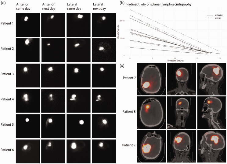
Many groups have reported lymphatic and glymphatic structures in animal and human brains, but tracer injection into the human brain to demonstrate real-time lymphatic drainage and mapping has not been described. We enrolled patients undergoing standard-of-care resection or stereotactic biopsy for suspected intracranial tumors. Patients received peritumoral injections of 99mTc-tilmanocept followed by planar or tomographic imaging. Fourteen patients with suspected brain tumors were enrolled. One was excluded from analysis because of tracer leakage during injection. There was no drainage of 99mTc-tilmanocept to regional lymph nodes in any of the patients. On average, after correcting for radioactive decay, 70.7% (95% CI: 59.9%, 81.6%) of the tracer in the injection site and 78.1% (95% CI: 71.1%, 85.1%) in the whole-head on the day of surgery remained the morning after, with variable radioactivity in the subarachnoid space. The retained fraction was much greater than expected based on the clearance rate from non-brain injection sites. In this pilot study, the lymphatic tracer 99mTc-tilmanocept was injected into the brain parenchyma, and there was no drainage outside the brain to the cervical lymph nodes. Our work demonstrates an inefficiency of drainage from peritumoral brain parenchyma and highlights a therapeutic opportunity to improve immunosurveillance of the brain.
Keywords: Brain; glymphatics; lymphatics; neuroimmunology; tilmanocept.
Conflict of interest statement
The author(s) declared no potential conflicts of interest with respect to the research, authorship, and/or publication of this article.
Figures


Similar articles
-
Within-patient comparison between [68Ga]Ga-tilmanocept PET/CT lymphoscintigraphy and [99mTc]Tc-tilmanocept lymphoscintigraphy for sentinel lymph node detection in oral cancer: a pilot study.Eur J Nucl Med Mol Imaging. 2022 May;49(6):2023-2036. doi: 10.1007/s00259-021-05645-0. Epub 2021 Dec 28. Eur J Nucl Med Mol Imaging. 2022. PMID: 34962582
-
Use of 99mTc-Tilmanocept as a Single Agent for Sentinel Lymph Node Identification in Breast Cancer: A Retrospective Pilot Study.J Nucl Med Technol. 2017 Sep;45(3):181-184. doi: 10.2967/jnmt.117.194415. Epub 2017 Jul 13. J Nucl Med Technol. 2017. PMID: 28705929
-
Comparing the hybrid fluorescent-radioactive tracer indocyanine green-99mTc-nanocolloid with 99mTc-nanocolloid for sentinel node identification: a validation study using lymphoscintigraphy and SPECT/CT.J Nucl Med. 2012 Jul;53(7):1034-40. doi: 10.2967/jnumed.112.103127. Epub 2012 May 29. J Nucl Med. 2012. PMID: 22645297
-
The application of sentinel node radiolocalization to solid tumors of the head and neck: a 10-year experience.Laryngoscope. 2004 Jan;114(1):2-19. doi: 10.1097/00005537-200401000-00002. Laryngoscope. 2004. PMID: 14709988 Review.
-
Anatomy and physiology of lymphatic drainage of the breast from the perspective of sentinel node biopsy.J Am Coll Surg. 2001 Mar;192(3):399-409. doi: 10.1016/s1072-7515(00)00776-6. J Am Coll Surg. 2001. PMID: 11245383 Review.
Cited by
-
Immunotherapy for Brain Tumors: Where We Have Been, and Where Do We Go From Here?Curr Treat Options Oncol. 2024 May;25(5):628-643. doi: 10.1007/s11864-024-01200-9. Epub 2024 Apr 23. Curr Treat Options Oncol. 2024. PMID: 38649630 Review.
-
Revolutionizing lymph node metastasis imaging: the role of drug delivery systems and future perspectives.J Nanobiotechnology. 2024 Mar 29;22(1):135. doi: 10.1186/s12951-024-02408-5. J Nanobiotechnology. 2024. PMID: 38553735 Free PMC article. Review.
References
-
- Bradbury MW, Cserr HF, Westrop RJ. Drainage of cerebral interstitial fluid into deep cervical lymph of the rabbit. Am J Physiol Renal Physiol 1981; 240: F329–F336. - PubMed
-
- Ling C, Sandor M, Fabry Z. In situ processing and distribution of intracerebrally injected OVA in the CNS. J Neuroimmunol 2003; 141: 90–98. - PubMed
Publication types
MeSH terms
Substances
Grants and funding
LinkOut - more resources
Full Text Sources
