. 2023 Apr 1;16(4):dmm049712.
doi: 10.1242/dmm.049712. Epub 2023 Apr 13.

Overexpression screen of chromosome 21 genes reveals modulators of Sonic hedgehog signaling relevant to Down syndrome

Affiliations

Overexpression screen of chromosome 21 genes reveals modulators of Sonic hedgehog signaling relevant to Down syndrome

Anna J Moyer et al. Dis Model Mech. .

Abstract

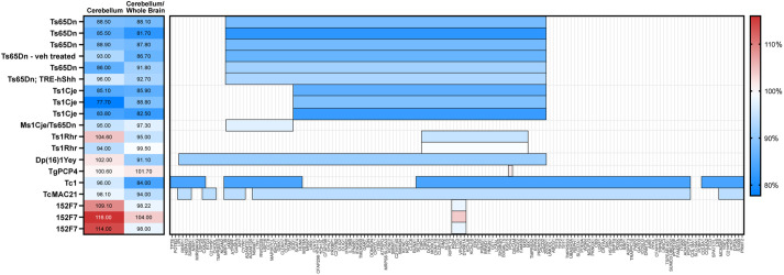
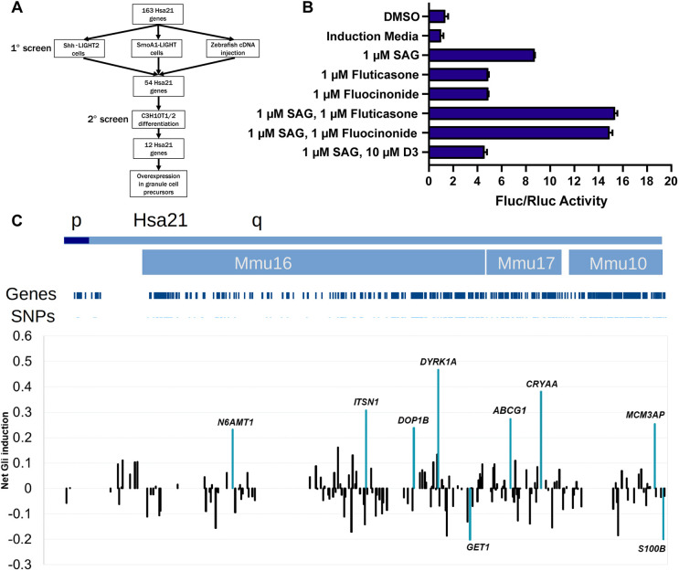
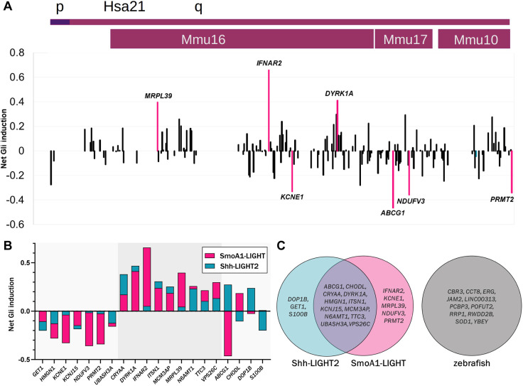
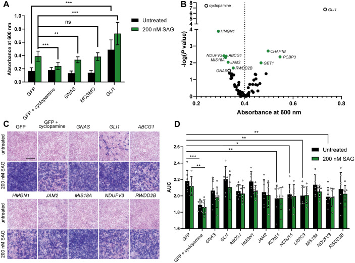
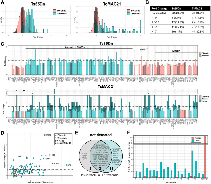
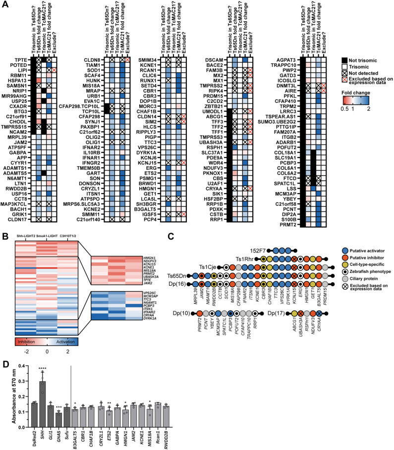
Trisomy 21 and mutations in the Sonic hedgehog (SHH) signaling pathway cause overlapping and pleiotropic phenotypes including cerebellar hypoplasia, craniofacial abnormalities, congenital heart defects and Hirschsprung disease. Trisomic cells derived from individuals with Down syndrome possess deficits in SHH signaling, suggesting that overexpression of human chromosome 21 genes may contribute to SHH-associated phenotypes by disrupting normal SHH signaling during development. However, chromosome 21 does not encode any known components of the canonical SHH pathway. Here, we sought to identify chromosome 21 genes that modulate SHH signaling by overexpressing 163 chromosome 21 cDNAs in a series of SHH-responsive mouse cell lines. We confirmed overexpression of trisomic candidate genes using RNA sequencing in the cerebella of Ts65Dn and TcMAC21 mice, model systems for Down syndrome. Our findings indicate that some human chromosome 21 genes, including DYRK1A, upregulate SHH signaling, whereas others, such as HMGN1, inhibit SHH signaling. Individual overexpression of four genes (B3GALT5, ETS2, HMGN1 and MIS18A) inhibits the SHH-dependent proliferation of primary granule cell precursors. Our study prioritizes dosage-sensitive chromosome 21 genes for future mechanistic studies. Identification of the genes that modulate SHH signaling may suggest new therapeutic avenues for ameliorating Down syndrome phenotypes.

Keywords: Aneuploidy; Down syndrome; Gene dosage effects; Genetic screen; Sonic hedgehog; Trisomy 21.


Conflict of interest statement

Competing interests M.O. is a CEO, employee and shareholder of Trans Chromosomics, Inc. Other authors declare no competing interests.

Figures

Fig. 1.
Fig. 1.
Comparison of cerebellar phenotypes in Down syndrome mouse models. (A) Previously published cerebellar volume or cross-sectional area and cerebellar volume or cross-sectional area normalized to that of the whole brain for mouse models are reported as a percentage of the corresponding values in euploid mice. Horizontal bars represent the human chromosome 21 or mouse orthologous regions that are trisomic in each model. Colors reflects the extent of cerebellar hypoplasia, where blue is the most affected and red is the least affected. Several additional studies quantifying cerebellar hypoplasia are generally consistent with these results but do not report these cerebellar measurements (Duchon et al., 2022, 2021; Garcia-Cerro et al., 2018; Ma et al., 2020). Publications referenced in this figure are listed in Table S1.
Fig. 2.
Fig. 2.
Overexpression of human chromosome 21 cDNAs in Shh-LIGHT2 cells. (A) Screening strategy for chromosome 21 cDNAs in Shh-LIGHT2 and SmoA1-LIGHT cell lines, zebrafish embryos, the C3H10T1/2 mesenchymal stem cell line, and primary granule cell precursors. (B) Fluc/Rluc activity in Shh-LIGHT2 cells exposed to SAG, the glucocorticoids fluocinonide and fluticasone, and vitamin D3, normalized to that of the induction media control (n=2 independent experiments with 12 technical replicates per treatment). All graphs show mean±s.d. unless otherwise noted. (C) Shh-LIGHT2 cells transfected with expression constructs for 163 chromosome 21 cDNAs and treated with SAG to induce SHH signaling (≥8 technical replicates per cDNA; see Table S4 for wells per cDNA). Averaged Fluc/Rluc activity for each gene across the Shh-LIGHT2 screen was scaled to 0 to show signal deflections from baseline. Values less than zero represent loci that decrease SAG-induced activation of the SHH signaling pathway. The net activity of the 8×GliBS reporter for each cDNA is plotted in chromosomal order according to the sequence along the proximal-distal length of human chromosome 21. Orthologous regions on mouse chromosomes 16, 17, and 10 are provided for additional context. The labeled cDNAs increased or decreased Fluc/Rluc activity by more than two standard deviations.
Fig. 3.
Fig. 3.
Overexpression of human chromosome 21 cDNAs in SmoA1-LIGHT cells. (A) SmoA1-LIGHT cells transfected with expression constructs for 163 human chromosome 21 cDNAs (≥8 technical replicates per cDNA; see Table S5 for wells per cDNA). Averaged Fluc/Rluc activity for each gene across the SmoA1-LIGHT screen was scaled to zero to show signal deflections from baseline. The labeled cDNAs increased or decreased Fluc/Rluc activity by more than two standard deviations. (B) Comparison of net reporter induction after overexpression of twenty cDNAs identified in SmoA-LIGHT and Shh-LIGHT2 screens. Sixteen cDNAs have the same direction of effect in both screens, whereas four cDNAs have opposite effects. The gray highlight indicates cDNAs that increase Fluc/Rluc activity in both cell lines. (C) Comparison of cDNAs identified in two luciferase assays and a previous screen in developing zebrafish embryos (Edie et al., 2018).
Fig. 4.
Fig. 4.
Overexpression of human chromosome 21 genes affects osteoblast differentiation of C3H10T1/2 cells. (A) Overexpression of GLI1 promotes osteoblast differentiation in the presence or absence of SAG, whereas treatment with cyclopamine or overexpression of GNAS inhibits SAG-induced osteoblast differentiation (n=20) (two-tailed unpaired Student's t-test). (B) Quantification of alkaline phosphatase activity in C3H10T1/2 cells transfected with human chromosome 21 cDNAs and treated with SAG (n=20). Multiple comparisons were corrected for by controlling the false discovery rate; green circles denote cDNAs with q<0.1; open circles denote controls (Kruskal–Wallis test followed by Dunn's post hoc test). (C) Representative images of alkaline phosphatase staining in C3H10T1/2 cells transfected with human chromosome 21 cDNAs and counterstained with Nuclear Fast Red (n=3). Scale bar: 100 μm. (D) MTT viability assay in C3H10T1/2 cells transfected with chromosome 21 cDNAs (n=7). The y-axis represents area under the curve (AUC) values of cell viability 48, 72 and 96 h (n=7 for each) after transfection (two-way ANOVA followed by Fisher's least significant difference test). Differences reported as statistically significant have q<0.05. ns, not significant; *P<0.05; **P<0.01; ***P<0.001.
Fig. 5.
Fig. 5.
Expression pattern of chromosome 21 genes and their mouse orthologs in Ts65Dn and TcMAC21 cerebellum. (A) Density histograms of disomic (salmon) and trisomic (teal) fold changes in Ts65Dn and TcMAC21 cerebella (n=4 Ts65Dn, 4 Ts65Dn euploid littermates, 4 TcMAC21, 4 TcMAC21 euploid littermates). The plots represent 13,807 detectable transcripts. (B) Trisomic gene fold changes binned by expression levels. (C) Fold changes of human chromosome 21 genes and their mouse orthologs arranged in chromosomal order from proximal to distal. Human chromosome 21 orthologs are located on mouse chromosome 16 (MMU16), MMU17 and MMU10. For TcMAC21, teal represents the proportion of length-normalized reads contributed by mouse copies and dark teal represents reads derived from the human chromosome. Four previously reported deletions are labeled ‘A’ through ‘D’. Five human genes that were detected in TcMAC21 but have no expression of mouse orthologs for normalization (POTED, BTG3, RUNX1, C21orf58 and TSPEAR-AS1) are excluded. (D) Scatterplot of log2(fold change) values for human chromosome 21 gene expression in TcMAC21 P6 cerebellum and P1 forebrain (Kazuki et al., 2020). Pearson correlation coefficient R=0.392 and P=2.3×10⁻⁵. (E) Human chromosome 21 transcripts not detected in the P6 cerebellum, P1 forebrain or both. (F) Chromosomal locations of differentially expressed genes in Ts65Dn and TcMAC21 cerebellum. Trisomic genes are located on MMU16 and MMU17 in Ts65Dn mice and Hsa21 in TcMAC21 mice.
Fig. 6.
Fig. 6.
Prioritization of candidate cDNAs and overexpression in primary granule cell precursors. (A) Summary of expression data in the developing cerebellum. Black boxes indicate genes that are not trisomic in Ts65Dn and TcMAC21 mouse models, and white boxes indicate genes that are trisomic in these models. Fold change in gene expression is indicated by color, with red signifying decreased expression and blue signifying increased expression. Transcripts with black crosses were not detected in our RNA-seq dataset, and transcripts with red crosses were excluded based on our expression data, expression in the BrainSpan Atlas of the Developing Human Brain and single cell RNA-seq data from euploid mouse granule cell precursors and granule cells. (B) Comparison of the effects of 54 human chromosome 21 cDNAs in Shh-LIGHT2, SmoA1-LIGHT and C3H10T1/2 screens. cDNAs are sorted by average z-score, with red signifying inhibition and blue signifying activation of the SHH pathway. The inset shows top- and bottom-ranked cDNAs. (C) Chromosomal locations of the mouse orthologs of candidate cDNAs in Down syndrome mouse models. LINC00313 (C21ORF84) and TPTE are not shown. LINC00313, which was identified in the zebrafish screen, is a human-specific gene and not present in the listed mouse models. TPTE is located on the short arm of human chromosome 21 and has a putative homolog on mouse chromosome 8. (D) Lentiviral overexpression of candidate genes inhibits proliferation of granule cell precursors treated with 6 nM SAG and pulsed with EdU for 24 h (n=4). Dark gray bars indicate control cDNAs and light gray bars indicate human chromosome 21 cDNAs. *P<0.05; **P<0.01; ***P<0.001; ****P<0.0001 (one-way ANOVA followed by Fisher's least significant difference test). Differences reported as statistically significant have q<0.05.

Similar articles

Cited by

References

    1. Aldridge, K., Reeves, R. H., Olson, L. E. and Richtsmeier, J. T. (2007). Differential effects of trisomy on brain shape and volume in related aneuploid mouse models. Am. J. Med. Genet. A 143A, 1060-1070. 10.1002/ajmg.a.31721 - DOI - PMC - PubMed
    1. Anders, S. and Huber, W. (2010). Differential expression analysis for sequence count data. Genome Biol. 11, R106. 10.1186/gb-2010-11-10-r106 - DOI - PMC - PubMed
    1. Andreu-Cervera, A., Catala, M. and Schneider-Maunoury, S. (2021). Cilia, ciliopathies and hedgehog-related forebrain developmental disorders. Neurobiol. Dis. 150, 105236. 10.1016/j.nbd.2020.105236 - DOI - PubMed
    1. Arbones, M. L., Thomazeau, A., Nakano-Kobayashi, A., Hagiwara, M. and Delabar, J. M. (2019). DYRK1A and cognition: a lifelong relationship. Pharmacol. Ther. 194, 199-221. 10.1016/j.pharmthera.2018.09.010 - DOI - PubMed
    1. Atas-Ozcan, H., Brault, V., Duchon, A. and Herault, Y. (2021). Dyrk1a from gene function in development and physiology to dosage correction across life span in down syndrome. Genes (Basel) 12, 1833. 10.3390/genes12111833 - DOI - PMC - PubMed

Publication types