Review
. 2023 Mar 30;6(1):21.
doi: 10.1186/s42155-023-00364-z.

Standardizing lymphangiography and lymphatic interventions: a preclinical in vivo approach with detailed procedural steps

Affiliations
Review

Standardizing lymphangiography and lymphatic interventions: a preclinical in vivo approach with detailed procedural steps

Feng Pan et al. CVIR Endovasc. .

Abstract

Purpose: To present a preclinical in vivo approach for standardization and training of lymphangiography and lymphatic interventions using a pictorial review.

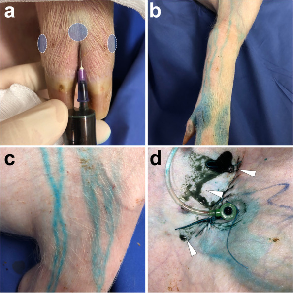
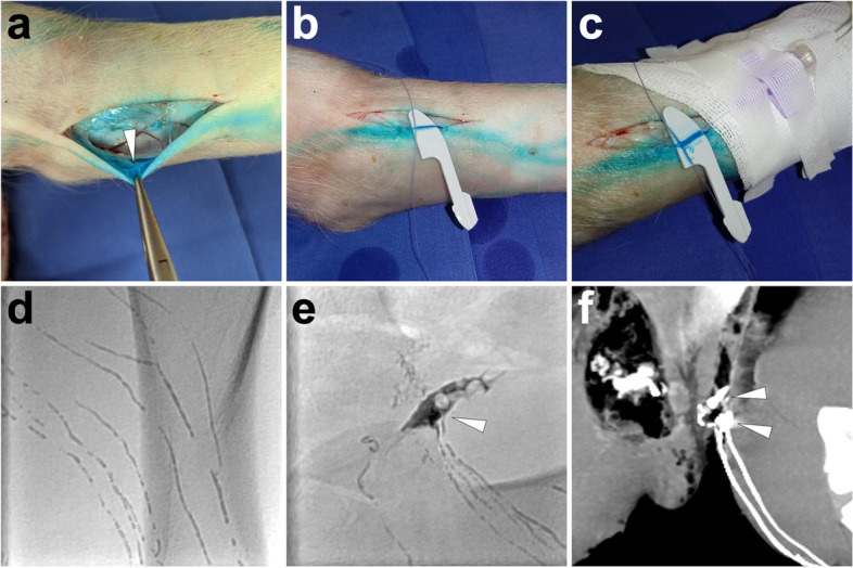
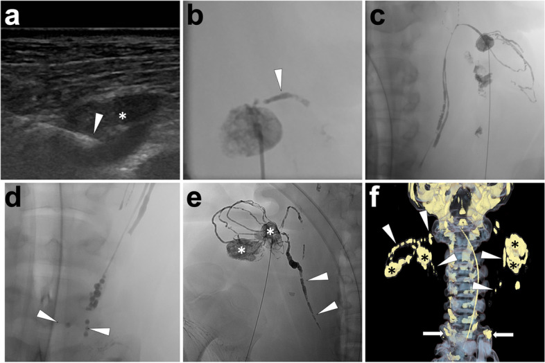
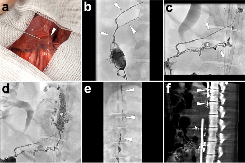
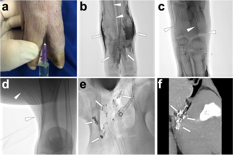
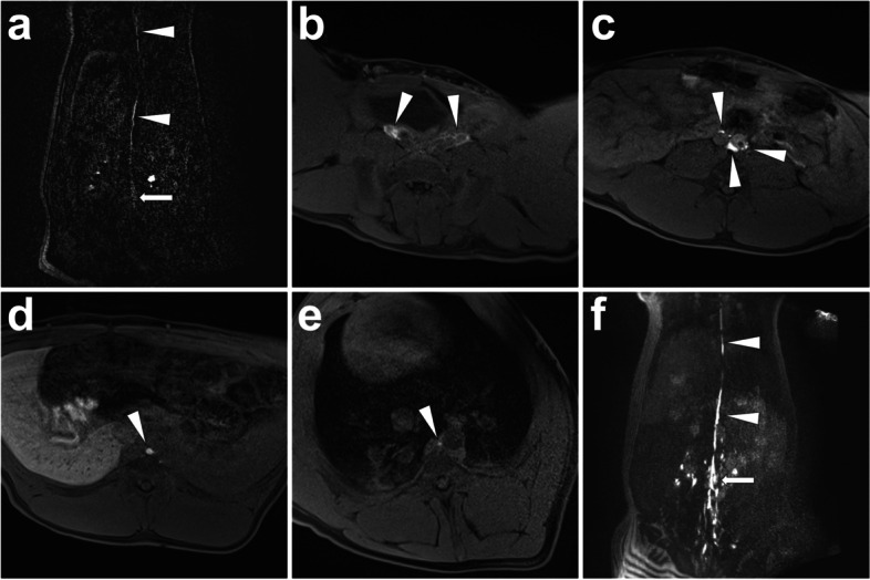
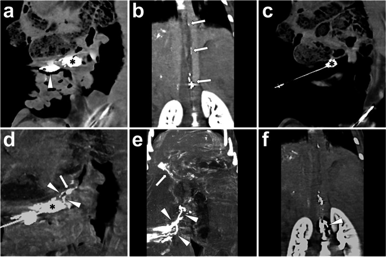
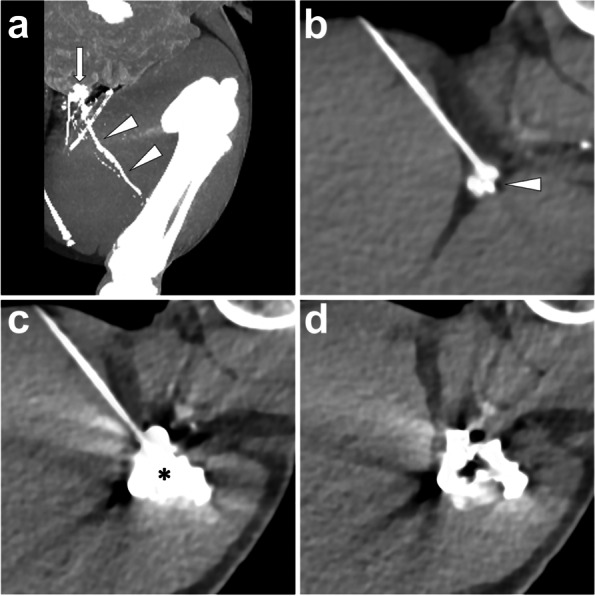
Materials and methods: Different lipiodol- and gadolinium-based lymphangiography and lymphatic interventions were performed in twelve (12) landrace pigs with a mean bodyweight of 34 ± 2 kg using various imaging and guiding modalities, similar to the procedures used in humans. The techniques used were explicitly introduced and illustrated. The potential applications of each technique in preclinical training were also discussed.

Results: By applying visual, ultrasonography, fluoroscopy, CT, cone-beam CT, and/or MRI examination or guidance, a total of eleven techniques were successfully implemented in twelve pigs. The presented techniques include inguinal postoperative lymphatic leakage (PLL) establishment, interstitial dye test, five types of lymphangiography [incl. lipiodol-based translymphatic lymphangiography (TL), lipiodol-based percutaneous intranodal lymphangiography (INL), lipiodol-based laparotomic INL, lipiodol-based interstitial lymphangiography, and interstitial magnetic resonance lymphangiography (MRL)], and four types of percutaneous interventions in the treatment of PLL [incl. thoracic duct embolization (TDE), intranodal embolization (INE), afferent lymphatic vessel sclerotherapy (ALVS), and afferent lymphatic vessel embolization (ALVE)].

Conclusion: This study provides a valuable resource for inexperienced interventional radiologists to undergo the preclinical training in lymphangiography and lymphatic interventions using healthy pig models.

Keywords: Animal Model; Lymphatic Intervention; Lymphography; Postoperative Lymphatic Leakage; Preclinical Training.


Conflict of interest statement

The corresponding author declares that this animal study was financially supported by Research & Innovation Department, Guerbet, Roissy, France.

Figures

Fig. 1
Fig. 1
PLL establishment in a healthy pig. After a transverse incision at the left groin, a 6-French vascular sheath accessed the left femoral artery under direct vision. Afterwards, the incision was sutured. The image was from Pig No.3. Abbreviation: PLL, postoperative lymphatic leakage
Fig. 2
Fig. 2
Interstitial dye test with the PLL identification. Fifteen minutes after the interdigital (imaginary circle) injection of Patent Blue V (A) at the right foot (1 ml per each interdigital space), the subcutaneous lymphatic vessels at the lower limb (B) and thigh (C) were dyed blue color which could be visually observed. With more time, PLL could be identified by the blue dye extravasation (white arrowheads) from the incision around the arterial sheath (D). Images were from Pig No. 3. Abbreviation: PLL, postoperative lymphatic leakage
Fig. 3
Fig. 3
Illustration of lipiodol-based TL in a PLL pig model. Fifteen minutes after Patent Blue V injection at feet, along with the dye, incise the skin at the left lower limb to identify an accessible lymphatic vessel (white arrowhead) as the target for cannulation (A). Then, separate the targeted lymphatic vessel with a plastic blade and a suture (B). Afterwards, access the lymphatic vessel with a 26-gauge trocar needle, which was later fixed and connected to an infusion tube (C). When manually injecting the lipiodol, linear lymphatic vessels in the lower limb were opacified under fluoroscopy (D). The lipiodol extravasation (white arrowhead) could be fluoroscopically observed at the left groin with lymphatic vessel disruptions (E). In the coronal MIP image of the sequential post-lymphangiography CT, lipiodol extravasation (white arrowheads) could also be delineated (F). Images were from Pig No. 1. Abbreviations: TL, translymphatic lymphangiography; PLL, postoperative lymphatic leakage; MIP, maximum intensity projection; CT, computed tomography
Fig. 4
Fig. 4
Illustration of lipiodol-based cervical INL in a pig model. In this case, lipiodol-based cervical INL was introduced. Under ultrasonography guidance, the tip of the needle (white arrowhead) was advanced until reaching the center of the target lymph node (*) (A). An efferent lymphatic vessel (white arrowhead) at the left neck was opacified after a small amount of lipiodol injection (B); then, continue the lipiodol injection. Over time, lipiodol gradually shifted towards the left jugular vein angle over time (C). The lipiodol injection ceased when oil-dropped lipiodol entered the left brachiocephalic vein (white arrowheads; D). As the same, the lymph nodes (*), efferent lymphatic vessels, and right lymphatic duct (white arrowheads) at the right side of the neck were gradually opacified after the lipiodol injection (E). After INL, remove the needles and carry out the volume rendering reconstruction of post-lymphangiography CT to demonstrate the three-dimensional lymphatic structure, such as lymph nodes (*), lymphatic vessels (white arrowheads), and bilateral confluences of jugular venous angles (white arrows; F). Images were from Pig No. 7. Abbreviation: INL, intranodal lymphangiography; CT, computed tomography
Fig. 5
Fig. 5
Illustration of lipiodol-based laparoscopic INL in a pig model. After laparotomic exposure of the mesentery, the translucent lymph node (white arrowhead) was punctured using a 22-gauge Chiba needle (A). Under fluoroscopy monitoring, a small amount of lipiodol (1 ml) was first injected. The opacification of the targeted lymph node and efferent lymphatic vessels (white arrowheads) indicated an ideal needle position (B). Afterwards, continuous injection of lipiodol visualized the mesenteric lymphatics (white arrowheads; C), mesenteric lymph nodes (*; C), cisterna chyli (*; D), and thoracic duct (white arrowheads; E). The MIP image of the following post-lymphangiography CT presented the details between lymphatic structures such as cisterna chyli (*) and thoracic duct (white arrowheads) to the surrounding tissue (F). Images were from Pig No. 4. Abbreviations: INL, intranodal lymphangiography; MIP, maximum intensity projection; CT, computed tomography
Fig. 6
Fig. 6
Illustration of lipiodol-based interstitial lymphangiography in a pig model. First, inject 2 ml lipiodol at each interdigital space of the left foot, similar to the interstitial dye test (A). After 15 min, the interstitial (white arrows) and surrounding lymphatic vessels (white arrowheads) were opacified under fluoroscopy (B). Over time, the lymphatic vessels (white arrowheads) at the ankle (C) and lower leg (D) were visually opacified under fluoroscopy. When the lipiodol reached the left groin region, evident extravasation of lipiodol (white arrows) was observed with the disruption of lymphatic vessels, indicating a typical PLL diagnosis (E). The coronal MIP image of the sequential post-lymphangiography CT demonstrated the subcutaneously sporadic distribution of lipiodol (white arrows) at the left groin (F). Images were from Pig No. 10. Abbreviations: PLL, postoperative lymphatic leakage; MIP, maximum intensity projection
Fig. 7
Fig. 7
Illustration of interstitial MRL in a pig model. After interstitial injection of gadoxetate disodium at the bilateral foot dorsal, a dynamic coronal 3D-TWIST scan was performed. Thirty minutes later, the definite thoracic duct (white arrowheads) started from cisterna chyli (white arrow) was visualized (A). Afterwards, a 3D-FLASH scan was performed, which presented the continuous lymphatic network surrounding bilateral iliac arteries and veins (white arrowheads; B) to retroperitoneal lymphatic vessels (white arrowheads; C) and thoracic duct (white arrowhead; D, E). Coronal 3D-FLASH MIP image demonstrated a clear structure of retroperitoneal lymphatic vessels, cisterna chyli (white arrow), and proximal thoracic duct (white arrowheads) (F). Images were from Pig No. 11. Abbreviations: MRL, magnetic resonance lymphangiography; 3D-TWIST, three-dimensional time-resolved angiography with interleaved stochastic trajectories; 3D-FLASH, three-dimensional fast low angle shot; MIP, maximum intensity projection
Fig. 8
Fig. 8
Illustration of TDE using coils and sequential NBCA/lipiodol mixture embolization. A 22-gauge Chiba needle was punctured into the cisterna chyli (white arrowhead) under fluoroscopy (A). Then, remove the stylet and coaxially exchange a 0.018-inch guidewire into the thoracic duct (B). Coaxially advance a microcatheter into the thoracic duct and conduct an angiography to delineate the complete thoracic duct (white arrowheads) using the water-soluble iodinated contrast (C). Afterwards, advance the microcatheter into the distal part of the thoracic duct with reference to the angiographic image and release four coils (4 mm*14 cm, Nester® Embolization Coil, Cook Medical, Bloomington, USA) followed by glue injection (in this pig, 4 ml 1:2 NBCA/lipiodol mixture was used) with the simultaneous withdrawal of the microcatheter under fluoroscopy (D). After the accomplishment of embolization, perform the fluoroscopy (E) and CT scan (MIP image, F) to demonstrate the embolized thoracic duct presenting as a linear opacity. Images were from Pig No. 8. Abbreviations: TDE, thoracic duct embolization; NBCA, N-butyl-2-cyanoacrylate; CT, computed tomography; MIP, maximum intensity projection
Fig. 9
Fig. 9
Illustration of cervical INE under fluoroscopy guidance. After prior lipiodol-based lymphangiography, the target lymph node in the right neck (*) was punctured using a 22-gauge Chiba needle under fluoroscopy (A). Make the needle tip at the center of the lymph node and remove the stylet; then, inject 1 ml lipiodol through the trocar to confirm the eligible needle position, where the efferent lymphatic vessel (white arrowhead) would be gradually visualized but without obvious peripheral extravasation (B). After confirmation, inject 4 ml NBCA/lipiodol mixture (1:4) until glue stasis (C). Afterwards, the CBCT was implemented to present the spatial details of the embolized lymph nodes (*) and efferent lymphatic vessels (white arrowheads) (D). Images were from Pig No. 10. Abbreviations: INE, intranodal embolization; NBCA, N-butyl-2-cyanoacrylate; CBCT, cone-beam computed tomography
Fig. 10
Fig. 10
Illustration of mesenteric INE under CT guidance. An hour after prior lipiodol-based laparotomic lymphangiography, the conventional CT was performed, which showed opacified mesenteric lymph nodes [A, incl. the cannulated lymph node in the previous lymphangiography (white arrowhead) and a surrounding lymph node as the target for following INE procedure (*)] and intermittent opacified thoracic duct (B, white arrows). In INE procedure, the target lymph node in the mesentery (*) was punctured using a 22-gauge Chiba needle under CT guidance (C). Make sure the needle tip is at the center of the lymph node and remove the stylet; then, inject 1 ml lipiodol into the target lymph node (*) to confirm the eligible needle position, where the efferent lymphatic vessels (white arrowheads) and distal lymph node (white arrow) were visualized but without obvious peripheral extravasation (D). After confirmation, inject 4 ml NBCA/lipiodol mixture (1:4); afterwards, the CT was re-implemented to present the embolized efferent mesenteric lymphatic vessels (E, white arrowheads) and hepatic lymphatic vessels (E, white arrow) owing to the glue reflux from embolized cisterna chyli (F, white arrowhead). Images were from Pig No. 4. Abbreviations: INE, intranodal embolization; CT, computed tomography; NBCA, N-butyl-2-cyanoacrylate; CBCT, cone-beam computed tomography
Fig. 11
Fig. 11
Illustration of percutaneous ALVS. In the PLL pig model, the post-lymphangiography CT scan showed a definite lipiodol extravasation (white arrow) at the left groin from the afferent lymphatic vessels (white arrowheads) at the left thigh (A). After confirming the afferent vessels responsible for the PLL, percutaneously advance a 22-gauge Chiba needle and make the needle tip close to the target lymphatic vessels (white arrowhead) at the left thigh (B). Inject 2 ml diluted iodinated contrast to show an ideal contrast dispersion completely covering the afferent vessels (*) (C). After sequential sclerotherapy by injecting 2 ml of 95% ethanol, remove the needle and carry out the CT scan presenting gas bubbles in the center of the dispersed iodinated contrast (D). Images were from Pig No. 5. Abbreviations: ALVS, afferent lymphatic vessel sclerotherapy; PLL, postoperative lymphatic leakage; CT, computed tomography
Fig. 12
Fig. 12
Illustration of percutaneous ALVE using a “cutting-off” technique. In a PLL pig model, puncture the needle targeting the afferent lymphatic vessel (white arrowhead) under fluoroscopy (A, paralleled flat detector position). Then, cut off the target lymphatic vessel (white arrowhead) (B, paralleled flat detector position) and inject 1 ml NBCA/lipiodol mixture (1:2) when simultaneously withdrawing the needle. Repeat the same operations to ensure all afferent lymphatic vessels are embolized (C). After the ALVE procedure, the volume rendering image of the CT scan demonstrated the relationship between the PLL site (white arrow) and the embolized region (*) (D). Images were from Pig No. 1. Abbreviations: ALVE, afferent lymphatic vessel embolization; PLL, postoperative lymphatic leakage; NBCA, N-butyl-2-cyanoacrylate
Fig. 13
Fig. 13
Histopathological demonstrations before and after lipiodol-based INL and INE. The upper row shows an unaffected lymph node structure with outer capsule and subcapsular sinuses, lymphoid follicles (*), and uniform extrafollicular regions (exFs) in the cortex with extended trabeculae and trabecular blood vessels; in each follicle, germinal center (GC) and mantle zone (MZ) are sharply demarcated; besides, there are contact sinuses and moderate histiocytosis beneath the capsule. The second row shows a parenchymal defect of the needle tract with surrounding hemorrhage and focal necrosis. The third row shows extensive empty spaces within the dilated sponge-like sinuses (black arrows) and dilated blood vessels (asterisks) after lipiodol-based INL due to the histological preprocessing. The fourth row shows the restoration of extensive empty spaces without any persistent parenchymal injury 6 days after lipiodol-based INL. The lowest row shows the glue (black arrowhead) injected in INE was seen both within the sinuses (black arrows) and trabecular blood vessels (TBVs), with mildly focal nuclear pyknosis and partial loss of intercellular borders after 6 days. All images were from Pig No. 7&8 under standard HE stains. Notes: *, lymphoid follicle; asterisk, blood vessel; black arrow, lymphatic sinus; black arrowhead, consolidated glue framework. Abbreviations: GC, germinal center; MZ, mantle zone; exF, extrafollicular zone; TBV: trabecular blood vessels; INL, intranodal lymphangiography; INE, intranodal embolization

References

    1. Baek Y, Won JH, Chang SJ, Ryu HS, Song SY, Yim B, Kim J. Lymphatic Embolization for the Treatment of Pelvic Lymphoceles: Preliminary Experience in Five Patients. J Vasc Interv Radiol. 2016;27(8):1170–1176. doi: 10.1016/j.jvir.2016.04.011. - DOI - PubMed
    1. Baek Y, Won JH, Kong TW, Paek J, Chang SJ, Ryu HS, Kim J. Lymphatic Leak Occurring After Surgical Lymph Node Dissection: A Preliminary Study Assessing the Feasibility and Outcome of Lymphatic Embolization. Cardiovasc Intervent Radiol. 2016;39(12):1728–1735. doi: 10.1007/s00270-016-1435-x. - DOI - PubMed
    1. Boffa DJ, Sands MJ, Rice TW, Murthy SC, Mason DP, Geisinger MA, Blackstone EH. A critical evaluation of a percutaneous diagnostic and treatment strategy for chylothorax after thoracic surgery. Eur J Cardiothorac Surg. 2008;33(3):435–439. doi: 10.1016/j.ejcts.2007.11.028. - DOI - PubMed
    1. Cope C, Kaiser LR. Management of unremitting chylothorax by percutaneous embolization and blockage of retroperitoneal lymphatic vessels in 42 patients. J Vasc Interv Radiol. 2002;13(11):1139–1148. doi: 10.1016/S1051-0443(07)61956-3. - DOI - PubMed
    1. Dargent M, Chassard JL, Pommateau E, Samim F. Failures and complications of lymphography with ultrafluid lipiodol. Lyon Chir. 1962;58:870–876. - PubMed

LinkOut - more resources