. 2023 Apr 25;42(4):112271.
doi: 10.1016/j.celrep.2023.112271. Epub 2023 Mar 7.

Rapid escape of new SARS-CoV-2 Omicron variants from BA.2-directed antibody responses

Collaborators, Affiliations

Rapid escape of new SARS-CoV-2 Omicron variants from BA.2-directed antibody responses

Aiste Dijokaite-Guraliuc et al. Cell Rep. .

Abstract

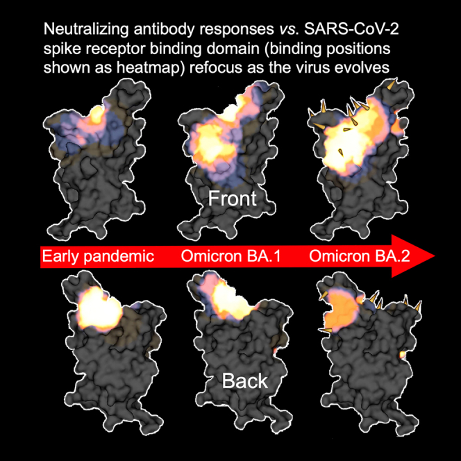
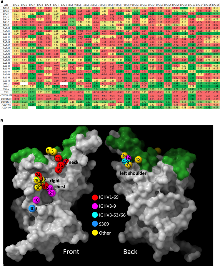
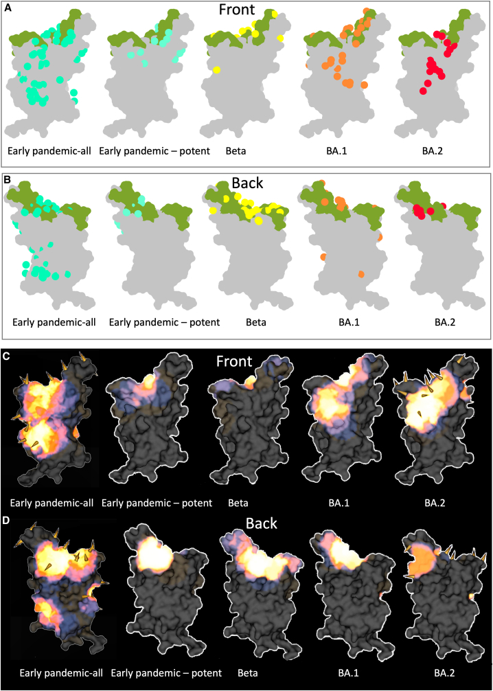
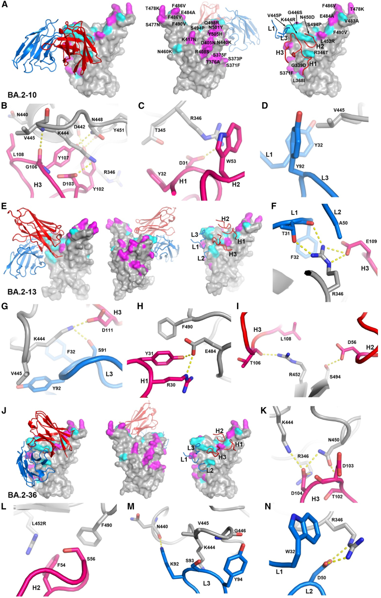
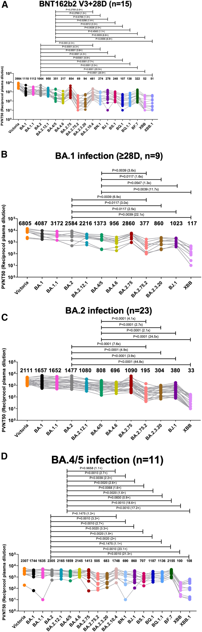
In November 2021, Omicron BA.1, containing a raft of new spike mutations, emerged and quickly spread globally. Intense selection pressure to escape the antibody response produced by vaccines or severe acute respiratory syndrome coronavirus 2 (SARS-CoV-2) infection then led to a rapid succession of Omicron sub-lineages with waves of BA.2 and then BA.4/5 infection. Recently, many variants have emerged such as BQ.1 and XBB, which carry up to 8 additional receptor-binding domain (RBD) amino acid substitutions compared with BA.2. We describe a panel of 25 potent monoclonal antibodies (mAbs) generated from vaccinees suffering BA.2 breakthrough infections. Epitope mapping shows potent mAb binding shifting to 3 clusters, 2 corresponding to early-pandemic binding hotspots. The RBD mutations in recent variants map close to these binding sites and knock out or severely knock down neutralization activity of all but 1 potent mAb. This recent mAb escape corresponds with large falls in neutralization titer of vaccine or BA.1, BA.2, or BA.4/5 immune serum.

Keywords: CP: Immunology; CP: Microbiology; SARS-CoV-2, BA.2, variant, mutation, RBD, antibodies, binding site, breakthrough, neutralizing, structure, COVID-19.


Conflict of interest statement

Declaration of interests G.R.S. sits on the GSK Vaccines Scientific Advisory Board, consults for AstraZeneca, and is a founding member of RQ Biotechnology. D.I.S. consults for AstraZeneca. Oxford University holds intellectual property related to SARS-CoV-2 mAbs discovered in G.R.S.’s laboratory. S.J.D. is a scientific advisor to the Scottish Parliament on COVID-19.

Figures

None
Graphical abstract
Figure 1
Figure 1
Phylogeny of BA.2 sub-variants (A) Alignments of mutated RBD amino acid substitutions; these are mutations formed on the BA.2 background, i.e., they are in addition to mutations found in BA.2. (B) Amino acid substitutions present in sub-variants positioned on the RBD surface. Coloring is according to the frequency count for the change in the various sub-variants, shown in (A). The ACE2 footprint is shown in green. (C) Phylogenetic tree of a selection of BA.2/4/5 sub-lineages. See also Tables S1 and S2.
Figure 2
Figure 2
Generation of BA.2 mAbs (A) Sorting of BA.2-specific B cells. (B) Proportion of BA.2 mAbs binding to RBD. (C) ACE2 receptor blocking activity of mAbs. (D) Gene usage in 25 potent BA.2 mAbs compared with potent antibodies produced following infection with early-pandemic virus, which have been previously reported. (E) Number of somatic mutations found in BA.2 mAbs compared with sets previously recovered from early-pandemic, Beta, and Omicron (BA.1) infections, which have been reported previously.,,
Figure 3
Figure 3
Heatmap of IC50 neutralization titers (A) BA.2 mAb panel. (B) Commercial mAbs. Pseudoviral neutralization IC50 titers for indicated mAb against a panel of pseudoviruses expressing variant S sequences. All assays have been done at least twice. Commercial mAbs against Victoria, BA.2, BA.2.75, BA.4/5, and BA.4.6 previously reported are included for comparison.,, See also Figures S3 and S4.
Figure 4
Figure 4
Result of BLI competition mapping of BA.2 mAbs (A) Competition ratio results (using Mabscape; see STAR Methods). Numbers close to 0 represent complete competition between pairs of antibodies, and numbers close to 1 mean no competition. Cells are colored as red, yellow, and green, with the values ranging from 0 to 1. Antibodies with known structures (Omi-12, Omi-42, EY6A, S309, COVOX-45, COVOX-58, COVOX-278, AZD1061, and AZD8895) were used as references. (B) RBD surface representation with ACE2-binding site in green and balls corresponding to center of gravity of mapped potent BA.2 mAbs colored according to variable gene usage. See also Figure S1.
Figure 5
Figure 5
Comparison of mAb binding to RBD (A and B) Front and back views of Mabscape antibody maps from early-pandemic, Beta, BA.1, and BA.2 antibody panels. Early pandemic all represents the full set of antibodies, irrespective of neutralization potency, and all other panels show potent mAbs (IC50 < 100 ng/mL). The RBDs are shown surface rendered (gray) with the ACE2 footprint in green. (C and D) Heatmaps of surface occupation of RBD by early-pandemic, Beta, BA.1, and BA.2 antibody panels by iron heat colors (black > blue > red > orange > yellow > white hot) according to the relative level of antibody contact, calculated for each surface vertex as the number of antibodies within a 10 Å radius. BA.1 mutations are shown by the spikes. Only the early-pandemic-all panels and the sub-variant substitutions discussed here are shown mapped onto BA.2. Early-pandemic, Beta, and BA.1 data are taken from Dejnirattisai et al., Nutalai et al., Liu et al. See also Figure S1.
Figure 6
Figure 6
Structures of Delta-RBD complexes with BA.2-10, BA.2-13, and BA.2-36 (A) Binding position and orientation of BA.2-10 viewed from the front (left panel) and back (middle panel) of the RBD, and positions of the CDRs that have contact with the RBD (right panel). Only Vh (red) and Vl (blue) domains of the Fab are shown as ribbons for clarity. RBD is drawn as gray surface representation with BA.4 mutation sites highlighted in magenta, and the additional mutation sites of all variants shown in Figure 1A are shown in cyan. (B–D) Details of BA.2-10 and RBD interactions. The side chains of the RBD and Fab Vh and Vl are shown as gray, red, and blue sticks, respectively. The yellow broken bonds represent hydrogen bonds or salt bridges. (E and J) Complex of (E) Delta-RBD/BA.2-13 and (J) Delta-RBD/BA.2-36. The drawing style and color scheme are as in (A). (F–I and K–N) Contact details between RBD and BA.2-13 and RBD and BA.2-36, respectively. The drawing style and color scheme are as in (B)–(D). Antibodies bind both Delta and BA.2 RBDs well; however, mutations from early pandemic to Delta are T478K and L452R (in BA.2, the T478K change is present, while L452R is not). Additional changes in BA.2 are G339D, S371F, S373P, S375F, T376A, D405N, R408S, K417N, N440K, S477N, T478K, E484A, Q493R, Q498R, N501Y, and Y505H. See also Figures S1 and S2 and Table S3.
Figure 7
Figure 7
Serum neutralization IC50 titers (fold dilution) of lentivirus pseudotyped with the S gene of the indicated BA.2 sub-lineages (A and B) Serum obtained 28 days following the third dose of BNT162b2 vaccine (n = 15) or following infection with (B) BA.1 (n = 10 all vaccinated) taken 28–55 days following diagnosis median 41.5. (C and D) BA.2 (n = 23 all vaccinated) taken 12–43 days following diagnosis median 29 or (D) BA.4/5 (n = 11 all but one vaccinated) taken 23–48 days following diagnosis median 38 days. Geometric mean titers are shown above each column. The single unvaccinated serum shows the lowest reactivity to BA.4/5 in (D). The Wilcoxon matched-pairs signed rank test (C and D) and Mann-Whitney test were used and two-tailed p values calculated. Data for BNT162b2-vaccinated sera and BA.1 infection sera against Victoria, BA.1, BA.1.1, BA.2, BA.4/5, BA.4.6, BA.2.75, and BA.2.12.1 previously reported are included for comparison.,,, All assays have been done with the number of biological replicates indicated in the brackets.

References

    1. Domingo E. Mechanisms of viral emergence. Vet. Res. 2010;41:38. doi: 10.1051/vetres/2010010. - DOI - PMC - PubMed
    1. Sender R., Bar-On Y.M., Gleizer S., Bernshtein B., Flamholz A., Phillips R., Milo R. The total number and mass of SARS-CoV-2 virions. Proc. Natl. Acad. Sci. USA. 2021;118 doi: 10.1073/pnas.2024815118. - DOI - PMC - PubMed
    1. Lan J., Ge J., Yu J., Shan S., Zhou H., Fan S., Zhang Q., Shi X., Wang Q., Zhang L., Wang X. Structure of the SARS-CoV-2 spike receptor-binding domain bound to the ACE2 receptor. Nature. 2020;581:215–220. doi: 10.1038/s41586-020-2180-5. - DOI - PubMed
    1. Dejnirattisai W., Zhou D., Ginn H.M., Duyvesteyn H.M.E., Supasa P., Case J.B., Zhao Y., Walter T.S., Mentzer A.J., Liu C., et al. The antigenic anatomy of SARS-CoV-2 receptor binding domain. Cell. 2021;184:2183–2200.e22. doi: 10.1016/j.cell.2021.02.032. - DOI - PMC - PubMed
    1. Cerutti G., Guo Y., Zhou T., Gorman J., Lee M., Rapp M., Reddem E.R., Yu J., Bahna F., Bimela J., et al. Potent SARS-CoV-2 neutralizing antibodies directed against spike N-terminal domain target a single supersite. Cell Host Microbe. 2021;29:819–833.e7. doi: 10.1016/j.chom.2021.03.005. - DOI - PMC - PubMed

Publication types

Supplementary concepts