Review
. 2023 Mar 30;19(3):e1011210.
doi: 10.1371/journal.ppat.1011210. eCollection 2023 Mar.

Same, same but different: Exploring Plasmodium cell division during liver stage development

Affiliations
Review

Same, same but different: Exploring Plasmodium cell division during liver stage development

Magali Roques et al. PLoS Pathog. .

Abstract

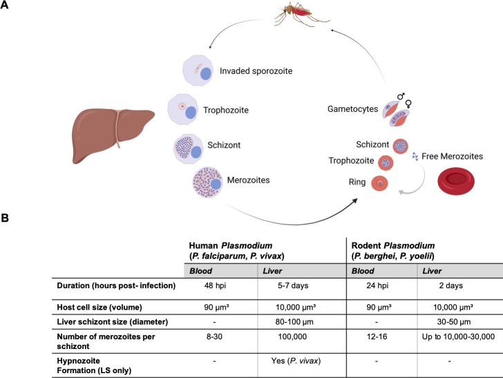
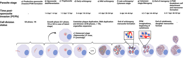
Plasmodium parasites have a complex life cycle alternating between a mosquito and a vertebrate host. Following the bite of an Anopheles female mosquito, Plasmodium sporozoites are transmitted from the skin to the liver; their first place of replication within the host. Successfully invaded sporozoites undergo a massive replication and growth involving asynchronous DNA replication and division that results in the generation of tens of thousands or even hundreds of thousands of merozoites depending on the Plasmodium species. The generation of a high number of daughter parasites requires biogenesis and segregation of organelles to finally reach a relatively synchronous cytokinesis event. At the end of liver stage (LS) development, merozoites are packed into merosomes and released into the bloodstream. They are then liberated and infect red blood cells to again produce merozoites by schizogony for the erythrocytic stage of the life cycle. Although parasite LS and asexual blood stage (ABS) differ in many respects, important similarities exist between the two. This review focuses on the cell division of Plasmodium parasite LS in comparison with other life cycle stages especially the parasite blood stage.


Conflict of interest statement

The authors have declared that no competing interests exist.

Figures

Fig 1
Fig 1. Replication of human and rodent Plasmodium parasites during exoerythrocytic and erythrocytic development.
(A) Plasmodium life cycle with emphasis on the exoerythrocytic (liver) and erythrocytic (red blood cell) stages within the vertebrate host. Parasite development in the mosquito is symbolized by the depicted mosquito but is not shown. In the scheme, important exoerythrocytic (invaded sporozoite, trophozoite, schizont, and merozoites) and erythrocytic stages (ring, trophozoite, schizont, free merozoites, and gametocytes) are indicated (created with BioRender.com). (B) The table summarizes the main characteristics and morphological features of human (P. falciparum, P. vivax) and rodent (P. berghei, P. yoelii) Plasmodium parasites. Information is from [36,89,90,126] and also referred in the 2.2.2 section. LS, liver stage.
Fig 2
Fig 2. Schematic representation of Plasmodium cell division and development during the LS.
Typical features and molecular details of P. falciparum and P. berghei (Pf/Pb) during the entire LS development are indicated as “Parasite stage” from (a) to (i) and the “Cell division status” section are indicated for each parasite stage. The duration of development “Time post-sporozoite invasion (Pf/Pb)” indicates the in vivo Plasmodium LS development. Horizontal brackets assemble drawings that represent the “Cell division status” of the parasite. (a) Upon productive invasion of a hepatocyte (light blue) the parasite (pink) resides within a PV surrounded by the PVM (light blue). Initially, the parasite remains in G0, 1N but expands in size. Parasite organelles are depicted as follows: nucleus (blue), apicoplast (green), and mitochondria (red). (b) The dedifferentiation phase starts when the crescent-shape sporozoite transforms into a bulbous-shaped form that finally becomes a circular EEF trophozoite expanding in size and entering the G1 phase (c). (c’) A proportion of P. vivax and P. cynomolgi parasites do not develop further into schizonts but remain dormant (hypnozoite) as in G0 phase. (d) Schizogony is characterized by nuclei multiplication, and alternated DNA synthesis/mitosis (S/M) phases are triggered (DNA >1N). (e) During the process of schizogony, mitochondrion and apicoplast grow into highly branched structures. (f) The cytomere stage initiates merogony (merozoite formation). The PPM invaginates and the still singular mitochondrion organizes as a finger-like structure. The apicoplast locates between the nuclei and the surrounding PPM within parasite cytoplasm (left panel in (f)). Then, the apicoplast begins to form regular constrictions resulting in the fission of the organelle (right panel in (f)). (g) At the end of the cytomere stage, mitochondrion fission occurs prior to cytokinesis as depicted in the zoomed section. (h) Tens of thousands individual merozoites are formed, depending on the Plasmodium species, with each containing an individual set of organelles; cytokinesis signifies the end of LS merogony. (i) Finally, PVM rupture liberates merozoites into the host cytosol followed by host cell detachment and merosome formation (indicated by the merozoite-filled vesicle on top of the detached cell (created with BioRender.com). EEF, exoerythrocytic form; LS, liver stage; PPM, parasite plasma membrane; PVM, parasitophorous vacuole membrane.
Fig 3
Fig 3. Parasite and host cell factors involved in sporozoite transformation into EEF represented as hierarchical events from mosquito to LS.
The dark gray circle summarizes parasite factors currently known to exert posttranscriptional and translational control on sporozoites within mosquito salivary glands. The light gray circle summarizes parasite and host cell factors shown to have an effect on sporozoite transformation into EEF within the host hepatocyte. Arrows indicate the hierarchical events happening from sporozoite latency in the mosquito to the early LS developmental gene expression within hepatocyte. Question marks show a hypothetical role of factors acting directly on the sporozoite to EEF transition. References are listed in Table 1: pumilio-2 (Puf2), sporozoite and LS asparagine-rich protein/sporozoite asparagine-rich protein (SLARP/SAP1), up-regulated in infective sporozoites 1/initiation factor 2 kinase (UIS1/IK2), up-regulated in infective sporozoites 2 (UIS2), eukaryotic translation initiation factor 2 subunit alpha (eIF2α), AP2 domain transcription factor (AP2-L), liver-specific protein 2 (LISP2) and here for up-regulated in infective sporozoite 3 (UIS3) [127], up-regulated in infective sporozoite 4 (UIS4) [67], and here for liver-specific protein 1 (LISP1) [128], exported protein 1 (EXP1) [129], temperature and metabolites [–30] (created with BioRender.com). EEF, exoerythrocytic form; LS, liver stage.
Fig 4
Fig 4. Schematic representation of CPs with PbCen-4-GFP and nucleus behavior during LS Plasmodium schizogony.
The scheme from (a) to (j) represents Plasmodium nuclear and CP duplication and division through LS schizogony. Individual intracellular Plasmodium EEF (pink, from (b) to (i); green light in (a) and (j)) surrounded by the PVM (light gray) within a hepatocyte (light blue) and the parasite CP (dark green) that is represented as a dot proximal to the parasite nucleus (blue). Corresponding live cell confocal images to this scheme represent prominent Plasmodium LS developmental stages (hpi: hours post-sporozoite infection into host cell): as early schizont (a’), progression of schizogony (mid-schizogony) from (b’) to (c’), late schizogony/beginning of cytomere stage from (d’) to (i’), and finally end of merogony (merozoites formation) (j’). These images represent endogenous tagging of PbCen-4 (PBANKA_0941400, CP marker) in fusion with a GFP fluorescent marker at its C-terminus (GFP, green) during LS development. Parasite and host cell nucleus are stained with Hoechst 33342 (blue). The transgenic parasite line has been generated previously [8]. HeLa cells were infected with PbCen-4-GFP expressing salivary gland sporozoites. Images represent the progression of Plasmodium nuclear division through schizogony. For a better comprehension, 2 S/M asynchronous rounds where an early schizont stage duplicates its CP have been presented (b); the 2 CPs move at the opposite part of the starting duplicating nucleus (c), (d), from (b’) to (c’) and from (d’) to (i’) to finally be present as a dot close to each individual separated nuclei (e). The S/M alternative phases (represented by the arrow) are then repeated from (f) to (i) (second round of S/M represented here) until cytomere stage formation and merozoite formation in (j) and (j’). The scale bar for the early schizont image (a’) is 10 μm and for the late schizonts (from (b’) to (c’); from (d’) to (i’)) and end of merozoites formation (j’) is 20 μm (created with BioRender.com). CP, centriolar plaque; EEF, exoerythrocytic form; LS, liver stage; PVM, parasitophorous vacuole membrane.

References

    1. Francia ME, Striepen B. Cell division in apicomplexan parasites. Nat Rev Microbiol. 2014;12:125–136. doi: 10.1038/nrmicro3184 - DOI - PubMed
    1. Gubbels MJ, Coppens I, Zarringhalam K, Duraisingh MT, Engelberg K. The Modular Circuitry of Apicomplexan Cell Division Plasticity. Front Cell Infect Microbiol. 2021:11. doi: 10.3389/fcimb.2021.670049 - DOI - PMC - PubMed
    1. Gubbels MJ, Keroack CD, Dangoudoubiyam S, Worliczek HL, Paul AS, Bauwens C, et al.. Fussing About Fission: Defining Variety Among Mainstream and Exotic Apicomplexan Cell Division Modes. Front Cell Infect Microbiol. 2020:10. doi: 10.3389/FCIMB.2020.00269 - DOI - PMC - PubMed
    1. van Dooren GG, Marti M, Tonkin CJ, Stimmler LM, Cowman AF, McFadden GI. Development of the endoplasmic reticulum, mitochondrion and apicoplast during the asexual life cycle of Plasmodium falciparum. Mol Microbiol. 2005;57:405–419. doi: 10.1111/j.1365-2958.2005.04699.x - DOI - PubMed
    1. Stanway RR, Witt T, Zobiak B, Aepfelbacher M, Heussler VT. GFP-targeting allows visualization of the apicoplast throughout the life cycle of live malaria parasites. Biol Cell. 2009;101:415–435. doi: 10.1042/BC20080202 - DOI - PubMed

Publication types