. 2023 Apr 12;31(4):604-615.e4.
doi: 10.1016/j.chom.2023.03.004. Epub 2023 Mar 29.

Characterization of the rotavirus assembly pathway in situ using cryoelectron tomography

Affiliations

Characterization of the rotavirus assembly pathway in situ using cryoelectron tomography

Pranav N M Shah et al. Cell Host Microbe. .

Abstract

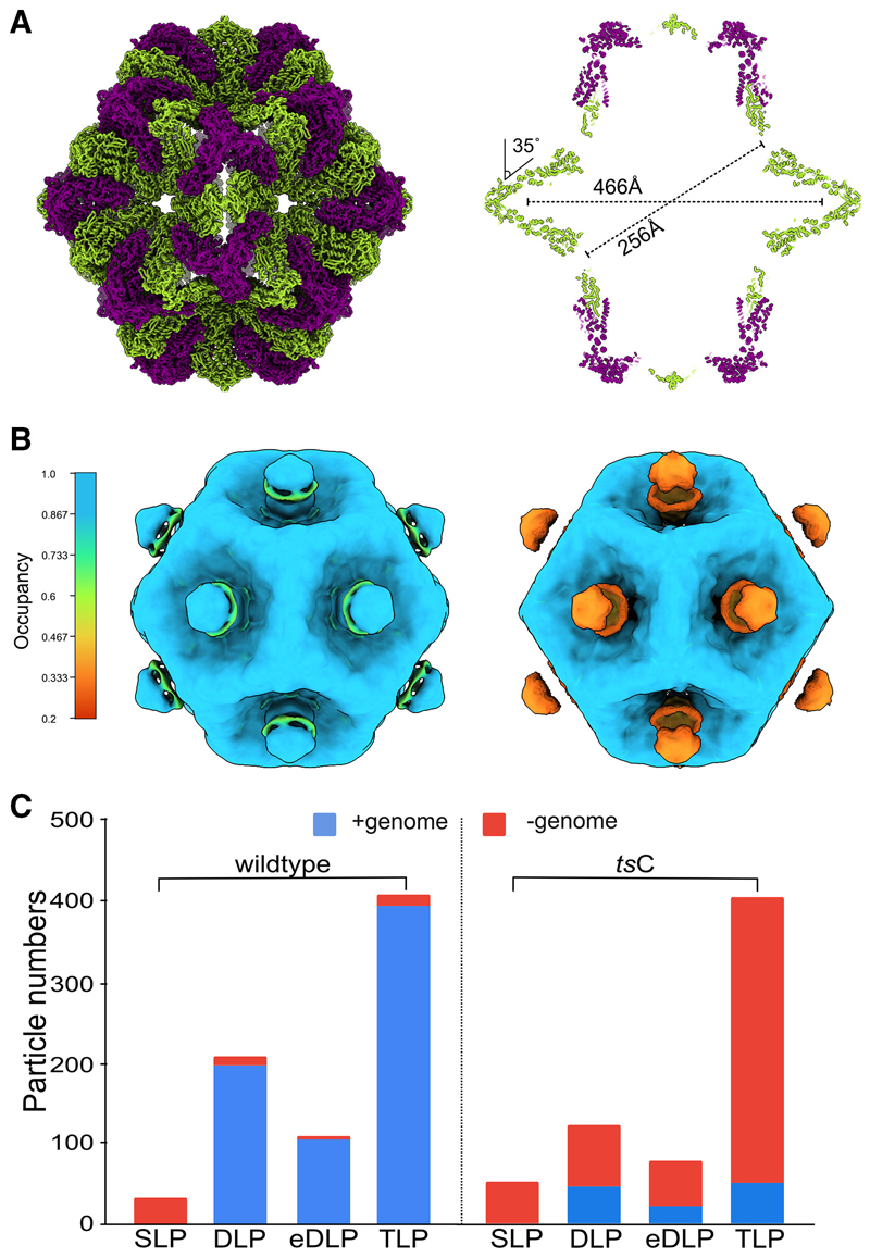
Rotavirus assembly is a complex process that involves the stepwise acquisition of protein layers in distinct intracellular locations to form the fully assembled particle. Understanding and visualization of the assembly process has been hampered by the inaccessibility of unstable intermediates. We characterize the assembly pathway of group A rotaviruses observed in situ within cryo-preserved infected cells through the use of cryoelectron tomography of cellular lamellae. Our findings demonstrate that the viral polymerase VP1 recruits viral genomes during particle assembly, as revealed by infecting with a conditionally lethal mutant. Additionally, pharmacological inhibition to arrest the transiently enveloped stage uncovered a unique conformation of the VP4 spike. Subtomogram averaging provided atomic models of four intermediate states, including a pre-packaging single-layered intermediate, the double-layered particle, the transiently enveloped double-layered particle, and the fully assembled triple-layered virus particle. In summary, these complementary approaches enable us to elucidate the discrete steps involved in forming an intracellular rotavirus particle.

Keywords: Reoviridae; cellular structural biology; cryoelectron tomography; dsRNA viruses; electron microscopy; in situ; rotavirus; subtomogram averaging; virus assembly; virus structure.


Conflict of interest statement

Declaration of interests The authors declare no competing interests.

Figures

Graphical abstract
Graphical abstract
Figure 1
Figure 1. Cryo-FIB milling of rotavirus-infected cells reveals virus assembly intermediates
(A) A cutaway view of a portion of the rotavirus capsid generated from this study is depicted as a liquorice cartoon. Protein subunits present in the virion are VP2 (light blue), VP6 (light green), VP7 (light yellow), and VP4 (salmon). The subunits that constitute the different assembly intermediates are indicated. (B) SEM image of a frozen vitrified MA104 cell infected with rotavirus prior to milling with milling windows indicated and viewed from above after milling. Scale bars, 15 μm. (C) Low-magnification overview of an infected cell with clusters of fully assembled virions in the ER. Scale bar, 100 nm. (D) Computational slice through a tomogram of an infected cell. The tomogram contrast has been enhanced using a deep learning-based denoising routine (STAR Methods). Scale bar, 100 nm. (E) Segmentation of the tomogram in (D) with virus particles in various stages of assembly. Color scheme indicates the different particle types TLP (green), eDLP (orange), DLP (purple), and SLP (red). (F–I) Close-up views of rotavirus assembly intermediates. Scale bars, 100 nm.
Figure 2
Figure 2. High-resolution subtomogram averages of rotavirus assembly intermediates
(A) TLP isosurface. Arrow indicates VP4 in its upright conformation. (B) eDLP isosurface with the membrane masked out. Arrow indicates the connection between the capsid and the membrane. (C) DLP isosurface. (D) SLP isosurface. Arrow indicates pronounced densities present at the 5-fold. (E) FSC traces for pentons of TLP (green), eDLP (orange), DLP (purple), and whole SLP (red). (F) Fit of the inner layer VP2 monomer (gray) in the maps obtained following 5-fold averaging of the pentons. Residues ranging from Arg694 to Ile676 are highlighted in the inset. All scale bars show 25 nm.
Figure 3
Figure 3. Localization of the VP4-C head domain on the TLP
(A) Architecture of the VP4 spike. Chains A and B are represented as surfaces (gray) and chain C as colored liquorice. Foot domain, blue; trunk domain, gold; head domain, red. Residue ranges of the domains are indicated. (B) Isosurface renderings ofVP4 maps. Top row, side views of EMD-21955 (left), in situ TLP (middle) and gradient purified TLP (right). Bottom row, top views. VP4 domains are colored as in (A), and the outer capsid layer VP7 is colored green. Insets highlight position of the head domain. (C) Comparison of the upright VP4-A chain with prone VP4-C. The head domain, represented as a Jones rainbow, is rotated by ~117°. The trunk domain is colored yellow. The z axis is perpendicular to the viewing direction. Inset, a least squares superposition of the trunk domains comparing VP4-A (gray) and VP4-C (yellow). (D) Electrostatic potential distribution on the surface of VP4-C. (E) As (D) except that the head and trunk domains are each rotated like an open book. Charge complementation between the two domains is indicated with dotted ovals.
Figure 4
Figure 4. Characterization of VP4 in the eDLP assembly intermediate
(A) Overview of the eDLP VP4 density. The VP6 layer is colored in orange with two of the subunits hidden to expose the foot domains of VP4. The membrane layer is represented as a cartoon. Insets show density in the lobes, struts, and foot regions. (B) Flexibility analysis of the pre-mature spike is depicted as central sections through each of the densities. The number of particles in each class is highlighted in the histogram. (C) Map of VP4 after focused classification following multibody analysis. Fit of one of the chains of the pre-mature VP4, colored as in Figure 3A. The membrane and two VP6 subunits have been masked out to aid visualization. The approximate location of the membrane is depicted as a cartoon (gray). (D) Liquorice diagram model of the pre-mature VP4 trimer. (E) Comparison of the pre-mature (left) and extended (right) VP4 molecules. VP4-A and B chains are rendered as surfaces, with VP4-C represented as a liquorice cartoon. (F) Comparison of VP4 from the entry intermediate form of the TLP (PDB: 6wxf, right) with the pre-mature VP4 seen on the eDLP (left). The different protein layers are represented as liquorice cartoons. Red triangle indicates the 3-fold axis.
Figure 5
Figure 5. Indented single-layered particle
(A) Model of the indented SLP is presented with the outer surface view (left) and a central section (right). The two chains of VP2 A and B are colored in green and purple, respectively. Long and short dimensions of the particle are indicated. The difference in the angle made by adjacent VP2 A in the indented form compared with their position in the expanded form adopted at later assembly stages, is indicated. (B) Isosurface representation of the SLP map from wild type (left) and tsC mutant (right) with the voxels colored by their occupancy. Occupancy of the blobs in the SLP maps was estimated in a resolution-independent manner using the relative contrast of the voxels within the map. (C) Comparison of the number of genome-containing and genome-less particles in the wild-type virus versus the conditionally lethal tsC mutant.
Figure 6
Figure 6. Assembly intermediates in the rotavirus replication cycle
Infection occurs following virus entry and the release of the incoming DLP into the cytoplasm where primary transcription occurs (1–4). Following viroplasm formation and synthesis of viral structural proteins, virus assembly begins with the indented SLPs which are converted to the DLP stage (5). The DLP acquires VP4 as it buds through the ER membrane gaining a transient envelope. Loss of the envelope and the rapid acquisition of the VP7 layer leads to the formation of the immature TLP in the ER lumen (6). The fully assembled virions are released into the extracellular medium where exposure to trypsin in the small intestine converts them to mature infectious virions (7).
Figure 7
Figure 7. Conformations of VP4 sampled during entry and maturation stages
Schematic of the transitions occurring in VP4 during maturation, egress, and entry beginning with (i) the insertion of VP4 into the VP6 layer in the transiently enveloped stage. (ii) The upright dimeric VP4 conformation occurs after the immature TLP is released into the ER lumen. (iii) Extracellular proteases present in the gut lumen cleave VP4 and render the TLP competent for entry. (iv and v) Following attachment VP4 undergoes gross conformational changes via an intermediate conformation in which the foot domain is present and culminating with the final entry-competent conformation in which the foot domain is disordered.

References

    1. Desselberger U. Rotaviruses. Virus Res. 2014;190:75–96. - PubMed
    1. Settembre EC, Chen JZ, Dormitzer PR, Grigorieff N, Harrison SC. Atomic model ofan infectious rotavirus particle. EMBO J. 2011;30:408–416. - PMC - PubMed
    1. Dormitzer PR, Greenberg HB, Harrison SC. Purified recombinant rotavirus VP7 forms soluble, calcium-dependent trimers. Virology. 2000;277:420–428. - PubMed
    1. Martinez MA, Lopez S, Arias CF, Isa P. Gangliosides have a functional role during rotavirus cell entry. J Virol. 2013;87:1115–1122. - PMC - PubMed
    1. Estes MK, Graham DY, Mason BB. Proteolytic enhancement of rotavirus infectivity: molecular mechanisms. J Virol. 1981;39:879–888. - PMC - PubMed

LinkOut - more resources