. 2023 May;617(7961):555-563.
doi: 10.1038/s41586-023-05948-2. Epub 2023 Mar 30.

Adeno-associated virus 2 infection in children with non-A-E hepatitis

Collaborators, Affiliations

Adeno-associated virus 2 infection in children with non-A-E hepatitis

Antonia Ho et al. Nature. 2023 May.

Abstract

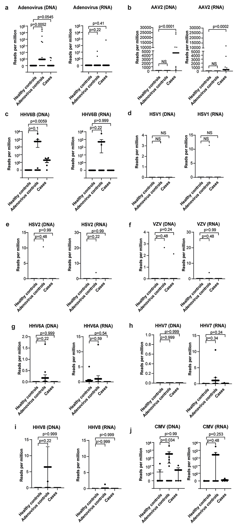
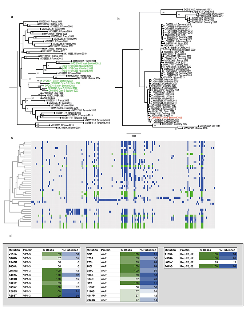
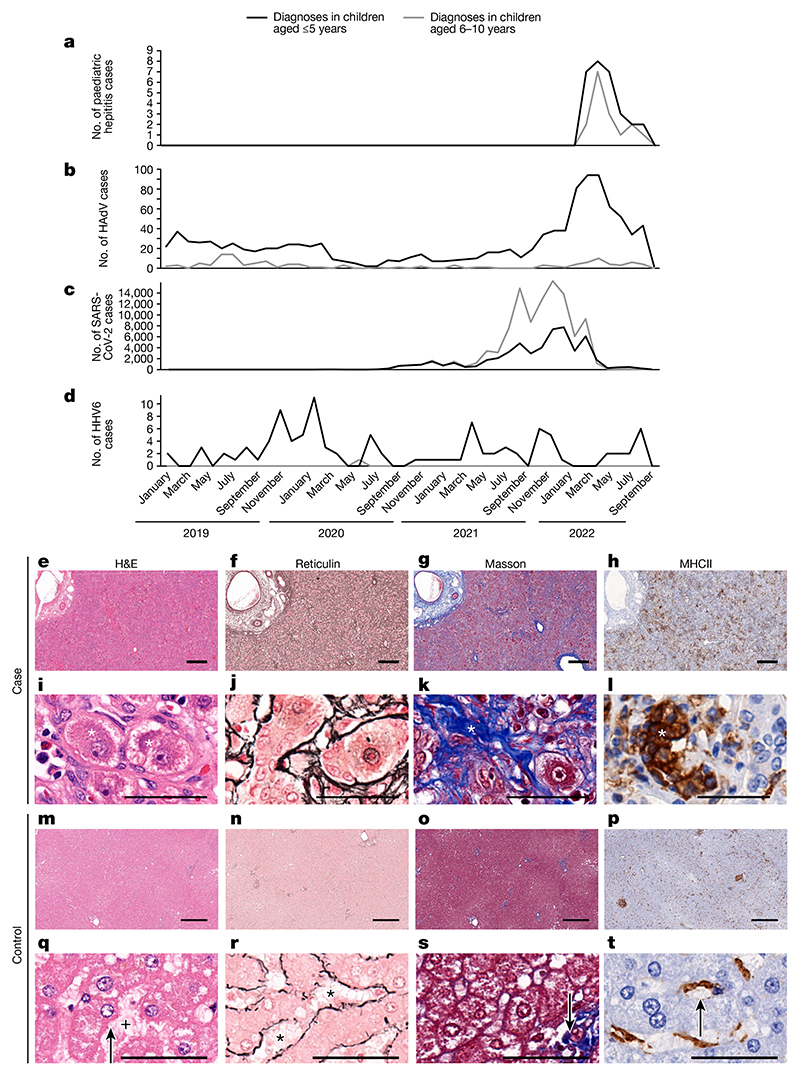
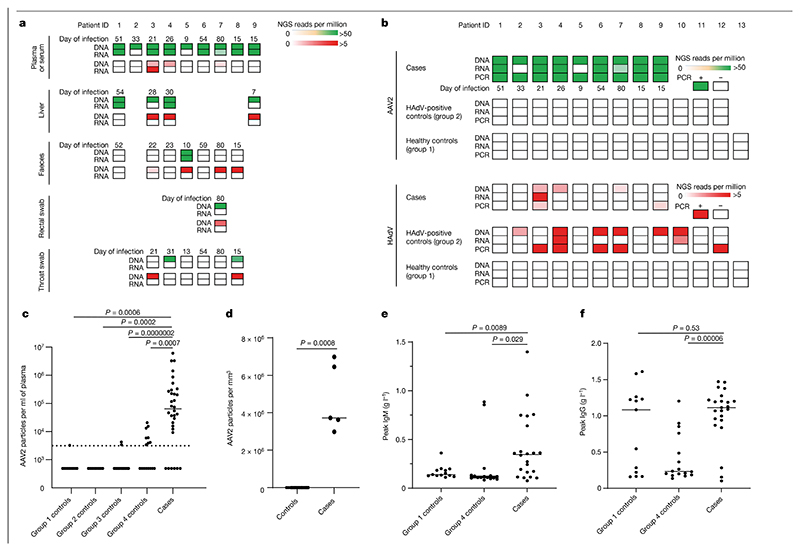
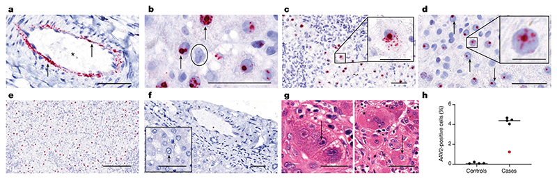
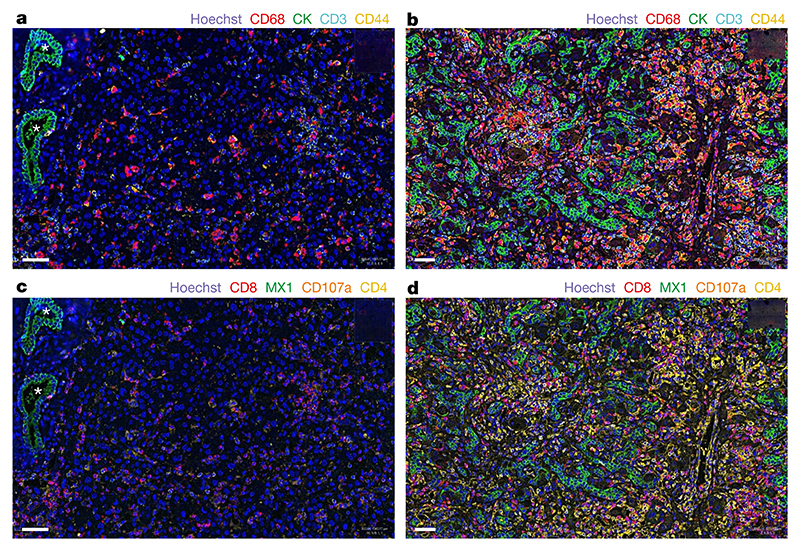
An outbreak of acute hepatitis of unknown aetiology in children was reported in Scotland1 in April 2022 and has now been identified in 35 countries2. Several recent studies have suggested an association with human adenovirus with this outbreak, a virus not commonly associated with hepatitis. Here we report a detailed case-control investigation and find an association between adeno-associated virus 2 (AAV2) infection and host genetics in disease susceptibility. Using next-generation sequencing, PCR with reverse transcription, serology and in situ hybridization, we detected recent infection with AAV2 in plasma and liver samples in 26 out of 32 (81%) cases of hepatitis compared with 5 out of 74 (7%) of samples from unaffected individuals. Furthermore, AAV2 was detected within ballooned hepatocytes alongside a prominent T cell infiltrate in liver biopsy samples. In keeping with a CD4+ T-cell-mediated immune pathology, the human leukocyte antigen (HLA) class II HLA-DRB1*04:01 allele was identified in 25 out of 27 cases (93%) compared with a background frequency of 10 out of 64 (16%; P = 5.49 × 10⁻¹²). In summary, we report an outbreak of acute paediatric hepatitis associated with AAV2 infection (most likely acquired as a co-infection with human adenovirus that is usually required as a 'helper virus' to support AAV2 replication) and disease susceptibility related to HLA class II status.


Conflict of interest statement

Competing interests The authors declare no competing interests.

Figures

Extended Data Fig. 1
Extended Data Fig. 1. AAV2, HAdV and human herpesvirus detection by target enrichment sequencing in cases and controls.
Read counts per million are plotted for a) HAdV; b) AAV2; c) HHV6B; d) HSV1; e) HSV2; f) VZV; g) HHV6A; h) HHV7; i) HHV8; and j) CMV in cases, Group 1 healthy controls and Group 2 controls (HAdV positive children with normal liver function). Statistical significance was estimated using a Mann-Whitney test (two-sided).
Extended Data Fig. 2
Extended Data Fig. 2. Phylogenetic and sequence analysis of AAV2 genomes.
a) Maximum likelihood phylogeny of AAV2 from hepatitis cases CVR1-9. The nine AAV2 genome sequences generated from the plasma samples via target enrichment (highlighted in green) were aligned with a range of the closest AAV GenBank sequences. AAV2 reference sequences are denoted by accession number, country and year of sampling b), Phylogeny of HAdV41 genome from case 5. The HAdV41 genome sequence from the faecal sample of patient 5 (red) was combined with complete genomes of HAdV41 from GenBank. Bootstrap values >70 are indicated. HAdV41 reference sequences are denoted by accession number, country and year of sampling; c), Key mutations and hierarchical clustering of AAV2 genomes. Mutations in published AAV2 sequences are highlighted in (blue) and case sequences (green); d) Mutations over-represented in hepatitis cases versus controls. Mutations in VP1-3, Rep78 and 52 and AAP are highlighted by % representation in case sequences (green) and published sequences (blue).
Extended Data Fig. 3
Extended Data Fig. 3. Reactivity of sera from paediatric hepatitis cases against human seasonal coronaviruses and SARS-CoV-2.
Sera from the paediatric hepatitis cases were screened for reactivity against spike proteins from a) seasonal coronaviruses 229E, OC43, NL63 and HKU1, and b) SARS-CoV-2 nucleocapsid (N), spike (S), and N-terminal domain (NTD) and receptor binding domain (RBD) of S by electrochemiluminescence (MSD-ECL). Reactivity of the 23 samples (Hepatitis) was compared with 16 sera from contemporaneous control samples from children (Group 4 Controls), and three groups of sera from adults of known SARS-CoV-2 status; Negatives (never tested positive for SARS-CoV-2; n = 30), Vaccinated two doses (n = 28) and Infected (n = 39).
Extended Data Fig. 4
Extended Data Fig. 4. Principal component analysis (PCA) plots.
PCA plots showing the first four genome-wide principal components to confirm genetic ancestry matching. a) Genomic PCA using full United Kingdom Biobank cohort as background population (grey), showing the subgroup of unrelated United Kingdom Biobank participants who were born in Scotland and of Caucasian ancestry (blue) and the hepatitis cases reported here (red). b) plots showing only the subgroup born in Scotland and of Caucasian ancestry.
Fig. 1
Fig. 1. Epidemiology and histological appearance of cases of paediatric hepatitis in Scotland.
a, The emergence of acute non-A–E hepatitis in children in March–September 2022 (ref. 3). bd, Cases of HAdV (b), SARS-CoV-2 (c) and HHV6 (d) infection in children aged ≤10 years in Scotland during the period January 2019 to September 2022. et, Histopathology of samples from cases of non-A–E hepatitis (el) and from healthy liver (mt). e,i,m,q, Serial sections of formalin-fixed and paraffin-embedded liver tissue sections (one section for each stain per patient sample) stained with haematoxylin and eosin (H&E). f,j,n,r, Reticulin staining highlighting structural organization. g,k,o,s, Masson staining highlighting collagen fibres. h,l,p,t, Staining for MHCII+ cells. mp, The regular lobular structure of the liver from a healthy individual (identifier 145783) is not recognisable in eh, which are sections collected from patient CVR35 who received a liver transplant. h, Immunohistochemistry showed an increase in MHCII+ cells in tissue samples from patient CVR35 compared with healthy liver (l,t). il, Higher magnification micrographs of eh showing details of liver histopathology. i,q, For patient CVR35 (i), enlarged (ballooned) and vacuolated hepatocytes (marked by asterisks) are evident compared with hepatocytes in healthy liver (q; from individual 145783) with regular morphology (indicated by the arrow) and regular sinus (indicated by the plus symbol). j,r, For the sample from patient CVR35 (j), reticulin staining shows destruction of the sinus structures and irregularly arranged fibres, whereas healthy liver (r) shows fibres lining the sinus (indicated by asterisks). k,s, For the sample from patient CVR35 (k), Masson staining shows an increase in collagen fibres (in blue, indicated by the asterisk) compared with minimal staining of fibres (indicated by the arrow) in healthy liver (s). l,t, High magnification image showing accumulation of MHCII+ cells in the liver (indicated by the asterisk) of patient CVR35 (l), whereas healthy liver (t), staining is limited to Kupffer cells (indicated by the arrow). Scale bars, 50 μm (il,qt) or 400 μm (eh,mp).
Fig. 2
Fig. 2. Detection of AAV2 in cases of paediatric hepatitis.
a, Heatmap of HAdV and AAV2 reads detected in cases of hepatitis by TE sequencing. Samples obtained for routine clinical investigation (plasma, liver, faeces, rectal swab and throat swab) were retrospectively sequenced following DNA or RNA extraction. AAV2 read counts are shown from 0 to >50 reads per million in green (top rows) and HAdV read counts are shown from 0 to >5 reads per million in red (bottom rows). b, Heatmap of viral reads of plasma samples from cases of hepatitis and of plasma or sera samples from controls. Plasma samples from cases of hepatitis (cases), and plasma or sera samples from children with HAdV infection (group 2 controls) and from age-matched healthy children (group 1 controls) were sequenced following DNA or RNA extraction. AAV2 read counts are shown from 0 to >50 reads per million in green and HAdV read counts are shown from 0 to >5 reads per million in red. The number of days between initial symptom onset and sample are indicated. c, AAV2 real-time RT–qPCR of serum or plasma samples from 32 cases of hepatitis (cases) and from 74 controls in four groups: 13 in group 1 (healthy controls); 12 in group 2 (HAdV-positive controls); 33 in group 3 (hepatitis controls); and 16 in group 4 (contemporaneous controls). The detection threshold of the assay (3,200 copies per ml) is shown as a dotted line. Values are shown as a scatter plot with a median line. d, AAV2 real-time RT–qPCR of liver biopsy samples from 5 cases of hepatitis and from 19 controls. e, IgM responses determined by ELISA in 22 cases of hepatitis and in 29 controls (13 in group 3, 16 in group 4). f, IgG responses determined by ELISA in 22 cases of hepatitis cases and in 29 controls (13 in group 3, 16 in group 4). For cf, statistical analysis was performed using Mann Whitney test (two-tailed), and experiments were performed in triplicate.
Fig. 3
Fig. 3. ISH of AAV2 in liver tissue.
ag, RNA ISH for the detection of AAV2 RNA in sections of formalin-fixed and paraffin-embedded liver tissues from children (one section per patient) with non A–E hepatitis. a, AAV2 RNA (red signal, indicated by an arrow) was detected in the endothelial cells of arteries in an explant liver section from patient CVR35. The vascular lumen is highlighted with by an asterisk. b, A positive AAV2 signal was detected in the nuclei of hepatocytes with vacuolated morphology from patient CVR4 (indicated by arrows) and in a negative cell (indicated by the circle). c,d, A liver section from patient CVR1 showed AAV2 RNA both in the nucleus and in the cytoplasm (c), whereas for patient CVR9 (d), AAV2 RNA was found only in the nucleus (indicated by arrows). e, A high percentage of hepatocytes with a positive signal for AAV2 was present predominantly in the nucleus of hepatocytes in the samples from patient CVR1. f, AAV2 was not detectable in liver sections from samples from healthy individuals in either the endothelial cells or hepatocytes. g, Samples from patient CVR35 showed inclusion bodies in hepatocytes. Left, small, dark basophilic intranuclear inclusions next to the nucleolus (indicated by arrows). Right, a large, pale basophilic, diffuse intranuclear inclusion body (suggestive of adenovirus infection; indicated by an arrow) next to a multinucleated giant cell in the liver (indicated by the asterisk). h, AAV2-positive cells were quantified using QuPath in biopsy samples from five patients with non-A–E hepatitis (cases) and from controls. Patient CVR35 (who received a liver transplant) is highlighted in red. Using the entire section, cells were segmented to identify the nuclei and cytoplasm, and the algorithm was tuned to detect red signals. All samples were analysed using the same algorithm. Scale bars, 25 μm (insets of c,d), 50 μm (ad,f,g) or 200 μm (e).
Fig. 4
Fig. 4. CODEX analysis of liver tissue.
ad, Images of liver tissue from patient CVR35 (b,d) and a liver sample from an unaffected individual (control; a,c) show differences in cellular composition (c,d). a, Regularly structured bile ducts in the liver biopsy from the control are highlighted by asterisks, and epithelial cells are stained green using cytokeratin (CK). Scattered macrophages (CD68, red), T cells (CD3, cyan) and activated T cells (CD44, yellow) are also present. b, By contrast, the explant liver from patient CVR35 shows prominent proliferation of epithelial cells throughout the liver tissue (green), with increased macrophages (red), T cells (cyan) and activated T cells (yellow). c, The control liver shows scattered cytotoxic T cells (CD8, red), CD107a-positive cells (brown) and CD4-positive cells (yellow) cells and low expression of the interferon-induced GTP-binding protein MX1 (green). d, High numbers of all cell types and high MX1 expression are observed in the explant liver from patient CVR35. One section of liver was stained per individual, and the entire area was manually outlined. Cells were segmented to identify the nuclei and cytoplasm, and the algorithm was tuned to detect the colour signal in the cells. All samples were analysed using the same algorithm for each stain. Scale bars, 50 μm.

Comment in

Similar articles

  • Acute hepatitis of unknown aetiology in children: evidence for and against causal relationships with SARS-CoV-2, HAdv and AAV2.
    Gurdasani D, Trent M, Ziauddeen H, Mnatzaganian E, Turville S, Chen X, Kunasekaran MP, Chughtai AA, Moa A, McEniery J, Greenhalgh T, MacIntyre CR. Gurdasani D, et al. BMJ Paediatr Open. 2024 Dec 9;8(1):e002410. doi: 10.1136/bmjpo-2023-002410. BMJ Paediatr Open. 2024. PMID: 39653515 Free PMC article. Review.
  • Adeno-associated virus type 2 in US children with acute severe hepatitis.
    Servellita V, Sotomayor Gonzalez A, Lamson DM, Foresythe A, Huh HJ, Bazinet AL, Bergman NH, Bull RL, Garcia KY, Goodrich JS, Lovett SP, Parker K, Radune D, Hatada A, Pan CY, Rizzo K, Bertumen JB, Morales C, Oluniyi PE, Nguyen J, Tan J, Stryke D, Jaber R, Leslie MT, Lyons Z, Hedman HD, Parashar U, Sullivan M, Wroblewski K, Oberste MS, Tate JE, Baker JM, Sugerman D, Potts C, Lu X, Chhabra P; Pediatric Hepatitis of Unknown Etiology Working Group; Ingram LA, Shiau H, Britt W, Gutierrez Sanchez LH, Ciric C, Rostad CA, Vinjé J, Kirking HL, Wadford DA, Raborn RT, St George K, Chiu CY. Servellita V, et al. Nature. 2023 May;617(7961):574-580. doi: 10.1038/s41586-023-05949-1. Epub 2023 Mar 30. Nature. 2023. PMID: 36996871 Free PMC article.
  • Case Report: HLA-DRB1 04:01 found in a child with adenovirus type 2 -linked hepatitis.
    Katsumi Y, Nishimura Y, Goto S, Ozawa S, Nishiura T, Kotera A, Kawahara Y, Higashikawa S, Iwasaki R, Toriiminami Y, Asai N, Fujita N. Katsumi Y, et al. Front Immunol. 2025 Feb 26;16:1544622. doi: 10.3389/fimmu.2025.1544622. eCollection 2025. Front Immunol. 2025. PMID: 40079016 Free PMC article.
  • Genomic investigations of unexplained acute hepatitis in children.
    Morfopoulou S, Buddle S, Torres Montaguth OE, Atkinson L, Guerra-Assunção JA, Moradi Marjaneh M, Zennezini Chiozzi R, Storey N, Campos L, Hutchinson JC, Counsell JR, Pollara G, Roy S, Venturini C, Antinao Diaz JF, Siam A, Tappouni LJ, Asgarian Z, Ng J, Hanlon KS, Lennon A, McArdle A, Czap A, Rosenheim J, Andrade C, Anderson G, Lee JCD, Williams R, Williams CA, Tutill H, Bayzid N, Martin Bernal LM, Macpherson H, Montgomery KA, Moore C, Templeton K, Neill C, Holden M, Gunson R, Shepherd SJ, Shah P, Cooray S, Voice M, Steele M, Fink C, Whittaker TE, Santilli G, Gissen P, Kaufer BB, Reich J, Andreani J, Simmonds P, Alrabiah DK, Castellano S, Chikowore P, Odam M, Rampling T, Houlihan C, Hoschler K, Talts T, Celma C, Gonzalez S, Gallagher E, Simmons R, Watson C, Mandal S, Zambon M, Chand M, Hatcher J, De S, Baillie K, Semple MG; DIAMONDS Consortium; PERFORM Consortium; ISARIC 4C Investigators; Martin J, Ushiro-Lumb I, Noursadeghi M, Deheragoda M, Hadzic N, Grammatikopoulos T, Brown R, Kelgeri C, Thalassinos K, Waddington SN, Jacques TS, Thomson E, Levin M, Brown JR, Breuer J. Morfopoulou S, et al. Nature. 2023 May;617(7961):564-573. doi: 10.1038/s41586-023-06003-w. Epub 2023 Mar 30. Nature. 2023. PMID: 36996872 Free PMC article.
  • Solved the enigma of pediatric severe acute hepatitis of unknown origin?
    Rodriguez-Frias F, Rando-Segura A, Quer J. Rodriguez-Frias F, et al. Front Cell Infect Microbiol. 2023 Sep 21;13:1175996. doi: 10.3389/fcimb.2023.1175996. eCollection 2023. Front Cell Infect Microbiol. 2023. PMID: 37808908 Free PMC article. Review.

Cited by

References

    1. Marsh K, et al. Investigation into cases of hepatitis of unknown aetiology among young children, Scotland, 1 January 2022 to 12 April 2022. Euro Surveill. 2022 doi: 10.2807/1560-7917.ES.2022.27.15.2200318. - DOI - PMC - PubMed
    1. Severe acute hepatitis of unknown aetiology in children—multi-country. World Health Organization; 2022. Jul 12, https://www.who.int/emergencies/disease-outbreak-news/item/2022-DON400 .
    1. Investigation into Acute Hepatitis of Unknown Aetiology in Children in England Technical Briefing 4. UK Health Security Agency; 2022.
    1. Gutierrez Sanchez LH, et al. A case series of children with acute hepatitis and human adenovirus infection. N Engl J Med. 2022 doi: 10.1056/NEJMoa2206294. - DOI - PMC - PubMed
    1. Karpen SJ. Acute hepatitis in children in 2022—human adenovirus 41? N Engl J Med. 2022 - PubMed

MeSH terms

Substances

Supplementary concepts