Review
. 2023 May 1:561:216150.
doi: 10.1016/j.canlet.2023.216150. Epub 2023 Mar 29.

Mucins as contrast agent targets for fluorescence-guided surgery of pancreatic cancer

Affiliations
Review

Mucins as contrast agent targets for fluorescence-guided surgery of pancreatic cancer

Kathryn M Muilenburg et al. Cancer Lett. .

Abstract

Pancreatic cancer is difficult to resect due to its unique challenges, often leading to incomplete tumor resections. Fluorescence-guided surgery (FGS), also known as intraoperative molecular imaging and optical surgical navigation, is an intraoperative tool that can aid surgeons in complete tumor resection through an increased ability to detect the tumor. To target the tumor, FGS contrast agents rely on biomarkers aberrantly expressed in malignant tissue compared to normal tissue. These biomarkers allow clinicians to identify the tumor and its stage before surgical resection and provide a contrast agent target for intraoperative imaging. Mucins, a family of glycoproteins, are upregulated in malignant tissue compared to normal tissue. Therefore, these proteins may serve as biomarkers for surgical resection. Intraoperative imaging of mucin expression in pancreatic cancer can potentially increase the number of complete resections. While some mucins have been studied for FGS, the potential ability to function as a biomarker target extends to the entire mucin family. Therefore, mucins are attractive proteins to investigate more broadly as FGS biomarkers. This review summarizes the biomarker traits of mucins and their potential use in FGS for pancreatic cancer.

Keywords: Biomarkers; Fluorescence; Intraoperative molecular imaging; Mucins; Optical surgical navigation.


Conflict of interest statement

Declaration of competing interest Kathryn M. Muilenburg: None. Carly C. Isder: None. Prakash Radhakrishnan: Equity interest in OncoCare Therapeutics, which has licensing rights to antibody AR9.6. NIH funding related mucins. Surinder K. Batra: Co-founders of Sanguine Diagnostics and Therapeutics, Inc., Omaha. Recipient of NIH funding related to mucins and fluorescence-guided surgery contrast agents that target mucins. Quan P. Ly: Co-investigator of NIH funding related pancreatic cancer and fluorescence-guided surgery. Stock ownership in Intuitive Surgical. Other stock information available upon request. Mark A. Carlson: Recent NIH funding related to fluorescence-guided surgery. Michael Bouvet: Consultant for Stryker, Inc. NIH and VA funding related anti-mucin antibodies for fluorescence-guided surgery. Michael A. Hollingsworth: Equity interest in OncoCare Therapeutics and Sanguine Diagnostics and Therapeutics. Recipient of NIH funding related to mucins and biomarkers for pancreatic cancer. Aaron M. Mohs: NIH funding related to fluorescence-guided surgery targeting mucins. Patents related to contrast agents and imaging systems for fluorescence-guided surgery.

Figures

Figure 1.
Figure 1.. Mucin expression pattern in malignant tissue compared to normal tissue.
Mucin expression displayed by organ. Mucins that are uniquely expressed in malignancy are written in brown. These mucins have optimal expression patterns as they are known to be expressed in malignant tissue with no known expression in normal tissue. Mucins in blue are also potential FGS biomarkers with upregulated expression in malignant tissue compared to normal tissue.
Figure 2.
Figure 2.. Heatmap of mucin expression in cancers.
Mucin expression in cancer is arranged alphabetically in each organ system. Mucin expression was scored by the intensity of stain and percent of expression tumors on a scale of −2 to 5. Legend: 0 represents similar mucin expression in malignant and normal tissue. 1 represents the weak intensity of staining in tumors and mucins with staining in a few tumors. 2 and 3 represent moderate mucin staining intensity in a few or many tumors. 4 and 5 represent high mucin staining intensity in few or many tumors. −1 and −2 represent malignancy mucin expression below normal tissue expression either slightly or dramatically. The white boxes indicate that the mucin was not found expressed in cancer or had unknown expression intensity.
Figure 3.
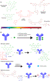
Figure 3.. FGS contrast agent components.
A. Examples of fluorescent dyes for conjugation to a biomolecular targeting agent are Alexa Fluor 488, fluorescein, Cy5.5, IRDye800CW, and ICG-sulfo-OSu. These dyes fluoresce in the visible or NIR channels. B. Antibody-dye conjugation reactions. Common methods, including NHS ester coupling, engineering of a cysteine amino acid, and maleimide coupling, are examples of methods to form a fluorescent dye-conjugated probe.
Figure 4.
Figure 4.. In vivo targeting of mucins for FGS.
A. In vivo imaging of subcutaneous CA19-9 positive and negative tumors of a fluorescent-labeled probe (left) and a dual-labeled probe (right). Adapted with permission from Houghton JL, Zeglis BM, Abdel-Atti D, et al., (2015) Site-specifically labeled CA19.9-targeted immunoconjugates for the PET, NIRF, and multimodal PET/NIRF imaging of pancreatic cancer. PNAS. B. Time-lapse imaging of the targeting ability of a conjugated MUC1 antibody compared to a conjugated IgG antibody in a subcutaneous pancreatic cancer model. Adapted with permission from Wu G, Maharjan S, Kim D, et al., (2018) A novel monoclonal antibody targets Mucin1 and attenuates growth in pancreatic cancer model. Int JMol Sci. C. In vivo imaging of antibody targeted to MUC5AC in an orthotopic pancreatic tumor model. The black arrow denotes pancreatic tumor fluorescence. The yellow arrow denotes a non-fluorescent normal pancreas. The white arrow denotes a fluorescent metastatic pancreatic tumor in the peritoneum. The red arrow denotes background fluorescence in the stomach. Adapted with permission from Turner MA, Hollandsworth HM, Nishino H, et al., (2022) Fluorescent Anti-MUC5AC brightly targets pancreatic cancer in a patient-derived orthotopic xenograft. In Vivo. D. Humanized antibody targeting MUC16 in a subcutaneous patient-derived xenograft tumor model of pancreatic cancer. Adapted with permission from Olson MT, Aguilar EN, Brooks CL et al., (2022) Preclinical evaluation of a humanized, near-infrared fluorescent antibody for fluorescence-guided surgery of MUC16-expressing pancreatic cancer. Mol Pharm. Copyright 2022 American Chemical Society.

References

    1. Sethi S, Ali S, Philip PA, Sarkar FH, Clinical advances in molecular biomarkers for cancer diagnosis and therapy, Int J Mol Sci, 14 (2013) 14771–14784. - PMC - PubMed
    1. Borrebaeck CA, Precision diagnostics: moving towards protein biomarker signatures of clinical utility in cancer, Nat Rev Cancer, 17 (2017) 199–204. - PubMed
    1. Srivastava A, Creek DJ, Discovery and Validation of Clinical Biomarkers of Cancer: A Review Combining Metabolomics and Proteomics, Proteomics, 19 (2019) e1700448. - PubMed
    1. Wojtynek NE, Mohs AM, Image-guided tumor surgery: The emerging role of nanotechnology, Wiley Interdiscip Rev Nanomed Nanobiotechnol, 12 (2020) e1624. - PMC - PubMed
    1. Olson MT, Ly QP, Mohs AM, Fluorescence Guidance in Surgical Oncology: Challenges, Opportunities, and Translation, Mol Imaging Biol, 21 (2019) 200–218. - PMC - PubMed

Publication types