. 2023 Mar 30;6(6):e202201589.
doi: 10.26508/lsa.202201589. Print 2023 Jun.

UPF3A is dispensable for nonsense-mediated mRNA decay in mouse pluripotent and somatic cells

Affiliations

UPF3A is dispensable for nonsense-mediated mRNA decay in mouse pluripotent and somatic cells

Chengyan Chen et al. Life Sci Alliance. .

Abstract

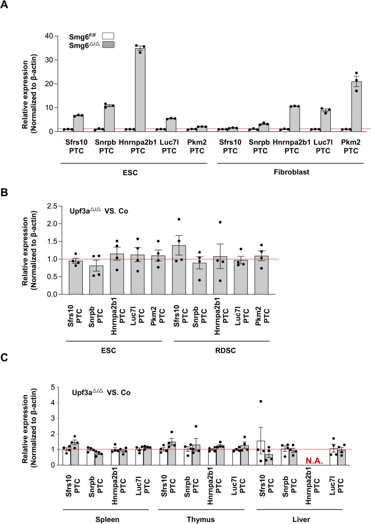
Nonsense-mediated mRNA decay (NMD) is a highly conserved regulatory mechanism of post-transcriptional gene expression in eukaryotic cells. NMD plays essential roles in mRNA quality and quantity control and thus safeguards multiple biological processes including embryonic stem cell differentiation and organogenesis. UPF3A and UPF3B in vertebrate species, originated from a single UPF3 gene in yeast, are key factors in the NMD machinery. Although UPF3B is a well-recognized weak NMD-promoting factor, whether UPF3A functions in promoting or suppressing NMD is under debate. In this study, we generated a Upf3a conditional knockout mouse strain and established multiple lines of embryonic stem cells and somatic cells without UPF3A. Through extensive analysis on the expressions of 33 NMD targets, we found UPF3A neither represses NMD in mouse embryonic stem cells, somatic cells, nor in major organs including the liver, spleen, and thymus. Our study reinforces that UPF3A is dispensable for NMD when UPF3B is present. Furthermore, UPF3A may weakly and selectively promote NMD in certain murine organs.


Conflict of interest statement

The authors declare that they have no conflict of interest.

Figures

Figure S1.
Figure S1.. UPF3A+UPF3B antibody characterization.
(A) Predicted molecular weight of mouse UPF3A, UPF3B, and GFP-tagged mouse UPF3A and UPF3B (GFP-mUpf3a and GFP-mUpf3b). (B) Western blot analysis on protein lysates from U2OS cells expressing GFP empty vector, GFP-mUpf3a, and GFP-mUpf3b. A UPF3A + UPF3B antibody (Abcam 269998, 1:1,000) is used to detect the fusion proteins (GFP-mUpf3a and GFP-mUpf3b). β-Actin is used as a loading control for Western blot. (C) Western blot analysis on protein lysates from ESC cells transfected with three siRNAs against mouse Upf3a and three siRNAs against mouse Upf3b. Protein samples from untreated ESCs (non-transfection), RNAimax (reagent)-treated ESCs, and non-targeting control (NC) siRNA-treated ESCs are used as negative controls for Upf3a or Upf3b knockdown. β-Actin is used as a loading control for Western blot. Source data are available for this figure.
Figure 1.
Figure 1.. Generation of Upf3a KO embryonic stem cells (ESCs) and somatic cells.
(A) Strategy to generate Upf3a conditional KO mouse. Exon 3 of Upf3a is chosen to be conditionally deleted by Cre recombinase in vitro and in vivo. Wt allele, floxed allele (F), and KO allele (△) are shown. The locations of genotyping primers (F1, R1, and M1) are marked on these Upf3a alleles. (B) Mating strategy to generate Upf3a-inducible KO mouse line (Upf3af/f Cre-ERT2+). Upf3af/f Cre-ERT2+ mice are further used to produce Upf3a-inducible KO embryonic stem cells and somatic cells from rib muscle (RDSCs). (C) PCR analysis on Upf3a locus (exon 3) deletion in Upf3af/f Cre-ERT2+ ESCs and RDSCs after 4-OHT treatment. WT allele, floxed allele (F), and KO allele (△) are marked. (D) Western blot analysis on UPF3A and UPF3B protein expressions in Upf3af/f Cre-ERT2+ ESCs and RDSCs after 4-OHT treatment. Protein lysates from U2OS cells expressing GFP-mUpf3a and GFP-mUpf3b are used to validate the UPF3A+UPF3B antibody. β-Actin is used as a loading control for Western blot. (E) Representative images of control ESCs/RDSCs (Upf3af/f) and Upf3a KO ESCs/RDSCs (Upf3a△/△). Source data are available for this figure.
Figure 2.
Figure 2.. UPF3A does not repress NMD targets expression.
(A, B) qPCR analysis on expressions of NMD targets in mESCs (A) and RDSCs (B). (A, B) Because of considerable variances on expression levels of these NMD target genes between individual ESC and RDSC line, each data point (A, B) represents the relative expression of indicated gene in Upf3a KO related to its parental cell line. (C, D, E) qPCR analysis on expressions of NMD targets in liver (C), thymus (D), and spleen (E) from control (Co: Upf3af/f Cre-ERT2− + TAM) and Upf3a KO (Upf3a△/△: Upf3af/f Cre-ERT2+ + TAM) mice. Note: *, P < 0.05; ***, P < 0.001; Unpaired t test is used.
Figure S2.
Figure S2.. Expression of truncated Upf3a mRNAs in Upf3a knockout ESCs and RDSCs.
(A) The primer design strategy to detect different exons of mouse Upf3a. 3 pairs of qPCR primers to detect Upf3a exons 1–2 (fragment A), exons 7–8 (fragment B), and exons 8–9 (fragment C) are designed in this study. (B) qPCR analysis on different exon fragments of Upf3a mRNAs in ESCs and RDSCs. Relative expression between control (Co, Upf3af/f Cre-ERT2+−OHT) and Upf3a KO (Upf3a△/△, Upf3af/f Cre-ERT2++OHT) samples is shown.
Figure S3.
Figure S3.. Generation and characterization of Upf3a KO ESCs and RDSCs.
(A) Western blot analysis showed complete knockout of UPF3A in ESCs (upper panel) and RDSCs (lower panel). Three independent ESC lines (ES1, ES2, ES3) and three independent RDSC lines (M1, M3, M7) are used for this analysis. (B) Proliferation of control (Co, Upf3af/f Cre-ERT2+−OHT) and Upf3a KO (Upf3a△/△, Upf3af/f Cre-ERT2++OHT) ESCs. ESCs derived from two independent ESC lines (ES1 and ES2) are shown. For each passage, 1 × 10⁶ ESCs are seeded in gelatin-coated 6-well dishes, and ESC numbers are counted every 3 d. Source data are available for this figure.
Figure S4.
Figure S4.. NMD targets and their NMD-inducing features.
Figure S5.
Figure S5.. NMD inhibition in Smg6 KO ESCs and fibroblasts.
(A, B) NMD target expression in Smg6 KO ESCs (A) and Smg6 KO fibroblasts (B). Error bars represent the variance of technical replicates in qPCR assay.
Figure S6.
Figure S6.. UPF3A is dispensable for NMD in ESCs.
(A) Expressions of PTC-containing isoforms of Sfrs10, Snrpb, Hnrnpa2b1, Luc7l, and Pkm2 in control (Smg6F/F) and Smg6 KO (Smg6△/△) ESCs and fibroblasts. Error bars represent the variance of technical replicates in qPCR assay. (B) Relative expressions of PTC-containing isoforms of Sfrs10, Snrpb, Hnrnpa2b1, Luc7l, and Pkm2 in the Upf3a-deficient (Upf3a△/△) and control (Upf3af/f) ESCs and RDSCs. (C) Relative expressions of PTC-containing isoforms of Sfrs10, Snrpb, Hnrnpa2b1, Luc7l, and Pkm2 in the Upf3a-deficient (Upf3a△/△) and control (Upf3af/f) spleen, thymus, and liver samples.
Figure 3.
Figure 3.. UPF3A deficiency does not affect AS-NMD in ESCs and RDSCs.
(A, B, C, D) RT–PCR analysis on AS-NMD in Upf3a KO ESCs (A, B) and RDSCs (C, D). (A, B, C, D) Accumulations of PTC+ isoforms generated with exon inclusion events (A, C) and exon skipping events (B, D) are analyzed by RT–PCR. ESC lines (ES1, ES2, ES3) and RDSC lines (M1, M3, M7) are used. AS-NMD defects in Smg6 KO ESCs and fibroblasts (Smg6) are used as a positive control of NMD inhibition for this analysis. Source data are available for this figure.
Figure S7.
Figure S7.. Co-depletions of UPF3A and UPF3B inhibit NMD in ESCs.
Three siRNAs against mouse Upf3b are used to knockdown UPF3B expression in the control (Con, Upf3af/f Cre-ERT2+−OHT) and Upf3a KO (Upf3a△/△, Upf3af/f Cre-ERT2++OHT) ESCs. Semi-quantitative RT–PCR analysis on AS-NMD events in control (Con, Upf3af/f Cre-ERT2+ −OHT) and Upf3a KO (Upf3a△/△, Upf3af/f Cre-ERT2+ +OHT) ESCs is shown. The Smg6 KO ESC (Smg6) is used as a positive control of AS-NMD inhibition. Two independent parental ESC lines with the genotype of Upf3af/f Cre-ERT2+ are used. RNAimax: reagent treatment; siRNA NC, non-targeting control siRNA treatment. Source data are available for this figure.
Figure S8.
Figure S8.. Efficiencies of Upf3a deletion in TAM-treated Upf3af/f Cre-ERT2+ mice.
(A) PCR analysis of Upf3a gene locus in different organs from a male Upf3af/f Cre-ERT2+ mouse (21 d after tamoxifen treatment). (B) Western blot analysis on UPF3A and UPF3B protein expression in the thymus, spleen, and liver from a pair of tamoxifen-treated male Upf3af/f Cre-ERT2− and Upf3af/f Cre-ERT2+ mice. Source data are available for this figure.
Figure S9.
Figure S9.. UPF3A deficiency does not affect AS-NMD in mouse livers and spleens.
(A, B, C, D) Semi-quantitative RT–PCR analysis on AS-NMD events in Upf3a KO livers (A, B) and spleens (C, D). (A, B, C, D) Accumulations of PTC+ isoforms from exon inclusion (A, C) and exon skipping (B, D) are analyzed by RT–PCR. AS-NMD defects in Smg6 KO ESC (Smg6) are used as a positive control of NMD inhibition for this analysis. Source data are available for this figure.

References

    1. Bufton JC, Powers KT, Szeto JYA, Toelzer C, Berger I, Schaffitzel C (2022) Structures of nonsense-mediated mRNA decay factors upf3b and upf3a in complex with upf2 reveal molecular basis for competitive binding and for neurodevelopmental disorder-causing mutation. Nucleic Acids Res 50: 5934–5947. 10.1093/nar/gkac421 - DOI - PMC - PubMed
    1. Chan WK, Huang L, Gudikote JP, Chang YF, Imam JS, MacLean JA 2nd, Wilkinson MF (2007) An alternative branch of the nonsense-mediated decay pathway. EMBO J 26: 1820–1830. 10.1038/sj.emboj.7601628 - DOI - PMC - PubMed
    1. Chan WK, Bhalla AD, Le Hir H, Nguyen LS, Huang L, Gecz J, Wilkinson MF (2009) A upf3-mediated regulatory switch that maintains RNA surveillance. Nat Struct Mol Biol 16: 747–753. 10.1038/nsmb.1612 - DOI - PubMed
    1. Gao Z, Wilkinson M (2017) An RNA decay factor wears a new coat: Upf3b modulates translation termination. F1000Res 6: 2159. 10.12688/f1000research.12704.1 - DOI - PMC - PubMed
    1. Han X, Wei Y, Wang H, Wang F, Ju Z, Li T (2018) Nonsense-mediated mRNA decay: A “nonsense” pathway makes sense in stem cell biology. Nucleic Acids Res 46: 1038–1051. 10.1093/nar/gkx1272 - DOI - PMC - PubMed

Publication types

MeSH terms