. 2023 May;44(5):505-510.
doi: 10.3174/ajnr.A7832. Epub 2023 Mar 30.

Erdheim-Chester Disease

Affiliations

Erdheim-Chester Disease

J C Benson et al. AJNR Am J Neuroradiol. 2023 May.

Abstract

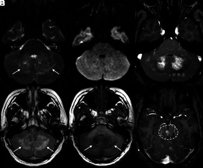
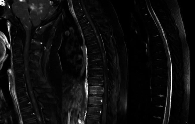
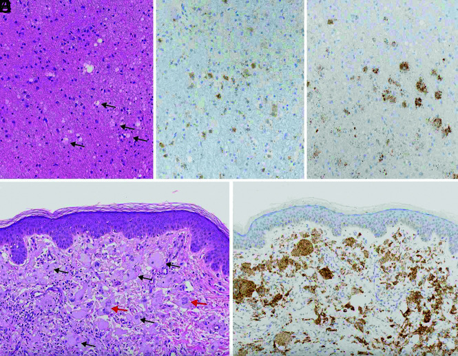
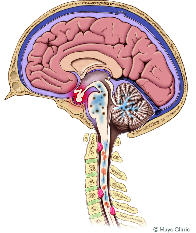
Erdheim-Chester disease is a rare non-Langerhans cell histiocytosis. The disease is widely variable in its severity, ranging from incidental findings in asymptomatic patients to a fatal multisystem illness. CNS involvement occurs in up to one-half of patients, most often leading to diabetes insipidus and cerebellar dysfunction. Imaging findings in neurologic Erdheim-Chester disease are often nonspecific, and the disease is commonly mistaken for close mimickers. Nevertheless, there are many imaging manifestations of Erdheim-Chester disease that are highly suggestive of the disease, which an astute radiologist could use to accurately indicate this diagnosis. This article discusses the imaging appearance, histologic features, clinical manifestations, and management of Erdheim-Chester disease.


Figures

FIG 1.
FIG 1.
MR images of the posterior fossa demonstrate multifocal abnormalities involving both cerebellar hemispheres, particularly located around the dentate nuclei. These regions are bright on T2 and FLAIR (arrows in A and B), lack restricted diffusion (C), are hypointense on T1 (arrows in D), and demonstrate patchy enhancement on postcontrast T1 fat-saturated images (E). Similar patchy enhancement is also noted in the midbrain (dashed oval on F).
FIG 2.
FIG 2.
MR imaging of the cervical (A) and thoracic (B and C) spine demonstrates numerous patchy areas of enhancement along the cervicothoracic cord (A and B). Corresponding fat-saturated STIR images of the thoracic spine demonstrate multifocal T2 hyperintensities in these regions (C).
FIG 3.
FIG 3.
H&E-stained sections of the cerebellar biopsy (A) demonstrate mildly hypercellular white matter with increased foamy macrophages (black arrows), highlighted by CD68 immunostain (B). The macrophages were cytologically bland but show expression of BRAF p.V600E (C), supporting the presence of a histiocytic neoplasm. A subsequent skin biopsy (D) shows more characteristic features of ECD, including large, foamy macrophages (black arrows) and Touton giant cells (red arrows), which were positive for BRAF p.V600E (E). Original magnification ×200 for all images.
FIG 4.
FIG 4.
Schematic showing the various types of involvement of ECD in the CNS. Intracranially, the most common findings are focal areas of enhancement in the brainstem and/or cerebellum, with surrounding edema. The pituitary infundibulum is commonly enlarged. In the spine, both intramedullary lesions and extramedullary lesions are observed, which are often associated with adjacent edema.

References

    1. Sedrak P, Ketonen L, Hou P, et al. . Erdheim-Chester disease of the central nervous system: new manifestations of a rare disease. AJNR Am J Neuroradiol 2011;32:2126–31 10.3174/ajnr.A2707 - DOI - PMC - PubMed
    1. Oliveira M, Monteiro S, Dos Santos J, et al. . Erdheim-Chester disease: a rare clinical entity. Eur J Case Rep Intern Med 2020;7:001630 10.12890/2020_001630 - DOI - PMC - PubMed
    1. Todisco A, Cavaliere C, Vaglio A, et al. . Erdheim-Chester disease: a challenging diagnosis for an effective therapy. Clin Neurol Neurosurg 2020;194:105841 10.1016/j.clineuro.2020.105841 - DOI - PubMed
    1. Sánchez-Villalobos JM, Jimeno-Almazán A, López-Peña C, et al. . Erdheim-Chester disease mimicking multiple sclerosis or a new association? Mult Scler Relat Disord 2019;30:94–97 10.1016/j.msard.2019.02.008 - DOI - PubMed
    1. Pegoraro F, Papo M, Maniscalco V, et al. . Erdheim-Chester disease: a rapidly evolving disease model. Leukemia 2020;34:2840–57 10.1038/s41375-020-0944-4 - DOI - PubMed

MeSH terms

LinkOut - more resources