. 2023 May;44(5):589-594.
doi: 10.3174/ajnr.A7840. Epub 2023 Mar 30.

Imaging the Tight Orbit: Radiologic Manifestations of Orbital Compartment Syndrome

Affiliations

Imaging the Tight Orbit: Radiologic Manifestations of Orbital Compartment Syndrome

C Maier et al. AJNR Am J Neuroradiol. 2023 May.

Abstract

Background and purpose: Orbital compartment syndrome is a sight-threatening emergency caused by rising pressure inside the orbit. It is usually diagnosed clinically, but imaging might help when clinical findings are inconclusive. This study aimed to systematically evaluate imaging features of orbital compartment syndrome.

Materials and methods: This retrospective study included patients from 2 trauma centers. Proptosis, optic nerve length, posterior globe angle, morphology of the extraocular muscles, fracture patterns, active bleeding, and superior ophthalmic vein caliber were assessed on pretreatment CT. Etiology, clinical findings, and visual outcome were obtained from patient records.

Results: Twenty-nine cases of orbital compartment syndrome were included; most were secondary to traumatic hematoma. Pathologies occurred in the extraconal space in all patients, whereas intraconal abnormalities occurred in 59% (17/29), and subperiosteal hematoma in 34% (10/29). We observed proptosis (affected orbit: mean, 24.4 [SD, 3.1] mm versus contralateral: 17.7 [SD, 3.1] mm; P < .01) as well as stretching of the optic nerve (mean, 32.0 [SD, 2.5] mm versus 25.8 [SD, 3.4] mm; P < .01). The posterior globe angle was decreased (mean, 128.7° [SD, 18.9°] versus 146.9° [SD, 6.4°]; P < .01). In 69% (20/29), the superior ophthalmic was vein smaller in the affected orbit. No significant differences were detected regarding the size and shape of extraocular muscles.

Conclusions: Orbital compartment syndrome is characterized by proptosis and optic nerve stretching. In some cases, the posterior globe is deformed. Orbital compartment syndrome can be caused by an expanding pathology anywhere within the orbit with or without direct contact to the optic nerve, confirming the pathophysiologic concept of a compartment mechanism.


Figures

FIG 1.
FIG 1.
Patient flow chart.
FIG 2.
FIG 2.
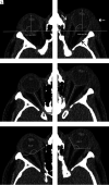
Representative case with traumatic left-sided OCS due to intraorbital hematoma in the extraconal space (white arrow) and in the subperiosteal space (black arrow). Imaging is helpful to differentiate intraorbital hematoma from periorbital hematoma (dashed white arrow), which is anterior to the orbital septum and therefore does not lead to compartment syndrome. Intraorbital hematoma in this case results in increased proptosis (A and B), optic nerve stretching (C and D), and a decreased posterior globe angle (E and F). Besides stretching of the optic nerve, subtle thinning of the nerve and loss of normal undulation can be observed. Note that the angulation is slightly different for each side because the planes of measurement were reconstructed separately relative to the respective anatomic landmarks.
FIG 3.
FIG 3.
Quantitative imaging data. Boxplots show the distribution of proptosis, defined as the distance between the interzygomatic line and the corneal apex (A); optic nerve length, defined as the distance between the orbital aperture of the optic canal and the scleral contour at the optic nerve head (B); and posterior globe angle, as measured by drawing tangents on the scleral contours at the optic nerve head (C). Each affected orbit is connected to its contralateral control by a thin line. The asterisk indicates P < .05.
FIG 4.
FIG 4.
Differences between the affected orbit and the contralateral orbit in patients with vision loss at discharge (n = 6) and in patients with preserved vision (n = 20) regarding proptosis (A), optic nerve length (B), and posterior globe angle (C). No significant differences were detected. For 3 patients, data on visual outcome were unavailable.

Comment in

References

    1. Lima V, Burt B, Leibovitch I, et al. . Orbital compartment syndrome: the ophthalmic surgical emergency. Surv Ophthalmol 2009;54:441–49 10.1016/j.survophthal.2009.04.005 - DOI - PubMed
    1. Bailey WK, Kuo PC, Evans LS. Diagnosis and treatment of retrobulbar hemorrhage. J Oral Maxillofac Surg 1993;51:780–82 10.1016/s0278-2391(10)80422-7 - DOI - PubMed
    1. Hargaden M, Goldberg SH, Cunningham D, et al. . Optic neuropathy following simulation of orbital hemorrhage in the nonhuman primate. Ophthalmic Plast Reconstr Surg 1996;12:264–72 10.1097/00002341-199612000-00009 - DOI - PubMed
    1. Vassallo S, Hartstein M, Howard D, et al. . Traumatic retrobulbar hemorrhage: emergent decompression by lateral canthotomy and cantholysis. J Emerg Med 2002;22:251–56 10.1016/s0736-4679(01)00477-2 - DOI - PubMed
    1. Ballard SR, Enzenauer RW, O'Donnell T, et al. . Emergency lateral canthotomy and cantholysis: a simple procedure to preserve vision from sight threatening orbital hemorrhage. J Spec Oper Med 2009;9:26–32 10.55460/1CLD-XJUV - DOI - PubMed

MeSH terms

LinkOut - more resources