

Site-Specifically Conjugated Single-Domain Antibody Successfully Identifies Glypican-3-Expressing Liver Cancer by Immuno-PET
- PMID: 36997331
- PMCID: PMC10315705
- DOI: 10.2967/jnumed.122.265171
Site-Specifically Conjugated Single-Domain Antibody Successfully Identifies Glypican-3-Expressing Liver Cancer by Immuno-PET
Abstract
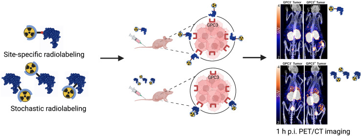
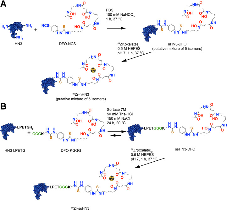
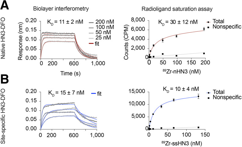
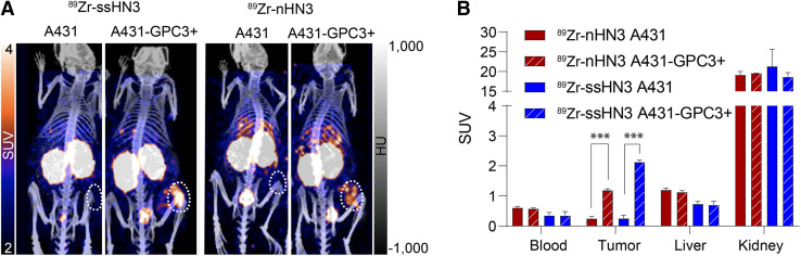
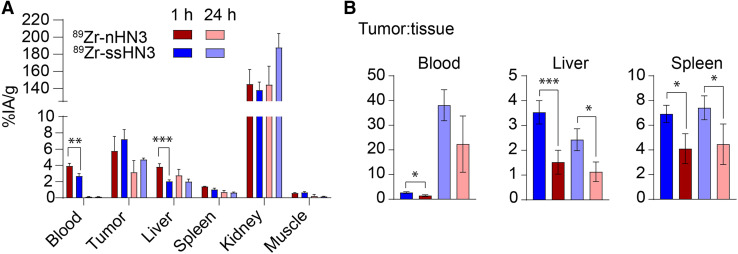
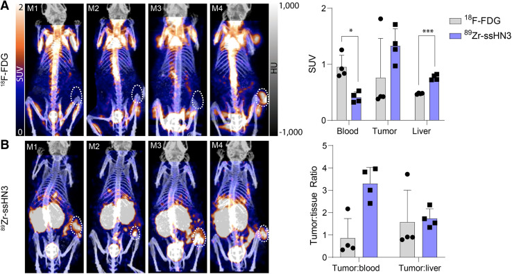
Primary liver cancer is the third leading cause of cancer-related deaths, and its incidence and mortality are increasing worldwide. Hepatocellular carcinoma (HCC) accounts for 80% of primary liver cancer cases. Glypican-3 (GPC3) is a heparan sulfate proteoglycan that histopathologically defines HCC and represents an attractive tumor-selective marker for radiopharmaceutical imaging and therapy for this disease. Single-domain antibodies are a promising scaffold for imaging because of their favorable pharmacokinetic properties, good tumor penetration, and renal clearance. Although conventional lysine-directed bioconjugation can be used to yield conjugates for radiolabeling full-length antibodies, this stochastic approach risks negatively affecting target binding of the smaller single-domain antibodies. To address this challenge, site-specific approaches have been explored. Here, we used conventional and sortase-based site-specific conjugation methods to engineer GPC3-specific human single-domain antibody (HN3) PET probes. Methods: Bifunctional deferoxamine (DFO) isothiocyanate was used to synthesize native HN3 (nHN3)-DFO. Site-specifically modified HN3 (ssHN3)-DFO was engineered using sortase-mediated conjugation of triglycine-DFO chelator and HN3 containing an LPETG C-terminal tag. Both conjugates were radiolabeled with 89Zr, and their binding affinity in vitro and target engagement of GPC3-positive (GPC3+) tumors in vivo were determined. Results: Both 89Zr-ssHN3 and 89Zr-nHN3 displayed nanomolar affinity for GPC3 in vitro. Biodistribution and PET/CT image analysis in mice bearing isogenic A431 and A431-GPC3+ xenografts, as well as in HepG2 liver cancer xenografts, showed that both conjugates specifically identify GPC3+ tumors. 89Zr-ssHN3 exhibited more favorable biodistribution and pharmacokinetic properties, including higher tumor uptake and lower liver accumulation. Comparative PET/CT studies on mice imaged with both 18F-FDG and 89Zr-ssHN3 showed more consistent tumor accumulation for the single-domain antibody conjugate, further establishing its potential for PET imaging. Conclusion: 89Zr-ssHN3 showed clear advantages in tumor uptake and tumor-to-liver signal ratio over the conventionally modified 89Zr-nHN3 in xenograft models. Our results establish the potential of HN3-based single-domain antibody probes for GPC3-directed PET imaging of liver cancers.
Keywords: GPC3; Nanobody; glypican-3; immuno-PET; liver cancer; molecular imaging.
© 2023 by the Society of Nuclear Medicine and Molecular Imaging.
Figures
References
Publication types
MeSH terms
Substances
Grants and funding
LinkOut - more resources
Full Text Sources
Other Literature Sources
Medical