. 2023 Mar 30;13(1):5162.
doi: 10.1038/s41598-023-32060-2.

Preclinical development of a first-in-class vaccine encoding HER2, Brachyury and CD40L for antibody enhanced tumor eradication

Affiliations

Preclinical development of a first-in-class vaccine encoding HER2, Brachyury and CD40L for antibody enhanced tumor eradication

Maria Hinterberger et al. Sci Rep. .

Abstract

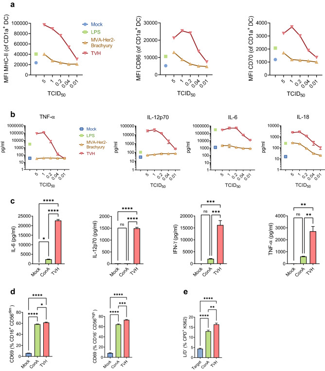
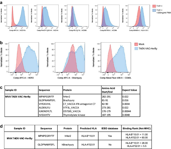
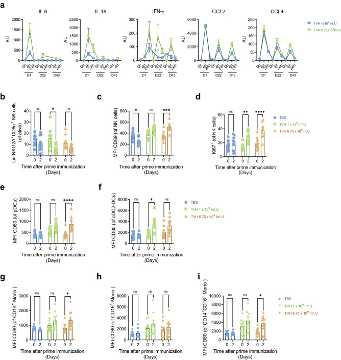
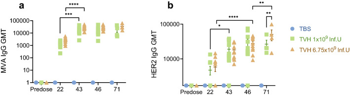
The induction of antiviral innate immunity by systemic immunization with live virus can be employed to positively impact the response to therapeutic vaccination. We previously demonstrated that systemic immunization with a non-replicating MVA encoding CD40 ligand (CD40L) enhances innate immune cell activation and function, and triggers potent antitumor CD8+ T cell responses in different murine tumor models. Antitumor efficacy was increased when combined with tumor targeting antibodies. Here we report the development of TAEK-VAC-HerBy (TVH), a first-in-class human tumor antibody enhanced killing (TAEK) vaccine based on the non-replicating MVA-BN viral vector. It encodes the membrane bound form of human CD40L, HER2 and the transcription factor Brachyury. TVH is designed for therapeutic use in HER2- or Brachyury-expressing cancer patients in combination with tumor targeting antibodies. To preclude possible oncogenic activities in infected cells and to prevent binding of vaccine-encoded HER2 by monoclonal antibodies trastuzumab and pertuzumab, genetic modifications of HER2 were introduced in the vaccine. Brachyury was genetically modified to prevent nuclear localization of the protein thereby inhibiting its transcriptional activity. CD40L encoded in TVH enhanced human leukocyte activation and cytokine secretion in vitro. Lastly, TVH intravenous administration to non-human primates was proven immunogenic and safe in a repeat-dose toxicity study. Nonclinical data presented here highlight TVH as a first-in-class immunotherapeutic vaccine platform currently under clinical investigation.


Conflict of interest statement

The authors enlisted are or have been employees of Bavarian Nordic.

Figures

Figure 1
Figure 1
Characterization of transgenes contained in TAEK-VAC-HerBy. (a) Schematic map of TAEK-VAC-HerBy showing the transgenes in the Intergenic Region (IGR) 88/89 of the MVA-BN genome. Expression of transgenes is controlled by different poxvirus promoters; (b) Transgene expression detected by flow cytometry. Briefly, HeLa cells were infected overnight either with mock or with TVH at a MOI of 4. Expression of vaccinia, HER2, Brachyury and CD40L (blue histogram) against mock control (red histogram) is shown; (c) Analysis of subcellular localization of Brachyury-ΔNLS and Brachyury-NLS by immunofluorescence staining. A31 cells were either mock infected or infected at a MOI of 2 with MVA-BN, MVA-Brachyury or TVH. At 17 h post infection, cells were fixed, permeabilized, and stained with anti-Brachyury antibody followed by PE-conjugated secondary antibody. Upper panel represents Brachyury staining, followed by clear field (middle panel) and overlay (lower panel). To better show subcellular localization of Brachyury-ΔNLS or Brachyury-NLS, inlays display higher magnification of representative cells indicated by arrow; (d) Differential binding of HER2 antibodies to mutated HER2 present in TVH. Briefly, CT26.WT cells were infected overnight with TVH at a MOI of 5. Then, infected CT26.WT cells as well as control CT26.HER2 cells were stained with different HER2 antibody clones, namely HER2 24D2, Herceptin® or Perjeta®. Data show HER2 expression detected by Herceptin® and Perjeta® normalized to HER2 24D2-mediated detection. Data shown in B-D are representative of several independent experiments.
Figure 2
Figure 2
TVH activates innate immune cells in vitro. (a, b) Activation and cytokine production of dendritic cells upon TVH infection. Monocyte-derived DCs (mo-DC) were generated after enrichment of CD14+ monocytes from human PBMCs and cultured for 7 days in the presence of GM-CSF and IL-4 as explained in Materials and Methods. Mo-DCs were infected overnight either with MVA-BN or TVH at different MOIs. LPS and mock infections were used as positive and negative controls, respectively. Supernatants were then collected, and cells were stained for flow cytometry analysis. (a) Mean Fluorescence Intensity (MFI) of MHC class II (MHC-II), CD86 and CD70 gated on CD1a+ DCs; (b) TNF-α, IL-12p70, IL-6 and IL-18 quantification in supernatants. A representative donor out of four tested is shown; (c-e) Cryopreserved PBMCs from the same donor were infected with TVH at a MOI of 0.5 or stimulated with ConA as a positive control. After overnight incubation, supernatants were collected, and cells were either stained for flow cytometry analysis or NK cells were magnetically sorted. Sorted NK cells were incubated with Cell Proliferation Dye (CPD) labelled K562 target cells in a 1:1 ratio (effector:target). (c) Cytokine secretion profile showing IL-6, IL-12p70, IFN-γ and TNF-α; (d) Frequency of CD69 positive cells on CD16+CD56low and CD16-CD56high NK cells detected by flow cytometry, indicating TVH-induced activity and cytotoxicity of NK cells. (e) Percentage of dead CPD K562 target cells after being incubated with sorted NK cells. Data are expressed as Mean ± SEM and are representative of two independent experiments. One-way ANOVA was performed on figures c-e. ns, non-significant; *, p < 0.05; **p < 0.01; ***p < 0.005; ****p < 0.001.
Figure 3
Figure 3
Brachyury and HER2 expressed in TVH are loaded onto HLA class I molecules. (a, b) TVH infection of THP-1-derived macrophages. THP-1 cells are cultured in the presence of 200 ng/ml PMA for 72 h. Then, cells are washed and rested in complete medium for additional 48 h until the start of the experiment. (a) Phenotype of THP-1-derived macrophages compared to undifferentiated THP-1 cells. Expression of hCD16, hCD14, hCD80, hCD86 and HLA-ABC is detected by flow cytometry. (b) Expression of HER2, Brachyury and CD40L on THP-1-derived macrophages. THP-1-derived macrophages were infected with TVH at Inf.U 4/cell. After overnight incubation, expression of HER2, Brachyury and CD40L was analysed by flow cytometry. (c-d) Analysis of peptides loaded onto HLA-ABC by mass spectrometry, and brief results of ProImmune ProPresent® MAPPS Antigen Presentation assay. Infected THP-1-derived macrophages are lysed, and HLA-peptide molecules recovered in an immune affinity step. Peptides are eluted from the HLA molecules and analysed by LC–MS/MS. Identified sequences are subjected to rigorous analysis to identify true positive peptides with high confidence. (c) Summary table of peptides detected by the ProPresent® Assay in TVH-infected macrophages. The likelihood of peptides to be real identities is described by their Expect Value. Peptides with an Expect Value ≤ 0.05 are indicative of identity; (d) Summary table for the features of TAA-derived peptides MPNPEGRYTF and GLDPNAMYSFL, showing predicted HLA-binding calculated using SYFPEITHI, whether they are listed in the IEDB database, and their binding rank to HLA-B*15:01 and HLA-A*02:01 defined by Net-MHC4.0.
Figure 4
Figure 4
Intravenous injection of TVH into cynomolgus monkeys induces innate immune cell driven cytokine release and activation. TVH administration scheme was defined in Table 1. Briefly, 3 homologous intravenous injections of either TBS, 1 × 109 Inf.U TVH or 6.75 × 109 Inf.U TVH were performed on days 1, 22 and 43. Blood was taken prior to each immunization, 4 h and 24 h after each immunization for cytokine and chemokine analysis. For analysis of innate immune cell subset activation, PBMCs were taken on day 0 and day 2 after the first immunization. (a) Quantification of IL-6, IL-18, IFN-γ, CCL2 and CCL4 in sera at 0 (predose), 4 and 24 h after each immunization by Luminex is indicated as arbitrary units (AU). (b) Frequency of NK cells; (c) Mean Fluorescence Intensity (MFI) of CD69 on NK cells; (d) Percentage of Ki67+ NK cells; (e) MFI of CD80 on pDCs; (f) on cDC2-DCs, (g) CD14+ monocytes, (h) CD16+ monocytes, and (i) CD14+ CD16+ monocytes are shown for the times only after prime immunization. Data in figures b-i shown for individual animals and as Mean (depicted by columns) ± SEM. Two-way ANOVA was performe to evaluate statistically significant differences. ns, non-significant; *p < 0.05; **p < 0.01; ***p < 0.005; ****p < 0.001.
Figure 5
Figure 5
Induction of HER2- and MVA-specific IgG upon systemic immunization of cynomolgus monkeys using TVH. TVH administration scheme was defined in Table 1. Briefly, 3 homologous intravenous injections of either TBS, 1 × 109 Inf.U TVH or 6.75 × 109 Inf.U TVH were performed on days 1, 22 and 43. Blood was collected prior to the first immunization (predose: Day -12 and on days 22, 43, 46 and 71; day 71 only in Recovery Group; n = 4). Sera were analyzed using (a) MVA-specific IgG ELISA and (b) HER2-specific IgG ELISA. In this graph, IgG titers for individual animals are shown as well as GMTs (Geometric Mean Titers) depicted by horizonal bars +/-SEM. Two-way ANOVA was performed on figures a-b. ns, non-significant; *p < 0.05; **p < 0.01; ***p < 0.005; ****p < 0.001.

References

    1. Pittman PR, et al. Phase 3 efficacy trial of modified vaccinia Ankara as a vaccine against smallpox. N. Engl. J. Med. 2019;381:1897–1908. doi: 10.1056/NEJMoa1817307. - DOI - PubMed
    1. Anywaine Z, et al. Safety and immunogenicity of a 2-dose heterologous vaccination regimen with Ad26.ZEBOV and MVA-BN-filo Ebola vaccines: 12-month data from a phase 1 randomized clinical trial in Uganda and Tanzania. J. Infect. Dis. 2019;220:46–56. doi: 10.1093/infdis/jiz070. - DOI - PMC - PubMed
    1. Jordan E, et al. Broad antibody and cellular immune response from a phase 2 clinical trial with a novel multivalent poxvirus-based respiratory syncytial virus vaccine. J. Infect. Dis. 2021;223:1062–1072. doi: 10.1093/infdis/jiaa460. - DOI - PubMed
    1. Gatti-Mays ME, et al. A phase 1 dose escalation trial of BN-CV301, a recombinant poxviral vaccine targeting MUC1 and CEA with costimulatory molecules. Clin. Cancer Res. 2019 doi: 10.1158/1078-0432.CCR-19-0183. - DOI - PMC - PubMed
    1. Frey SE, et al. Safety and immunogenicity of IMVAMUNE(R) smallpox vaccine using different strategies for a post event scenario. Vaccine. 2013;31:3025–3033. doi: 10.1016/j.vaccine.2013.04.050. - DOI - PMC - PubMed