. 2023 Mar 30;13(1):5194.
doi: 10.1038/s41598-023-31992-z.

Phylogenetic and functional characterization of water bears (Tardigrada) tubulins

Affiliations

Phylogenetic and functional characterization of water bears (Tardigrada) tubulins

Kamila Novotná Floriančičová et al. Sci Rep. .

Abstract

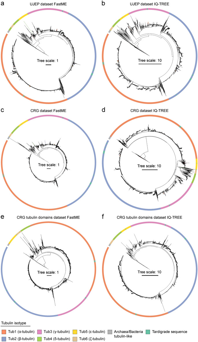
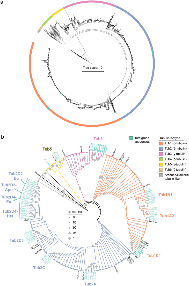
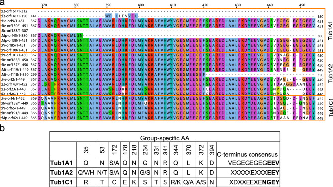
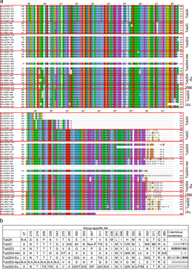
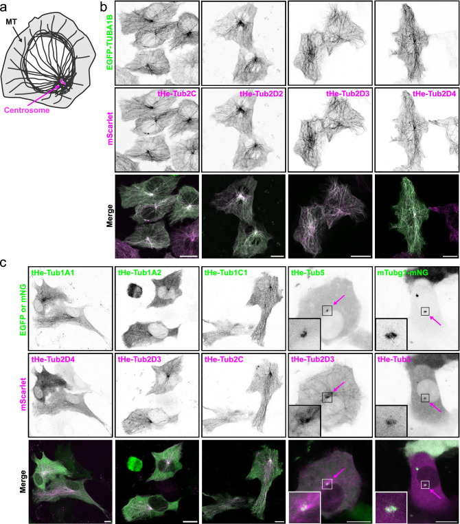
Tardigrades are microscopic ecdysozoans that can withstand extreme environmental conditions. Several tardigrade species undergo reversible morphological transformations and enter into cryptobiosis, which helps them to survive periods of unfavorable environmental conditions. However, the underlying molecular mechanisms of cryptobiosis are mostly unknown. Tubulins are evolutionarily conserved components of the microtubule cytoskeleton that are crucial in many cellular processes. We hypothesize that microtubules are necessary for the morphological changes associated with successful cryptobiosis. The molecular composition of the microtubule cytoskeleton in tardigrades is unknown. Therefore, we analyzed and characterized tardigrade tubulins and identified 79 tardigrade tubulin sequences in eight taxa. We found three α-, seven β-, one γ-, and one ε-tubulin isoform. To verify in silico identified tardigrade tubulins, we also isolated and sequenced nine out of ten predicted Hypsibius exemplaris tubulins. All tardigrade tubulins were localized as expected when overexpressed in mammalian cultured cells: to the microtubules or to the centrosomes. The presence of a functional ε-tubulin, clearly localized to centrioles, is attractive from a phylogenetic point of view. Although the phylogenetically close Nematoda lost their δ- and ε-tubulins, some groups of Arthropoda still possess them. Thus, our data support the current placement of tardigrades into the Panarthropoda clade.


Conflict of interest statement

The authors declare no competing interests.

Figures

Figure 1
Figure 1
Phylogenetic analysis of tubulin homologs found in tardigrades. (a-f) Phylogenetic trees computed from alignments of tardigrade tubulin homologous sequences with Findeisen et al. dataset. (a, c, e) Minimum evolution method (FastMe). (b, d, f) Maximum likelihood method (IQ-TREE). (a, b) UJEP dataset—full tubulin sequences, (c, d) CRG dataset—full tubulin sequences, (e–f) CRG dataset—extracted tubulin domains only. All phylogenetic trees are rooted by an archaeal tubulin-like group. The legend describes color coding. See also Supplementary data 1 and Supplementary Data 2.
Figure 2
Figure 2
Identification of tardigrade tubulin isoforms. (a, b) Different visualizations of an identical phylogenetic tree computed from an alignment of the curated tardigrade tubulin dataset with Findeisen et al. dataset. (a) Maximum likelihood method (IQ-TREE). (b) Maximum likelihood method (IQ-TREE), non-tardigrade tubulins branches collapsed and branch lengths ignored to simplify visualization. Grey circles indicate SH-aLRT test score. The legend describes color coding. See also Supplementary data 3 and Supplementary Table 1.
Figure 3
Figure 3
Specific sequence signatures of tardigrade α-tubulins. (a) An alignment of the C-terminal region of tardigrade α-tubulins. Orange boxes indicate individual isoforms. Note the very C-terminus. (b) Specific AA differences among tardigrade α-tubulin isoforms. See also Supplementary data 4.
Figure 4
Figure 4
Specific sequence signatures of tardigrade β-tubulins. (a) An alignment of the C-terminal region of tardigrade β-tubulins. Red boxes indicate individual isoforms. (b) Specific AA differences among tardigrade β-tubulin isoforms. See also Supplementary data 4.
Figure 5
Figure 5
Tardigrade tubulins localize to microtubules or to the centrosomes in mammalian cultured cells. (a) A schematic of a mammalian cell with microtubules and the centrosome. (b) Maximum intensity projections (MIP) of living U87-MG cells overexpressing indicated constructs. Human EGFP-tagged α-tubulin (TUBA1B) used as a control. Note that all tardigrade β-tubulins localize to microtubules. (c) MIP of living hTert-RPE-1 cells overexpressing indicated constructs. Note that tardigrade γ- and ε-tubulins localize to the centrosome (magenta arrows). Scale bars 10 μm.

Similar articles

Cited by

References

    1. Fleming JF, Arakawa K. Systematics of tardigrada: A reanalysis of tardigrade taxonomy with specific reference to Guil et al. (2019) Zool. Scr. 2021;50:376–382. doi: 10.1111/zsc.12476. - DOI
    1. Møbjerg N, Jørgensen A, Kristensen RM, Neves RC. Morphology and functional anatomy. In: Schill RO, editor. Water Bears: The Biology of Tardigrades. Springer International Publishing; 2018. pp. 57–94.
    1. Halberg KA, Jørgensen A, Møbjerg N. Desiccation tolerance in the tardigrade Richtersius coronifer relies on muscle mediated structural reorganization. PLoS ONE. 2013;8:e85091. doi: 10.1371/journal.pone.0085091. - DOI - PMC - PubMed
    1. Czerneková M, Jönsson KI, Chajec L, Student S, Poprawa I. The structure of the desiccated Richtersius coronifer (Richters, 1903) Protoplasma. 2017;254:1367–1377. doi: 10.1007/s00709-016-1027-2. - DOI - PubMed
    1. Czerneková M, Vinopal S. The tardigrade cuticle. Limnol. Rev. 2021;21:127–146. doi: 10.2478/limre-2021-0012. - DOI

Publication types