

MRI-based training model for left atrial appendage closure
- PMID: 36997829
- PMCID: PMC10589139
- DOI: 10.1007/s11548-023-02870-w
MRI-based training model for left atrial appendage closure
Abstract
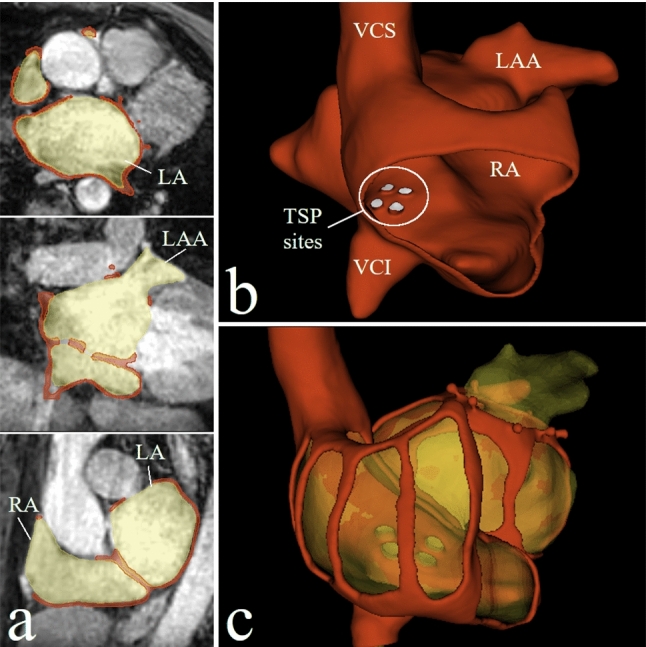
Purpose: Percutaneous closure of the left atrial appendage (LAA) reduces the risk of embolic stroke in patients with atrial fibrillation. Thereby, the optimal transseptal puncture (TSP) site differs due to the highly variable anatomical shape of the LAA, which is rarely considered in existing training models. Based on non-contrast-enhanced magnetic resonance imaging (MRI) volumes, we propose a training model for LAA closure with interchangeable and patient-specific LAA enabling LAA-specific identification of the TSP site best suited.
Methods: Based on patient-specific MRI data, silicone models of the LAAs were produced using a 3D-printed cast model. In addition, an MRI-derived 3D-printed base model was set up, including the right and left atrium with predefined passages in the septum, mimicking multiple TSP sites. The various silicone models and a tube mimicking venous access were connected to the base model. Empirical use of the model allowed the demonstration of its usability.
Results: Patient-specific silicone models of the LAA could be generated from all LAA patient MRI datasets. The influence of various combinations regarding TSP sites and LAA shapes could be demonstrated as well as the technical functionality of the occluder system. Via the attached tube mimicking the venous access, the correct handling of the deployment catheter even in case of not optimal puncture site could be practiced.
Conclusion: The proposed contrast-agent and radiation-free MRI-based training model for percutaneous LAA closure enables the pre-interventional assessment of the influence of the TSP site on the access of patient-specific LAA shapes. A straightforward replication of this work is measured by using clinically available imaging protocols and a widespread 3D printer technique to build the model.
Keywords: LAA closure; Training model; Transseptal puncture.
© 2023. The Author(s).
Conflict of interest statement
The authors declare that the research was conducted in the absence of any commercial or financial relationships that could be construed as a potential conflict of interest.
Figures


References
-
- Holmes DR, Kar S, Price MJ, Whisenant B, Sievert H, Doshi SK, Huber K, Reddy VY. Prospective randomized evaluation of the watchman left atrial appendage closure device in patients with atrial fibrillation versus long-term warfarin therapy: the PREVAIL trial. J Am Coll Cardiol. 2014;64:1–12. doi: 10.1016/j.jacc.2014.04.029. - DOI - PubMed
MeSH terms
Grants and funding
LinkOut - more resources
Full Text Sources
Medical
Miscellaneous
