

Septin-3 autoimmunity in patients with paraneoplastic cerebellar ataxia
- PMID: 36997937
- PMCID: PMC10061979
- DOI: 10.1186/s12974-023-02718-9
Septin-3 autoimmunity in patients with paraneoplastic cerebellar ataxia
Abstract
Background: Septins are cytoskeletal proteins with filament forming capabilities, which have multiple roles during cell division, cellular polarization, morphogenesis, and membrane trafficking. Autoantibodies against septin-5 are associated with non-paraneoplastic cerebellar ataxia, and autoantibodies against septin-7 with encephalopathy with prominent neuropsychiatric features. Here, we report on newly identified autoantibodies against septin-3 in patients with paraneoplastic cerebellar ataxia. We also propose a strategy for anti-septin autoantibody determination.
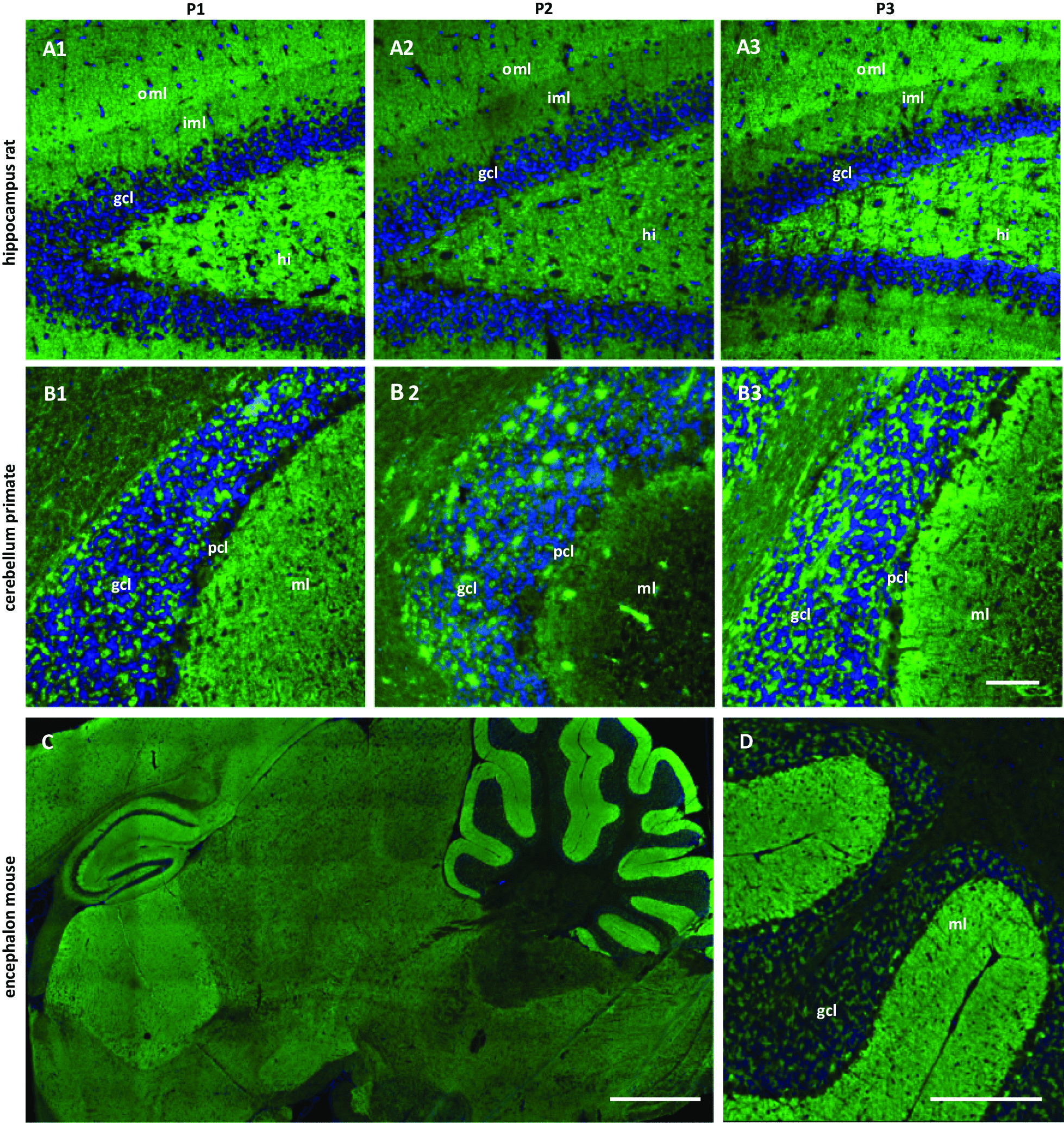
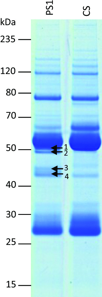
Methods: Sera from three patients producing similar immunofluorescence staining patterns on cerebellar and hippocampal sections were subjected to immunoprecipitation followed by mass spectrometry. The identified candidate antigens, all of which were septins, were expressed recombinantly in HEK293 cells either individually, as complexes, or combinations missing individual septins, for use in recombinant cell-based indirect immunofluorescence assays (RC-IIFA). Specificity for septin-3 was further confirmed by tissue IIFA neutralization experiments. Finally, tumor tissue sections were analyzed immunohistochemically for septin-3 expression.
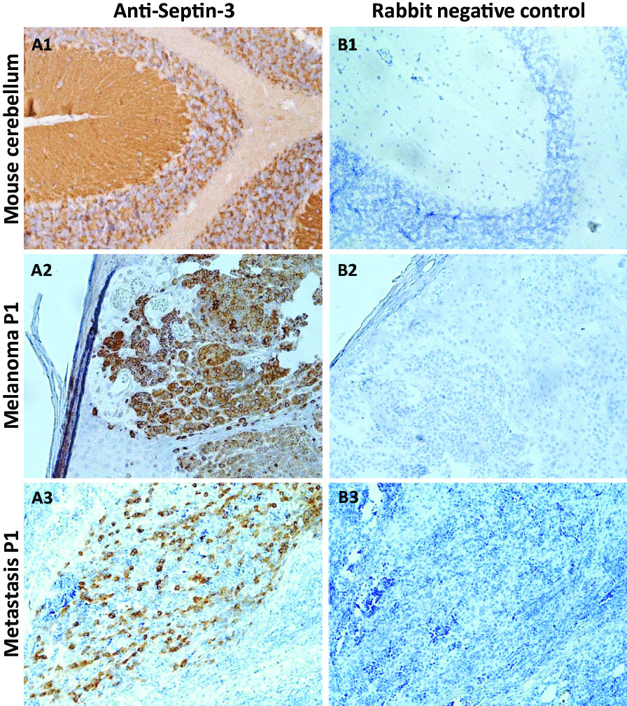
Results: Immunoprecipitation with rat cerebellum lysate revealed septin-3, -5, -6, -7, and -11 as candidate target antigens. Sera of all three patients reacted with recombinant cells co-expressing septin-3/5/6/7/11, while none of 149 healthy control sera was similarly reactive. In RC-IIFAs the patient sera recognized only cells expressing septin-3, individually and in complexes. Incubation of patient sera with five different septin combinations, each missing one of the five septins, confirmed the autoantibodies' specificity for septin-3. The tissue IIFA reactivity of patient serum was abolished by pre-incubation with HEK293 cell lysates overexpressing the septin-3/5/6/7/11 complex or septin-3 alone, but not with HEK293 cell lysates overexpressing septin-5 as control. All three patients had cancers (2 × melanoma, 1 × small cell lung cancer), presented with progressive cerebellar syndromes, and responded poorly to immunotherapy. Expression of septin-3 was demonstrated in resected tumor tissue available from one patient.
Conclusions: Septin-3 is a novel autoantibody target in patients with paraneoplastic cerebellar syndromes. Based on our findings, RC-IIFA with HEK293 cells expressing the septin-3/5/6/7/11 complex may serve as a screening tool to investigate anti-septin autoantibodies in serological samples with a characteristic staining pattern on neuronal tissue sections. Autoantibodies against individual septins can then be confirmed by RC-IIFA expressing single septins.
Keywords: Autoantibodies; Autoimmune cerebellar ataxia; Autoimmune encephalitis; Autoimmunity; Cerebellitis; Cerebellum; Immunoglobulin G; Immunoprecipitation; Melanoma; Paraneoplastic cerebellar degeneration; Paraneoplastic neurological syndrome; Septin-11; Septin-3; Septin-5; Septin-6; Septin-7; Septins; Small-cell lung cancer.
© 2023. The Author(s).
Conflict of interest statement
KB, BT, NR, SJ, KG, and ID have nothing to disclose. MS, RM, YD, CP, and LK are employees of EUROIMMUN. MS, RM, YD, and LK have septin-3 and -7 patent applications pending. KR received research support from Novartis Pharma, Merck Serono, German Ministry of Education and Research, European Union (821283-2), Stiftung Charité (BIH Clinical Fellow Program), Arthur Arnstein Foundation and Guthy Jackson Charitable Foundation. BW received grants from the German Ministry of Education and Research, Deutsche Forschungsgemeinschaft, Dietmar Hopp Foundation, Klaus Tschira Foundation, grants and personal fees from Merck, and personal fees from Alexion, Bayer, Biogen, Roche, none related to this work.
Figures


References
-
- Herrero San Martin A, Amarante Cuadrado C, Gonzalez Arbizu M, Rabano-Suarez P, Ostos-Moliz F, Naranjo L, Sabater L, Martinez Hernandez E, Ruiz Garcia R, Toledo Alfocea D. Autoimmune septin-5 disease presenting as spinocerebellar ataxia and nystagmus. Neurology. 2021;97:291–292. doi: 10.1212/WNL.0000000000012240. - DOI - PubMed
MeSH terms
Substances
LinkOut - more resources
Full Text Sources
