. 2023 Mar 30;42(1):77.
doi: 10.1186/s13046-023-02640-1.

Protein disulfide-isomerase A4 confers glioblastoma angiogenesis promotion capacity and resistance to anti-angiogenic therapy

Affiliations

Protein disulfide-isomerase A4 confers glioblastoma angiogenesis promotion capacity and resistance to anti-angiogenic therapy

Zewei Tu et al. J Exp Clin Cancer Res. .

Abstract

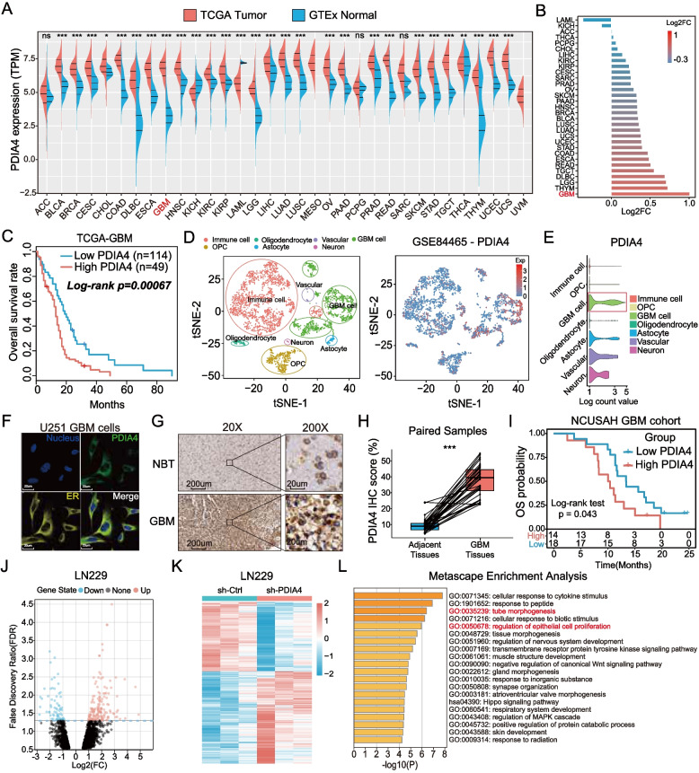
Introduction: Increasing evidence has revealed the key activity of protein disulfide isomerase A4 (PDIA4) in the endoplasmic reticulum stress (ERS) response. However, the role of PDIA4 in regulating glioblastoma (GBM)-specific pro-angiogenesis is still unknown.

Methods: The expression and prognostic role of PDIA4 were analyzed using a bioinformatics approach and were validated in 32 clinical samples and follow-up data. RNA-sequencing was used to search for PDIA4-associated biological processes in GBM cells, and proteomic mass spectrum (MS) analysis was used to screen for potential PDIA4 substrates. Western blotting, real-time quantitative polymerase chain reaction (RT-qPCR), and enzyme-linked immunosorbent assays (ELISA) were used to measure the levels of the involved factors. Cell migration and tube formation assays determined the pro-angiogenesis activity of PDIA4 in vitro. An intracranial U87 xenograft GBM animal model was constructed to evaluate the pro-angiogenesis role of PDIA4 in vivo.

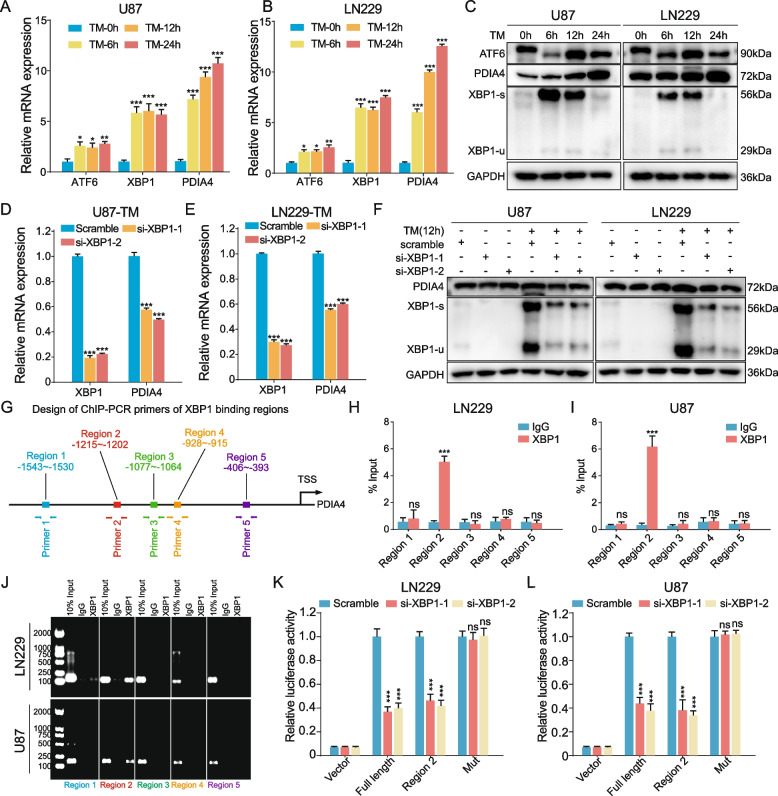
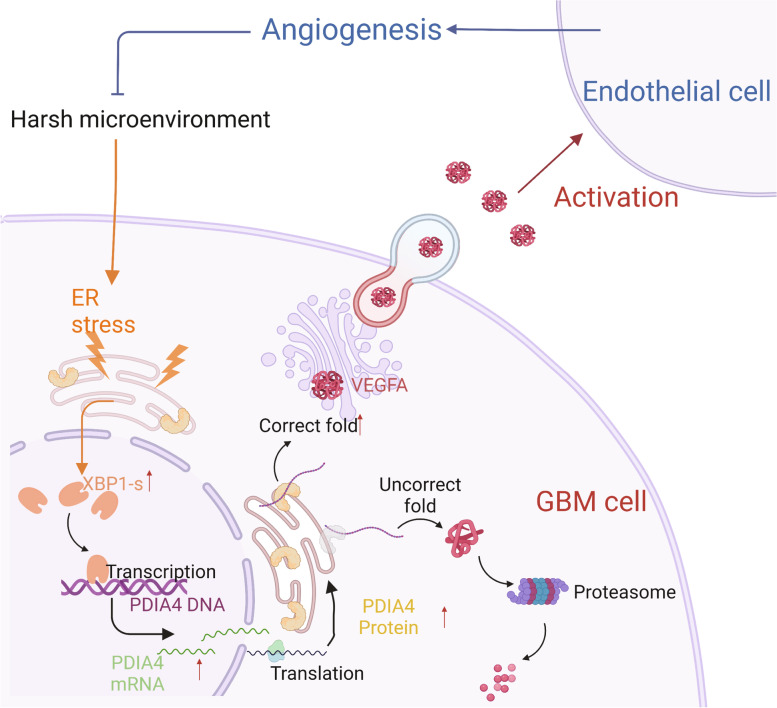
Results: Aberrant overexpression of PDIA4 was associated with a poor prognosis in patients with GBM, although PDIA4 could also functionally regulate intrinsic GBM secretion of vascular endothelial growth factor-A (VEGF-A) through its active domains of Cys-X-X-Cys (CXXC) oxidoreductase. Functionally, PDIA4 exhibits pro-angiogenesis activity both in vitro and in vivo, and can be upregulated by ERS through transcriptional regulation of X-box binding protein 1 (XBP1). The XBP1/PDIA4/VEGFA axis partially supports the mechanism underlying GBM cell survival under ER stress. Further, GBM cells with higher expression of PDIA4 showed resistance to antiangiogenic therapy in vivo.

Conclusions: Our findings revealed the pro-angiogenesis role of PDIA4 in GBM progression and its potential impact on GBM survival under a harsh microenvironment. Targeting PDIA4 might help to improve the efficacy of antiangiogenic therapy in patients with GBM.

Keywords: Angiogenesis; Endoplasmic reticulum stress (ERS); Glioblastoma (GBM); Protein disulfide-isomerase A4 (PDIA4); X-box binding protein 1 (XBP1).


Conflict of interest statement

No competing interest exists.

Figures

Fig. 1
Fig. 1
PDIA4 is extremely upregulated and correlates with worse prognosis of GBMs. A-B PDIA4 is overexpressed in 28 cancers, especially in GBM. ns P > 0.05; *P < 0.05; **P < 0.01; ***P < 0.001. C GBM patients with higher PDIA4 expression show poorer clinical prognosis in the TCGA cohort. D Scatter plots of the single-cell datset (GSE84465) showing the PDIA4 expressing distributions of different cell types in GBM microenvironment. E The violin plots of GSE84465 dataset showed that GBM cells expressed the highest PDIA4 in GBM microenvironment. F Immunofluorescence of U251 GBM cells indicates the subcellular localization in the ER of PDIA4 protein (Human Protein Atlas database). G Immunohistochemical staining of PDIA4 in clinical GBM samples and adjacent tissues showing distinct expression levels of PDIA4 protein. H Paired t-test was applied to compare the IHC scores of PDIA4 between adjacent tissues and GBM samples, and visualized in the box plots. ***P < 0.001. I The Kaplan–Meier survival analysis of GBM patients in neurosurgery department of NCUSAH verifies that higher PDIA4 expressions correlates worse clinical prognosis of GBM patients. J The volcano plot shows the RNA-seq analysis results of the different expressed genes (DEGs) between sh-Ctrl and sh-PDIA4 LN229 cells. K DEGs between sh-Ctrl and sh-PDIA4 LN229 cells were also visualized in this heatmap. L The Metascape enrichment analysis represents the gene ontology (GO) and KEGG pathway terms which these DEGs enriched in
Fig. 2
Fig. 2
Identification of secretory protein substrates of PDIA4 in GBM cells. A The general view of the construction of PDIA4 mutant plasmids, co-immunoprecipitation assay, and mass spectrum (MS) identification of PDIA4 potential substrates in U87 cells. B The Venn diagram shows twenty-eight proteins were identified as the secretary substrates of PDIA4 in GBM by intersecting 816 PDIA4 substrates and 878 human secretory proteins identified in Tao et al. Cell Chem Bio 2017. C Gene set enrichment analysis (GSEA) indicates the hallmark of angiogenesis is significantly enriched in high-PDIA4 GBM subgroup in the TCGA-GBM cohort. D Co-immunoprecipitation and western blot assays show the direct interaction between Flag-mPDIA4 and HA-VEGFA in U87 and LN229 GBM cells. E PDIA4 upregulation in U87 cells increases while knock-down of PDIA4 in LN229 cells decreases the VEGFA secretion in ELISA assay. ***P < 0.001. F Overexpression of mPDIA4 in U87 and LN229 GBM cells didn’t influence VEGFA secretion. ns P > 0.05. G-J Transwell migratory and tube formation assays of co-cultured HUVECs shows upregulation of PDIA4 in U87 cells can promote HUVEC migratory and tube formation capacities, which can be blocked by bevacizumab. *P < 0.05; **P < 0.01; ***P < 0.001 K-N Transwell migratory and tube formation assays of co-cultured HUVECs shows downregulation of PDIA4 in LN229 cells can decrease HUVEC migratory and tube formation capacities, which can be rescued by additional exogenous human VEGFA. **P < 0.01; ***P < 0.001
Fig. 3
Fig. 3
ER stress induced XBP1 upregulates PDIA4 expression transcriptionally. A-B RT-qPCR quantified the relative mRNA expressions of PDIA4, XBP1 and ATF6 in TM-induced ER stressed U87 (A) and LN229 (B) cells at 0, 6, 12 and 24 h. *P < 0.05; **P < 0.01; ***P < 0.001. C Western blot assay showed the ATF6, XBP1-s, XBP1-u and PDIA4 protein expressions were all upregulated under TM-induced ER stress in GBM cells. D-E Blocking XBP1 in ER stressed GBM cells by siRNAs significantly downregulates PDIA4 mRNA expressions. ***P < 0.001. F Upregulation of PDIA4 protein under ER stress was intercepted by blocking XBP1 expressions in GBM cells. G Design of ChIP-PCR primers of XBP1 binding regions. H-I The ChIP-PCR results showed Region 2 sequence (-1215 ~ -1202) on PDIA4 promoter were captured by XBP1 protein immunoprecipitation in U87 (H) and LN229 (I) cells. ns P > 0.05; ***P < 0.001. J The DNA gel electrophoresis shows the abundance of PCR produced DNA sequences of ChIP assay. K The luciferase activities of the vector, full length, region 2 and mutant PDIA4 promoter sequence transfected LN229 cells with or without XBP1 knock-down under 10 μg/mL TM induced ER stress. ns P > 0.05; ***P < 0.001. L The luciferase activities of the vector, full length, region 2 and mutant PDIA4 promoter sequence transfected U87 cells with or without XBP1 knock-down under 10 μg/mL TM induced ER stress. ns P > 0.05; ***P < 0.001
Fig. 4
Fig. 4
ER-stress induces XBP1/PDIA4/VEGFA regulatory axis in GBM. A The Pearson correlation analysis between XBP1 and PDIA4 IHC scores of clinical GBM samples reveals a strong positive correlation. B The Pearson correlation analysis between XBP1 expression and GSVA score of angiogenesis hallmark of GBM samples in the TCGA cohort. C The VEGFA secretion levels of LV-Ctrl and LV-PDIA4 U87 GBM cells with or without 10 μg/mL TM treatment. **P < 0.01. D The VEGFA secretion levels of sh-Ctrl and sh-PDIA4 LN229 GBM cells with or without 10 μg/mL TM treatment. ns P > 0.05; **P < 0.01. E–F The VEGFA secretion levels of XBP1 knock-down U87 (E) and LN229 (F) cells with or without 10 μg/mL TM treatment. ns P > 0.05; **P < 0.01. G The VEGFA secretion levels of XBP1 knock-down in LV-Ctrl and LV-PDIA4 U87 cells under 10 μg/mL TM induced ER stress. *P < 0.05; **P < 0.01. H The VEGFA secretion levels of XBP1 knock-down in sh-Ctrl and sh-PDIA4 LN229 cells under 10 μg/mL TM induced ER stress. ns P > 0.05; ***P < 0.001. I PDIA4 expression in high pro-operation edema GBMs is higher than low pro-operation edema GBMs. *P < 0.05
Fig. 5
Fig. 5
PDIA4 facilitated GBM growth, poor prognosis, and angiogenesis in vivo. A In vivo xenograft U87 GBM tumors were imaged at 7, 14, 21, and 28 days after the intracranial tumor was implanted using the IVIS system. B Representative HE staining images show the distinct size of xenograft U87 GBM with different PDIA4 expression. C The line chart shows the weight variation of GBM-bearing nude mice in two groups. **P < 0.01. D Recorded total flux at each time point indicates growing xenograft tumor size of each nude mice group dynamically. **P < 0.01. E Kaplan–Meier survival analysis showed nude mice with overexpression PDIA4 xenograft had poorer outcomes. F Representative VEGFA IHC images in two mice groups. G IHC scores of VEGFA in high-PDIA4 xenograft GBMs are significantly higher than low-PDIA4 xenograft GBMs. **P < 0.01. H Representative CD31 IHC images in two mice groups. I CD31-positive cell number in high-PDIA4 xenograft GBMs are significantly higher than low-PDIA4 xenograft GBMs. **P < 0.01. J Immunofluorescence of PDIA4 (green), CD31 (red) and DAPI (blue) in three cases of GBM samples
Fig. 6
Fig. 6
PDIA4 confers glioblastoma resistance to anti-angiogenic therapy. A The workflow of animal therapy study. B In vivo GBMs were imaged at 7, 14, 21, and 28 days after the intracranial tumor was implanted using the IVIS system. C Representative HE staining images show the tumor size of xenograft GBM in each group. D Recorded total flux at each time point present tumor size of each nude mice group dynamically. ns P > 0.05; *P < 0.05. E The line chart exhibits the weight variation of nude mice in each group. ns P > 0.05; **P < 0.01. F Kaplan–Meier survival analysis showed the prognosis of each mice group. ns P > 0.05; *P < 0.05; **P < 0.01; ***P < 0.001. G The number of anti-angiogenic therapy responder in each group were compared (Chi-square test, p = 0.018). H Representative VEGFA IHC images of each xenograft GBM mice group with different treatment. I Bar plots show the IHC scores of VEGFA in each xenograft GBM mice group with different treatment. ns P > 0.05. J Representative CD31 IHC image of each xenograft GBM mice group with different treatment. K Bar plots show the CD31 + cell number in each xenograft GBM mice group with different treatment. ns P > 0.05; *P < 0.05
Fig. 7
Fig. 7
Mechanism diagram of XBP1/PDIA4/VEGFA axis in GBM. Under the extracellular stress, ER stress of GBM cell would start, and activates the unfolded protein pathway (URP), which will upregulate the X-box binding protein 1 (XBP1), and then transcriptionally upregulated PDIA4 expression. As an ER stress effect protein, PDIA4 could increase the expression and secretion of VEGFA protein in GBM cells, and activate the tumor associated endothelial cell. This mechanism elicits the pro-angiogenesis function of PDIA4 of GBM

References

    1. Killock D. CNS cancer: molecular classification of glioma. Nat Rev Clin Oncol. 2015;12:502. doi: 10.1038/nrclinonc.2015.111. - DOI - PubMed
    1. Jiang T, Nam DH, Ram Z, Poon WS, Wang J, Boldbaatar D, et al. Clinical practice guidelines for the management of adult diffuse gliomas. Cancer Lett. 2021;499:60–72. doi: 10.1016/j.canlet.2020.10.050. - DOI - PubMed
    1. Brennan CW, Verhaak RG, McKenna A, Campos B, Noushmehr H, Salama SR, et al. The somatic genomic landscape of glioblastoma. Cell. 2013;155:462–477. doi: 10.1016/j.cell.2013.09.034. - DOI - PMC - PubMed
    1. Aldape K, Zadeh G, Mansouri S, Reifenberger G, von Deimling A. Glioblastoma: pathology, molecular mechanisms and markers. Acta Neuropathol. 2015;129:829–848. doi: 10.1007/s00401-015-1432-1. - DOI - PubMed
    1. Xu S, Sankar S, Neamati N. Protein disulfide isomerase: a promising target for cancer therapy. Drug Discov Today. 2014;19:222–240. doi: 10.1016/j.drudis.2013.10.017. - DOI - PubMed

Substances