. 2023 Mar 31;9(1):6.
doi: 10.1186/s42234-023-00108-3.

Hepatic stellate cell activation markers are regulated by the vagus nerve in systemic inflammation

Affiliations

Hepatic stellate cell activation markers are regulated by the vagus nerve in systemic inflammation

Osman Ahmed et al. Bioelectron Med. .

Abstract

Background: The liver is an important immunological organ and liver inflammation is part of the pathophysiology of non-alcoholic steatohepatitis, a condition that may promote cirrhosis, liver cancer, liver failure, and cardiovascular disease. Despite dense innervation of the liver parenchyma, little is known about neural regulation of liver function in inflammation. Here, we study vagus nerve control of the liver response to acute inflammation.

Methods: Male C57BL/6 J mice were subjected to either sham surgery, surgical vagotomy, or electrical vagus nerve stimulation followed by intraperitoneal injection of the TLR2 agonist zymosan. Animals were euthanized and tissues collected 12 h after injection. Samples were analyzed by qPCR, RNAseq, flow cytometry, or ELISA.

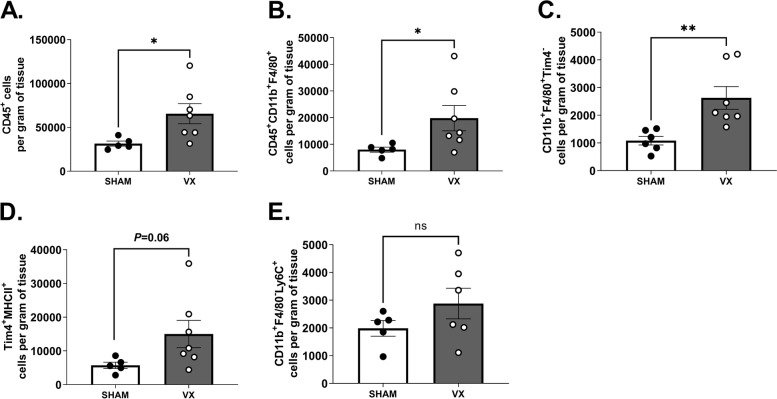
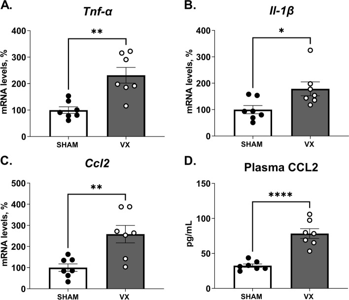
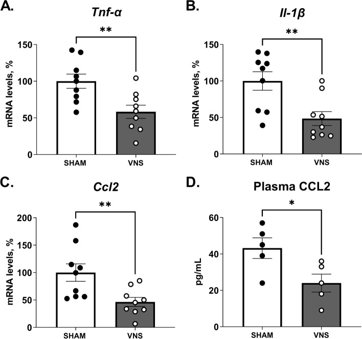
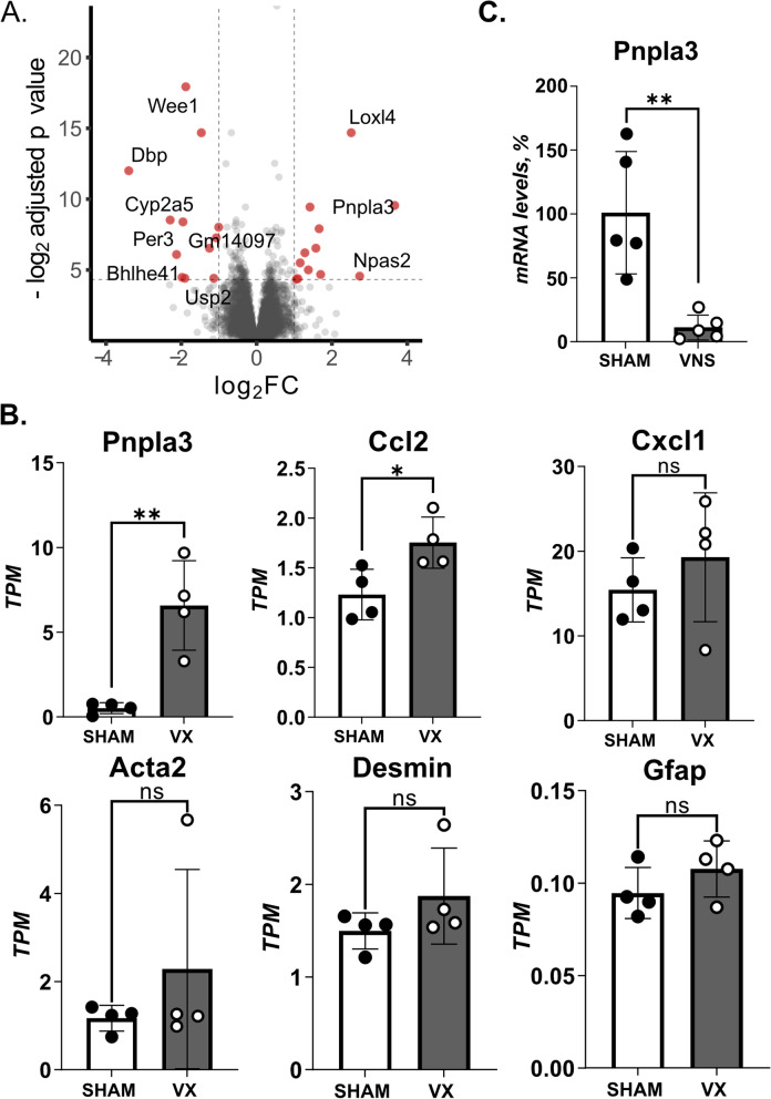
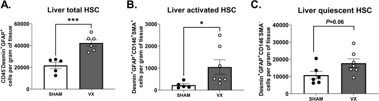
Results: Hepatic mRNA levels of pro-inflammatory mediators Ccl2, Il-1β, and Tnf-α were significantly higher in vagotomized mice compared with mice subjected to sham surgery. Differences in liver Ccl2 levels between treatment groups were largely reflected in the plasma chemokine (C-C motif) ligand 2 (CCL2) concentration. In line with this, we observed a higher number of macrophages in the livers of vagotomized mice compared with sham as measured by flow cytometry. In mice subjected to electrical vagus nerve stimulation, hepatic mRNA levels of Ccl2, Il1β, and Tnf-α, and plasma CCL2 levels, were significantly lower compared with sham. Interestingly, RNAseq revealed that a key activation marker for hepatic stellate cells (HSC), Pnpla3, was the most significantly differentially expressed gene between vagotomized and sham mice. Of note, several HSC-activation associated transcripts were higher in vagotomized mice, suggesting that signals in the vagus nerve contribute to HSC activation. In support of this, we observed significantly higher number of activated HSCs in vagotomized mice as compared with sham as measured by flow cytometry.

Conclusions: Signals in the cervical vagus nerve controlled hepatic inflammation and markers of HSC activation in zymosan-induced peritonitis.

Keywords: Kupffer cells; Liver; Non-alcoholic fatty liver disease; PNPLA3.


Conflict of interest statement

PSO is a founder and shareholder of Emune AB. OA is a co-founder and shareholder of Lipoprotein Research Stockholm AB. The remaining authors declare no competing interests.

Figures

Fig. 1
Fig. 1
Elevated peritonitis-induced liver leukocyte content in vagotomized mice. Wild-type C57BL/6 J mice were subjected to either left cervical unilateral vagotomy (VX, grey bars) or sham surgery (SHAM, white bars). After 7 days, mice were injected intraperitoneally with zymosan (0.1 mg/mouse) and euthanized 12 h thereafter. Liver homogenates were analyzed by flow cytometry for (A) CD45+, (B) CD45+CD11b+F4/80+, (C) CD45+CD11b+F4/80+Tim4, (D) CD45+CD11b+F4/80+Tim4+MHCII+, and (E) CD45+CD11b+F4/80Ly6C+ cell subsets. Results were normalized to liver biopsy mass (g). n = 6–7 per group. Results are expressed as mean ± SEM. ns = not significant; * p < 0.05; ** p < 0.01 (two-sided Student´s t-test; Welch’s correction was applied as appropriate)
Fig. 2
Fig. 2
Elevation of peritonitis-induced pro-inflammatory mediators in liver homogenates and blood from vagotomized mice. Wild-type C57BL/6J mice were subjected to either left cervical vagotomy (VX, grey bars) or sham surgery (SHAM, white bars). After 7 days, mice were injected intraperitoneally with zymosan (0.1 mg/mouse) and euthanized after 12 h. Liver and blood samples were collected and analyzed by qPCR and ELISA. (A-C) Transcripts of pro-inflammatory mediators in liver homogenates were measured by qPCR. Bars show mean ± SEM, dots represent individual mice. Transcript levels were normalized to Ppia and expressed relative to SHAM (100%). (D) Plasma CCL2 was measured by ELISA. n = 7 mice per group. Bars show mean ± SEM, dots represent individual mice. * p < 0.05; ** p < 0.01; *** p < 0.001; **** p < 0.0001. (two-sided Student´s t-test)
Fig. 3
Fig. 3
Reduced levels of peritonitis-induced pro-inflammatory mediators in livers from mice subjected to electrical vagus nerve stimulation. Wild-type C57BL/6J mice were subjected to either left cervical unilateral vagus nerve stimulation (VNS, grey bars) or sham surgery (SHAM, white bars) followed by intraperitoneal injection of zymosan (0.1 mg/mouse) 1 h thereafter. Liver and blood samples were collected 12 h after zymosan injection and analyzed by qPCR and ELISA. (A-C) Transcripts of pro-inflammatory mediators were measured in liver homogenates by qPCR (two independent experiments, n = 5 or 4 per group, respectively). Bars show mean ± SEM, dots represent individual mice. Transcript levels were normalized to Ppia and expressed relative to SHAM (100%). (D) Plasma CCL2 was measured by ELISA (n = 5 mice per group). Bars show mean ± SEM, dots represent individual mice. * p < 0.05; ** p < 0.01 (two-sided Student´s t-test)
Fig. 4
Fig. 4
HSC activation-markers in inflammation controlled by the vagus nerve. (A-B) Wild-type C57BL/6J mice were subjected to either left cervical unilateral vagotomy (VX) (n = 4) or sham surgery (n = 4). After 7 days, mice were injected with zymosan (0.1 mg/mouse) and euthanized after 12 h. Liver biopsies were collected and analyzed by RNA sequencing. (A) Volcano plot of differentially regulated genes between VX and sham mice. Red color indicates significant fold change (FC) outside limits shown by dashed lines (FC ≥ 1, FDR-adjusted p < 0.05) (B) Levels of select inflammation- and hepatic stellate cell (HSC)-associated transcripts were plotted. (C) Wild-type C57BL/6J mice were subjected to either left cervical unilateral vagus nerve stimulation (VNS, grey bars) or sham surgery (SHAM, white bars) followed by intraperitoneal injection of zymosan (0.1 mg/mouse) 1 h thereafter. Liver and blood samples were collected 12 h after zymosan injection and analyzed by qPCR (n = 5 per group). Bars show mean ± SEM, dots represent individual mice. PNPLA3 mRNA level were normalized to Ppia and expressed relative to SHAM (100%). * p < 0.05; ** p < 0.01 (two-sided Student´s t-test)
Fig. 5
Fig. 5
Higher activation of hepatic stellate cells in acute inflammation in vagotomized mice. Wild-type C57BL/6J mice were subjected to either left cervical unilateral vagotomy (VX, grey bars) or sham surgery (SHAM, white bars). After 7 days, mice were injected intraperitoneally with zymosan (0.1 mg/mouse) and euthanized 12 h thereafter. Liver homogenates were analyzed for (A) CD45Desmin+GFAP+ HSCs, (B) CD45Desmin+GFAP+CD146+SMA+ activated HSCs, and (C) CD45Desmin+GFAP+CD146SMA. quiescent HSCs, and normalized by liver biopsy mass (g). n = 6–7 per group. Results are shown as mean ± SEM. ns = not significant; * p < 0.05, *** p < 0.001 (two-sided Student t-test; Welch’s test correction was applied when variances were significantly different)

References

    1. Adori C, Daraio T, Kuiper R, Barde S, Horvathova L, Yoshitake T, et al. Disorganization and degeneration of liver sympathetic innervations in nonalcoholic fatty liver disease revealed by 3D imaging. Sci Adv. 2021;7(30):eabg5733. doi: 10.1126/sciadv.abg5733. - DOI - PMC - PubMed
    1. Baeck C, Wehr A, Karlmark KR, Heymann F, Vucur M, Gassler N, et al. Pharmacological inhibition of the chemokine CCL2 (MCP-1) diminishes liver macrophage infiltration and steatohepatitis in chronic hepatic injury. Gut. 2012;61(3):416–426. doi: 10.1136/gutjnl-2011-300304. - DOI - PubMed
    1. Bonaz B, Sinniger V, Hoffmann D, Clarençon D, Mathieu N, Dantzer C, et al. Chronic vagus nerve stimulation in Crohn’s disease: a 6-month follow-up pilot study. Neurogastroenterol Motil. 2016;28(6):948–953. doi: 10.1111/nmo.12792. - DOI - PubMed
    1. Bonnardel J, T’Jonck W, Gaublomme D, Browaeys R, Scott CL, Martens L, et al. Stellate Cells, Hepatocytes, and Endothelial Cells Imprint the Kupffer Cell Identity on Monocytes Colonizing the Liver Macrophage Niche. Immunity. 2019;51(4):638–654.e9. doi: 10.1016/j.immuni.2019.08.017. - DOI - PMC - PubMed
    1. Borovikova LV, Ivanova S, Zhang M, Yang H, Botchkina GI, Watkins LR, et al. Vagus nerve stimulation attenuates the systemic inflammatory response to endotoxin. Nature. 2000;405(May):458–462. doi: 10.1038/35013070. - DOI - PubMed

LinkOut - more resources