. 2023 Mar 30;23(1):31.
doi: 10.1186/s40644-023-00546-x.

Intraoperative MRI-derived volumetric ablation margins and initial correlation with local outcome after MRI-guided cryoablation of renal tumors

Affiliations

Intraoperative MRI-derived volumetric ablation margins and initial correlation with local outcome after MRI-guided cryoablation of renal tumors

Nienke S de Jager et al. Cancer Imaging. .

Abstract

Purpose: To assess volumetric ablation margins derived from intraoperative pre- and post-ablation MRI after magnetic resonance imaging (MRI)-guided percutaneous cryoablation of renal tumors and explore its correlation with local treatment success.

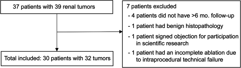
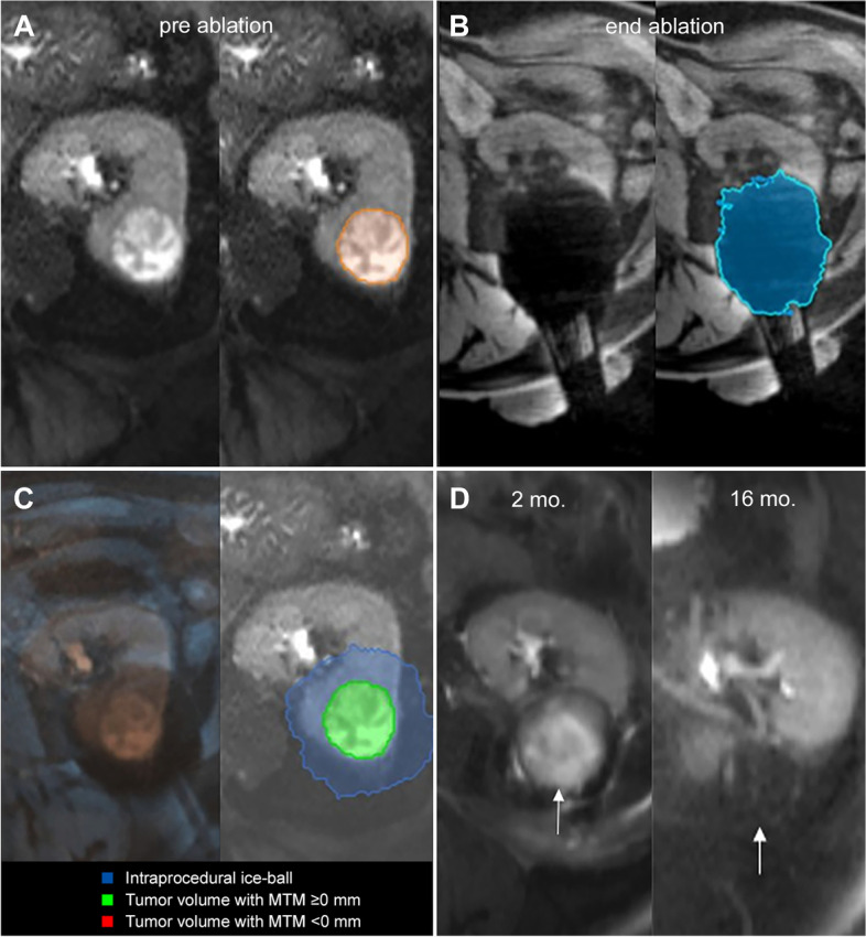
Methods: Retrospective analysis was performed on 30 patients (mean age 69y) who underwent percutaneous MRI-guided cryoablation between May 2014 and May 2020 for 32 renal tumors (size: 1.6-5.1 cm). Tumor and ice-ball volumes were segmented on intraprocedural pre- and post-ablation MR images using Software Assistant for Interventional Radiology (SAFIR) software. After MRI-MRI co-registration, the software automatically quantified the minimal treatment margin (MTM),defined as the smallest 3D distance between the tumor and ice-ball surface. Local tumor progression (LTP) after cryoablation was assessed on follow-up imaging.

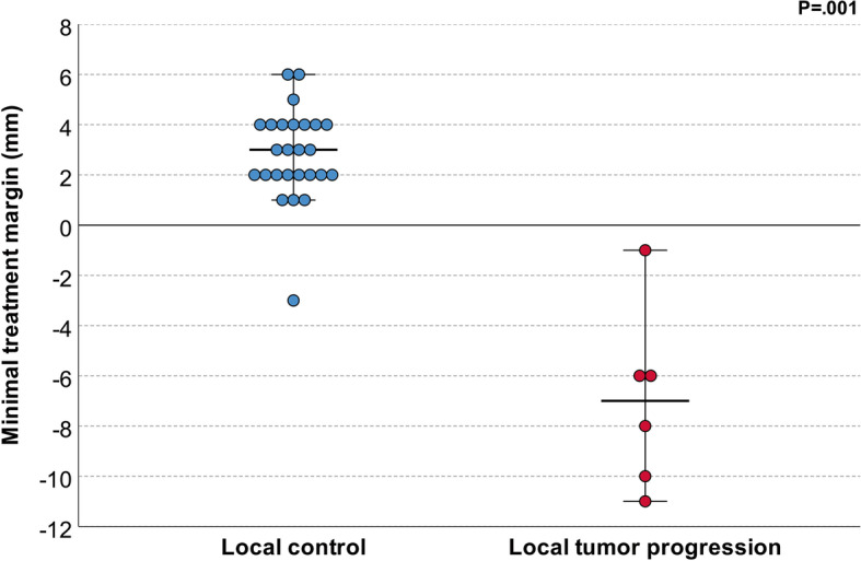
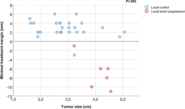
Results: Median follow-up was 16 months (range: 1-58). Local control after cryoablation was achieved in 26 cases (81%) while LTP occurred in 6 (19%). The intended MTM of ≥5 mm was achieved in 3/32 (9%) cases. Median MTM was significantly smaller for cases with (- 7 mm; IQR:-10 to - 5) vs. without LTP (3 mm; IQR:2 to 4) (P < .001). All cases of LTP had a negative MTM. All negative treatment margins occurred in tumors > 3 cm.

Conclusions: Determination of volumetric ablation margins from intraoperative MRI was feasible and may be useful in predicting local outcome after MRI-guided renal cryoablation. In our preliminary data, an intraoperative MRI-derived minimal margin extending at least 1 mm beyond the MRI-visible tumor led to local control and this was more difficult to achieve in tumors > 3 cm. Ultimately, online margin analysis may be a valuable tool to intraoperatively assess therapy success, but larger prospective studies are needed to establish a reliable threshold for clinical use.

Keywords: Local recurrence; MRI; Percutaneous cryoablation; Renal tumor; Treatment margin.


Conflict of interest statement

The authors declare that they have no competing interests.

Figures

Fig. 1
Fig. 1
Schematic overview of patient selection
Fig. 2
Fig. 2
Overview of Software Assistant for Interventional Radiology (SAFIR) layout. Available image series and tools are shown in bars on the left. Image views from left to right, top to bottom: (1) 3D view of annotated ice ball (blue) and tumor (green) overlap, (2) tumor divided in octants along the left-right, cranio-caudal and anterior-posterior axes showing color-coded subareas of total tumor volume with complete coverage including user-defined treatment margin (green), complete coverage but insufficient margin (yellow) and incomplete coverage (red)., (3) pre-ablation and (4) post-ablation T2-haste coronal MRI-images showing the tumor (green) and co-registered ice ball (blue) volumes. Side bar on the left shows calculated 3D ablation margin metrics
Fig. 3
Fig. 3
Minimal treatment margin for cases with local tumor control and local tumor progression
Fig. 4
Fig. 4
A Segmentation (orange outline) of a 3.5 cm diameter tumor located interpolar in the left kidney on pre-ablation axial T2-weighted MR imaging. B Segmentation (blue outline) of corresponding end ablation ice-ball volume on post-ablation axial T1-weighted MR imaging. C Overlay of pre-ablation and post-ablation images after co-registration (left) and resulting 3D margin calculation (right). Volumetric assessment shows complete tumor coverage (green) by the end-ablation ice-ablation ice-ball (blue) with MTM of 4 mm. D Ablation zone (white arrows) without tumor recurrence on follow-up axial T2-weighted MR imaging 2 months (left) and 16 months (right) after cryoablation
Fig. 5
Fig. 5
A Segmentation of a 4.2 cm diameter tumor located in the lower pole of the right kidney (outlined in orange) on pre-ablation axial T2-weighted MR imaging. B Segmentation of the corresponding ice ball (outlined in blue) on post-ablation axial T1-weighted MR imaging. C Overlay of pre-ablation and post-ablation images after co-registration (left) and resulting 3D margin calculation (right). Volumetric assessment shows predicted incomplete ablation at medial side (red) with MTM of − 6 mm. D Follow-up axial T2-weighted MR imaging at 2 months (left) after cryoablation shows ablation zone (white arrows) with suspected incomplete coverage on medial side (red arrows). Six months follow-up MRI (right) confirms local tumor progression (red arrow) at the medial side
Fig. 6
Fig. 6
Minimal treatment margin plotted against tumor size for cases of local control and local tumor progression

References

    1. Prins FM, Kerkmeijer LGW, Pronk AA, Vonken EPA, Meijer RP, Bex A, et al. Renal cell carcinoma: alternative nephron-sparing treatment options for small renal masses, a systematic review. J Endourol. 2017;31(10):963–975. doi: 10.1089/end.2017.0382. - DOI - PubMed
    1. Ljungberg B, Albiges L, Abu-Ghanem Y, Bensalah K, Dabestani S, Fernandez-Pello S, et al. European Association of Urology guidelines on renal cell carcinoma: the 2019 update. Eur Urol. 2019;75(5):799–810. doi: 10.1016/j.eururo.2019.02.011. - DOI - PubMed
    1. Escudier B, Porta C, Schmidinger M, Rioux-Leclercq N, Bex A, Khoo V, et al. Renal cell carcinoma: ESMO clinical practice guidelines for diagnosis, treatment and follow-up. Ann Oncol. 2016;27(suppl 5):v58–v68. doi: 10.1093/annonc/mdw328. - DOI - PubMed
    1. Krokidis ME, Kitrou P, Spiliopoulos S, Karnabatidis D, Katsanos K. Image-guided minimally invasive treatment for small renal cell carcinoma. Insights Imaging. 2018;9(3):385–390. doi: 10.1007/s13244-018-0607-4. - DOI - PMC - PubMed
    1. Mogami T, Harada J, Kishimoto K, Sumida S. Percutaneous MR-guided cryoablation for malignancies, with a focus on renal cell carcinoma. Int J Clin Oncol. 2007;12(2):79–84. doi: 10.1007/s10147-006-0654-6. - DOI - PubMed