

Case Reports
doi: 10.1093/jscr/rjad153.
eCollection 2023 Mar.
Giant pedunculated lipofibroma of the thigh
Affiliations
- PMID: 36998262
- PMCID: PMC10049795
- DOI: 10.1093/jscr/rjad153
Item in Clipboard
Case Reports
Giant pedunculated lipofibroma of the thigh
J Surg Case Rep.
.
Abstract
Pedunculated lipofibroma is a rare form of nevus lipomatous cutaneous superficialis. They are usually solitary lesions found around the thighs, buttocks and trunk; thought to have a predilection for pressure areas. There are two types of lipofibromas; sessile or pedunculated. They are mostly asymptomatic but can cause symptoms as they grow larger affecting daily activities. Treatment is not indicated in smaller lesions, except for cosmetic purposes. Herein we present this rare benign lesion with an unusually large size.
Published by Oxford University Press and JSCR Publishing Ltd. © The Author(s) 2023.
Conflict of interest statement
The authors declare they have no competing interests.
Figures
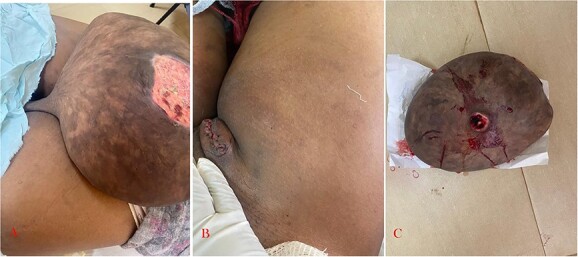
(A) Clinical photograph showing giant lipofibroma arising from the inguinal region with a stalk. (B) Post complete excision. (C) Resected (giant) lipofibroma.
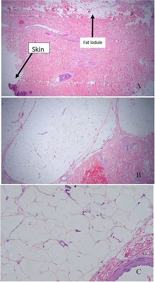
(A) Ulcerated skin and the lobules of mass with fat tissue (×20 magnification). It is also showing ulceration on the adjacent skin. (B) Lobules of fat tissue compressing the adjacent dermal collagenous tissue. There are groups and strands of mature adipocytes extending between collagen bundles of the dermal layer of the skin (×20 magnification). (C) High-power field of adipose tissue with mature adipocytes and without atypia. Adipocytes have small eccentric nuclei with minimal atypia in a perivascular space (×200 magnification).
References
-
- Nogita T, et al. Pedunculated lipofibroma. J Am Acad Dermatol 1994; 31:235–40. - PubMed
-
- Mehregan AH, Tavafoghi V, Ghandchi A. Nevus lipomatosus cutaneus superficialis (Hoffmann-Zurhelle). J Cutaneous Pathol 1975;2:307–13. - PubMed
-
- Buch AC, Panicker NK, Karve PP. Solitary nevus lipomatosus cutaneous superficialis. J Postgrad Med 2005;51:47–8. - PubMed
Publication types
LinkOut - more resources
Full Text Sources
