. 2023 Mar 14:16:1119164.
doi: 10.3389/fnmol.2023.1119164. eCollection 2023.

Systematic identification of potential key microRNAs and circRNAs in the dorsal root ganglia of mice with sciatic nerve injury

Affiliations

Systematic identification of potential key microRNAs and circRNAs in the dorsal root ganglia of mice with sciatic nerve injury

Youfen Yu et al. Front Mol Neurosci. .

Abstract

Background: Neuropathic pain (NeP) is a pathological condition arising from a lesion or disease affecting the somatosensory system. Accumulating evidence has shown that circular RNAs (circRNAs) exert critical functions in neurodegenerative diseases by sponging microRNAs (miRNAs). However, the functions and regulatory mechanisms of circRNAs as competitive endogenous RNAs (ceRNAs) in NeP remain to be determined.

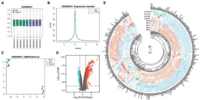
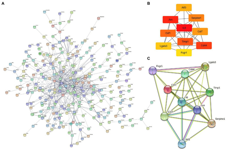
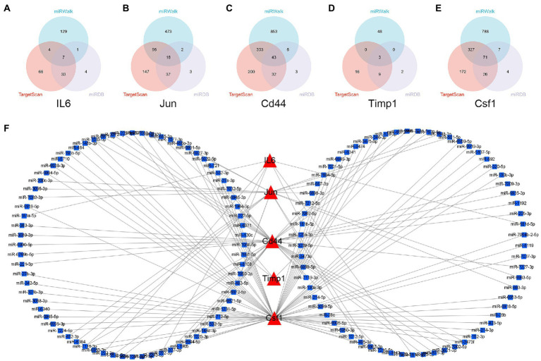
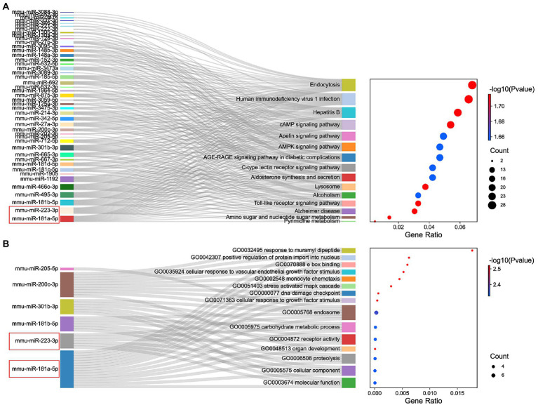
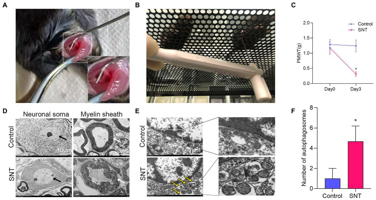
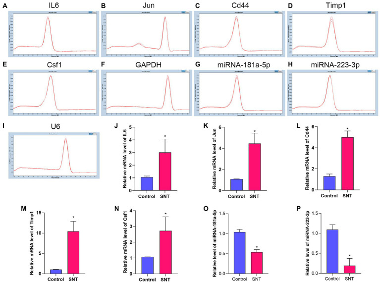
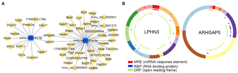
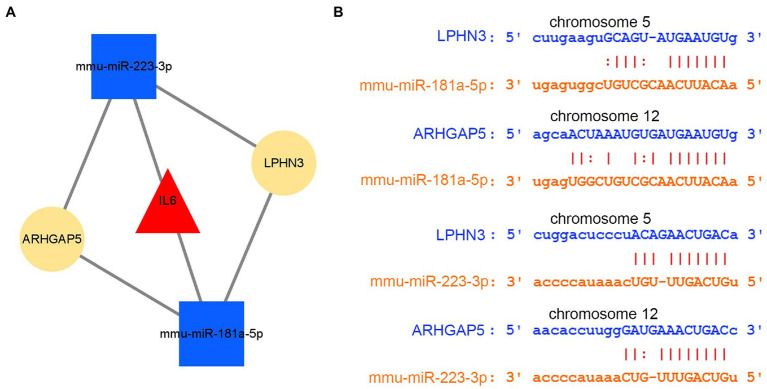
Methods: The sequencing dataset GSE96051 was obtained from the public Gene Expression Omnibus (GEO) database. First, we conducted a comparison of gene expression profiles in the L3/L4 dorsal root ganglion (DRG) of sciatic nerve transection (SNT) mice (n = 5) and uninjured mice (Control) (n = 4) to define the differentially expressed genes (DEGs). Then, critical hub genes were screened by exploring protein-protein interaction (PPI) networks with Cytoscape software, and the miRNAs bound to them were predicted and selected and then validated by qRT-PCR. Furthermore, key circRNAs were predicted and filtered, and the network of circRNA-miRNA-mRNA in NeP was constructed.

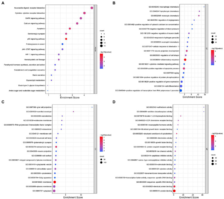
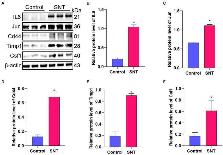
Results: A total of 421 DEGs were identified, including 332 upregulated genes and 89 downregulated genes. Ten hub genes, including IL6, Jun, Cd44, Timp1, and Csf1, were identified. Two miRNAs, mmu-miR-181a-5p and mmu-miR-223-3p, were preliminarily verified as key regulators of NeP development. In addition, circARHGAP5 and circLPHN3 were identified as key circRNAs. Gene Ontology (GO) and Kyoto Encyclopedia of Genes and Genomes (KEGG) analysis demonstrated that these differentially expressed mRNAs and targeting miRNAs were involved in signal transduction, positive regulation of receptor-mediated endocytosis and regulation of neuronal synaptic plasticity. These findings have useful implications for the exploration of new mechanisms and therapeutic targets for NeP.

Conclusion: These newly identified miRNAs and circRNAs in networks reveal potential diagnostic or therapeutic targets for NeP.

Keywords: ceRNA; circRNA; dorsal root ganglia; miR-181a-5p; miR-223-3p; microRNA; neuropathic pain.


Conflict of interest statement

The authors declare that the research was conducted in the absence of any commercial or financial relationships that could be construed as a potential conflict of interest.

Figures

Figure 1
Figure 1
Differentially expressed profile of mRNAs and characterization between the SNT group and the control. (A) Boxplot shows the distribution of the values of the DRG samples, abscissa represents sample name and ordinate represents the normalized intensity values, the upper and lower sides of the rectangular box mean minimum and maximum values, the upper and lower lines of the error bars mean interquartile range, the line inside the rectangular box means median value. (B) The density map of GSE96051 shows the expression of each sample. (C) The UMAP shows the distribution relationships between the samples. (D) Volcano plot of DEmRNAs. The vertical lines correspond to twice the fold changes in upregulation or downregulation; the horizontal line represents p-adjusted = 0.05; red points indicate mRNAs with upregulation and blue points indicate mRNAs with downregulation. (E) Hierarchical cluster analysis of DEmRNAs. Blue and red color indicate down-regulation and up-regulation of gene expression, respectively.
Figure 2
Figure 2
Gene Ontology (GO) and Kyoto Encyclopedia of Genes and Genomes (KEGG) analyses for DEmRNAs. (A) KEGG pathway analysis shows the significantly enriched pathways and their gene counts. (B–D) The GO enrichment results of DEmRNAs, including GO-BP (B), GO-CC (C), and GO-MF (D).
Figure 3
Figure 3
The construction of protein–protein interaction network and identification of hub genes. (A) PPI network is established by 421 DEmRNA-encoded proteins using the STRING website. (B) Relationship network diagram of hub genes from PPI network using cytoscape. (C) Relationship network of 10 hub genes that extracted from the PPI network and the structure of proteins they encoded.
Figure 4
Figure 4
Verification of the top 5 hub genes. (A) The expression of the IL6, Jun, Cd44, Timp1 and Csf1 in DRG tissues of mice induced by SNT and the control by Western blot. (B–F) The expression analysis of the IL6, Jun, Cd44, Timp1 and Csf1. Data are presented as means ± SD (n = 3; *p < 0.05).
Figure 5
Figure 5
Construction of miRNA-mRNA network. (A–E) Overlap analysis for the miRNAs binding to the IL6, Jun, Cd44, Timp1, and Csf1 from the TargetScan, miRWalk, and miRDB database. (F) The miRNAs-hub genes network.
Figure 6
Figure 6
Functional enrichment analysis of miRNAs. (A) KEGG pathways enriched by the shared miRNAs between 3 different databases, TargetScan, miRWalk, and miRDB. (B) The GO enrichment analysis shows the top 15 significant items and their gene counts.
Figure 7
Figure 7
Changes of pain threshold and DRG ultrastructure after SNT. (A) The right sciatic nerve of mice was exposed and transected. (B) Behavioral test of mice using Von Frey. (C) SNT induced reduction of PMWT in mice. (D) Electron microscope analysis of the ultrastructural changes in L3/L4 DRGs on the 3rd day after SNT in different groups. The morphology of neuronal somata and myelin sheaths of DRG neurons in two groups. The neuron nucleus is indicated by the black arrow, Scale bar = 10 μm; magnification ×1,000; The myelin sheath is indicated by the white arrow, Scale bar = 5 μm; magnification ×2,000. (E) Representative electron microscope views of autophagosomes (yellow arrow) in DRG of the two groups. Scale bar = 1 μm; magnification ×12,000. (F) The number of autophagosomes of the two groups in per vision was determined on the 3rd day after SNT. Data are presented as means ±SD (n = 3; *p < 0.05).
Figure 8
Figure 8
Amplification and identification of differentially expressed mRNAs and miRNAs. (A–I) The melt curves of the identified mRNAs and miRNAs. (J–P) The results of qRT-PCR show the expression levels of DEGs between the two groups. Data are presented as means ± SD (n = 3; *p < 0.05).
Figure 9
Figure 9
The analysis of circRNA-miRNA networks. (A) The competing endogenous RNA networks of DEmiRNAs. (B) Structural patterns of circLPHN3 and circARHGAP5. The colored circle represents the circRNAs that consist of exons. The numbers on the circRNAs mean the exon number. The red,blue, and green regions inside the circRNA molecule, respectively represent MRE (microRNA response element), RBP (RNA binding protein), and ORF (open reading frame).
Figure 10
Figure 10
The analysis of circRNA-miRNA-mRNA network. (A) The circRNA–miRNA-hub gene network. (B) The interaction of novel circRNAs/DEmiRNAs.

Similar articles

Cited by

References

    1. Agarwal V., Bell G. W., Nam J.-W., Bartel D. P. (2015). Predicting effective microRNA target sites in mammalian mRNAs. elife 4:5005. doi: 10.7554/eLife.05005, PMID: - DOI - PMC - PubMed
    1. Arvola O., Kaidonis G., Xu L., Griffiths B., Stary C. M. (2019). Hippocampal sub-regional differences in the microRNA response to forebrain ischemia. Mol. Cell. Neurosci. 98, 164–178. doi: 10.1016/j.mcn.2019.05.003, PMID: - DOI - PubMed
    1. Berger A. A., Liu Y., Possoit H., Rogers A. C., Moore W., Gress K., et al. . (2021). Dorsal root ganglion (DRG) and chronic pain. Anesth. Pain Med. 11:e113020. doi: 10.5812/aapm.113020, PMID: - DOI - PMC - PubMed
    1. Bjersing J. L., Lundborg C., Bokarewa M. I., Mannerkorpi K. (2013). Profile of cerebrospinal microRNAs in fibromyalgia. PLoS One 8:e78762. doi: 10.1371/journal.pone.0078762, PMID: - DOI - PMC - PubMed
    1. Cai L., Liu X., Guo Q., Huang Q., Zhang Q., Cao Z. (2020). MiR-15a attenuates peripheral nerve injury-induced neuropathic pain by targeting AKT3 to regulate autophagy. Genes Genom. 42, 77–85. doi: 10.1007/s13258-019-00881-z - DOI - PubMed