Review
. 2023 Mar 24:17:953-967.
doi: 10.2147/OPTH.S388292. eCollection 2023.

Diagnostic and Management Strategies of Bietti Crystalline Dystrophy: Current Perspectives

Affiliations
Review

Diagnostic and Management Strategies of Bietti Crystalline Dystrophy: Current Perspectives

Ali Osman Saatci et al. Clin Ophthalmol. .

Abstract

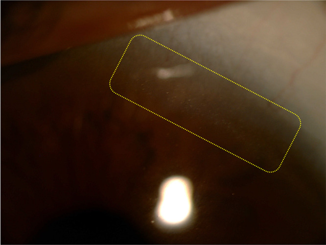
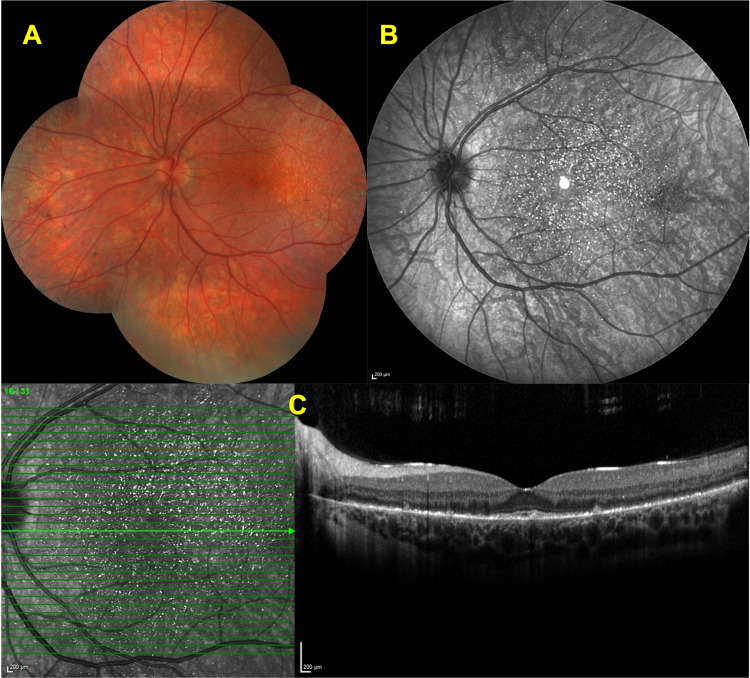
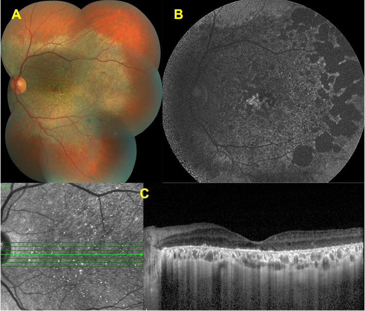
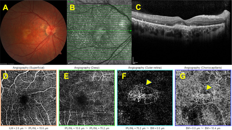
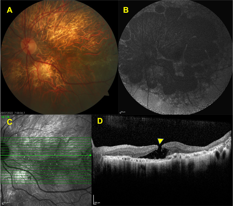
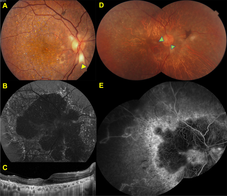
Bietti crystalline dystrophy (BCD) is a rare, genetically determined chorioretinal dystrophy presenting with intraretinal crystalline deposits and varying degrees of progressive chorioretinal atrophy commencing at the posterior pole. In some cases, there can be concomitant corneal crystals noted first in the superior or inferior limbus. CYP4V2 gene, a member of the cytochrome P450 family is responsible for the disease and more than 100 mutations have been defined thus far. However, a genotype-phenotype correlation has not been established yet. Visual impairment commonly occurs between the second and third decades of life. By the fifth or sixth decade of life, vision loss can become so severe that the patient may potentially become legally blind. Multitudes of multimodal imaging modalities can be utilized to demonstrate the clinical features, course, and complications of the disease. This present review aims to reiterate the clinical features of BCD, update the clinical perspectives with the help of multimodal imaging techniques, and overview its genetic background with future therapeutic approaches.

Keywords: Bietti crystalline dystrophy; chorioretinal dystrophy; corneal crystals; inherited disorder; vision loss.


Conflict of interest statement

The authors report no conflicts of interest in this work.

Figures

Figure 1
Figure 1
Corneal crystals. Slit- lamp biomicroscopy photo showing the crystalline deposits located at the limbus.
Figure 2
Figure 2
Features of advanced disease stage. Color fundus (A) and IR-R images. (B) of a left eye showing the marked diffuse chorioretinal atrophy and choroidal sclerosis with some pigment clumping areas. The horizontal SD-OCT section through the fovea (C) revealing the loss of EZ and RPE line with choroidal thinning.
Figure 3
Figure 3
Features of early disease stage. Color fundus image of a left eye (A)showing scattered intraretinal crystals throughout the posterior pole. Infrared reflectance (IR-R) image (B) depicting the several bright reflective spots at posterior pole. Spectral domain-optical coherence tomography (SD-OCT) section (C) delineating the hyperreflective deposits at the retina pigment epithelium -Bruch’s membrane complex and ellipsoid zone (EZ) disruption in the left eye (C).
Figure 4
Figure 4
Features of intermediate disease stage. Color fundus image of a left eye (A) showing the chorioretinal atrophy extending from the posterior pole and glistening crystals at the macula. Fundus autofluorescence image (B) revealing the well-demarcated hypofluorescent patches corresponding to the atrophy. Spectral domain-optical coherence tomography (SD-OCT) section (C) demonstrating the hyperreflective deposits at the RPE – Bruch’s membrane complex, together with the disruption and loss of the EZ band.
Figure 5
Figure 5
Associated macular neovascularization. Right eye, Color fundus image showing the hypopigmented looking well-demarcated lesion at the macula (A). The horizontal SD-OCT section demonstrating the macular neovascularization (B,C). Optical coherence tomography angiographic slabs demonstrating the presence of quiescent choroidal neovascularization (yellow arrows) (DG).
Figure 6
Figure 6
Associated macular edema. Left eye, early venous (A) and late composite venous (B) fluorescein angiographic images revealing the increasing macular hyperfluorescence. SD-OCT section showing the cystoid macular changes corresponding to macular edema (C).
Figure 7
Figure 7
Associated macular hole. Left eye, color fundus image (A) delineating the extensive chorioretinal atrophy and choroidal sclerosis. FAF image (B) revealing the multiple hypofluorescent patches corresponding to the posterior pole. SD-OCT section showing the presence of full -thickness hole (yellow arrow) (C,D).
Figure 8
Figure 8
Incidental findings Right eye, Color fundus image (A) showing the scattered glistening crystals at the posterior pole and peripapillary myelinated nerve fiber (yellow arrow), FAF image (B) demonstrating the patchy hypofluorescent areas with ill-demarcated hyperfluorescent borders at the posterior pole in 2011. SD-OCT section (C) revealing the hyperreflective deposits at the RPE – Bruch’s membrane complex, disruption, and loss of the EZ band. Composite color fundus image (D) demonstrating the marked chorioretinal atrophy and attenuated retinal vessels with the optociliary shunt (green arrows) in 2021. Composite venous fluorescein angiographic image (E) revealing the nonperfused areas related to old central retinal vein occlusion detected in fluorescein angiography image shows extensive ischemic areas throughout posterior pole.

References

    1. Bietti G. On the familial occurrence of “retinitis punctata albescens” (associated with “dystrophia marginalis cristallinea cornea)”, glistening, vitreous humor and other degenerative ocular changes. Augenvera¨Nderungen Klin Monatsbl Augenheilkd. 1937;99:737–756.
    1. Welch RB. Bietti’s tapetoretinal degeneration with marginal corneal dystrophy crystalline retinopathy. Trans Am Ophthalmol Soc. 1977;75:164–179. - PMC - PubMed
    1. Osman Saatci A, Can Doruk H. An overview of rare and unusual clinical features of Bietti’s crystalline dystrophy. Med Hypothesis Discov Innov Ophthalmol. 2014;3:51–56. - PMC - PubMed
    1. Bagolini B, Ioli-Spada G. Bietti’s tapetoretinal degeneration with marginal corneal dystrophy. Am J Ophthalmol. 1968;65:53–60. doi: 10.1016/0002-9394(68)91028-3 - DOI - PubMed
    1. Furusato E, Cameron JD, Chan CC. Evolution of cellular inclusions in Bietti’s crystalline dystrophy. Ophthalmol Eye Dis. 2010;2010:9–15. doi: 10.4137/OED.S2821 - DOI - PMC - PubMed