. 2023 Mar 14:17:1026939.
doi: 10.3389/fnins.2023.1026939. eCollection 2023.

Oxytocin receptors in the Magel2 mouse model of autism: Specific region, age, sex and oxytocin treatment effects

Affiliations

Oxytocin receptors in the Magel2 mouse model of autism: Specific region, age, sex and oxytocin treatment effects

Valentina Gigliucci et al. Front Neurosci. .

Abstract

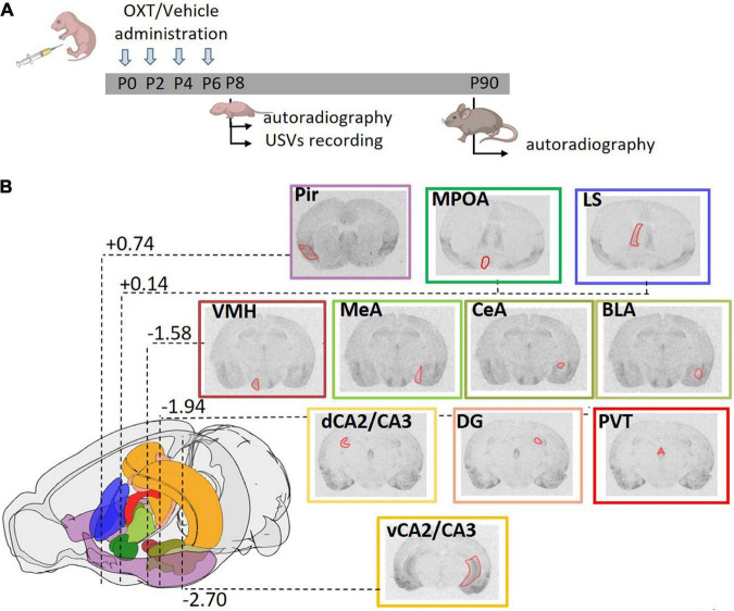
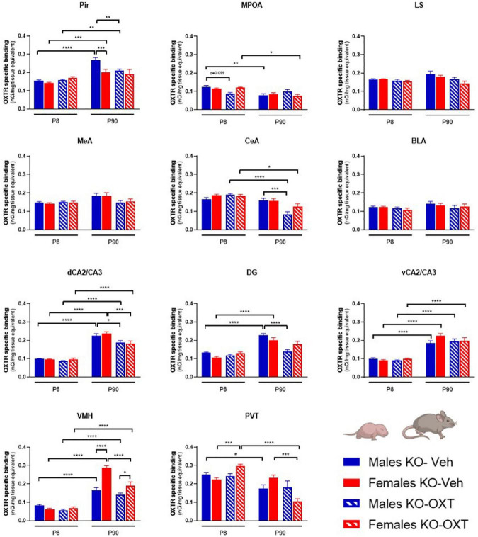
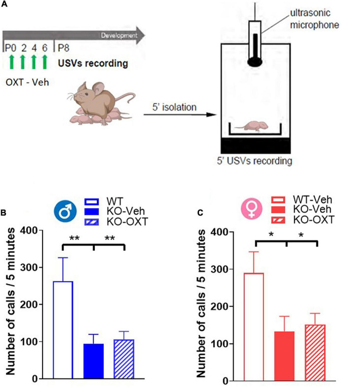
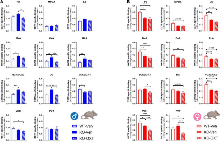
The neurohormone oxytocin (OXT) has been implicated in the regulation of social behavior and is intensively investigated as a potential therapeutic treatment in neurodevelopmental disorders characterized by social deficits. In the Magel2-knockout (KO) mouse, a model of Schaaf-Yang Syndrome, an early postnatal administration of OXT rescued autistic-like behavior and cognition at adulthood, making this model relevant for understanding the actions of OXT in (re)programming postnatal brain development. The oxytocin receptor (OXTR), the main brain target of OXT, was dysregulated in the hippocampus of Magel2-KO adult males, and normalized upon OXT treatment at birth. Here we have analyzed male and female Magel2-KO brains at postnatal day 8 (P8) and at postnatal day 90 (P90), investigating age, genotype and OXT treatment effects on OXTR levels in several regions of the brain. We found that, at P8, male and female Magel2-KOs displayed a widespread, substantial, down-regulation of OXTR levels compared to wild type (WT) animals. Most intriguingly, the postnatal OXT treatment did not affect Magel2-KO OXTR levels at P8 and, consistently, did not rescue the ultrasonic vocalization deficits observed at this age. On the contrary, the postnatal OXT treatment reduced OXTR levels at P90 in male Magel2-KO in a region-specific way, restoring normal OXTR levels in regions where the Magel2-KO OXTR was upregulated (central amygdala, hippocampus and piriform cortex). Interestingly, Magel2-KO females, previously shown to lack the social deficits observed in Magel2-KO males, were characterized by a different trend in receptor expression compared to males; as a result, the dimorphic expression of OXTR observed in WT animals, with higher OXTR expression observed in females, was abolished in Magel2-KO mice. In conclusion, our data indicate that in Magel2-KO mice, OXTRs undergo region-specific modifications related to age, sex and postnatal OXT treatment. These results are instrumental to design precisely-timed OXT-based therapeutic strategies that, by acting at specific brain regions, could modify the outcome of social deficits in Schaaf-Yang Syndrome patients.

Keywords: Prader-Willi Syndrome (PWS); Schaaf-Yang Syndrome; neurodevelopmental disorders (NDD); oxytocin receptor expression; postnatal oxytocin treatment.


Conflict of interest statement

The authors declare that the research was conducted in the absence of any commercial or financial relationships that could be construed as a potential conflict of interest.

Figures

FIGURE 1
FIGURE 1
Experimental strategy. (A) Schematic diagram representing the treatment regime administered to the mice and the paradigm of the analysis performed. Mice were subcutaneously injected with OXT or vehicle (single injection/day) in the first week of life at P0, P2, P4, and P6. Brain autoradiography was performed at P8 or P90. A separate group of animals at P8 was tested for ultrasonic vocalizations (USVs). (B) Schematic representation of the mouse brain showing the stereotactic coordinates of the coronal planes in which OXTR were analyzed. Distance from bregma, reported in millimeters according to Franklin and Paxinos mouse brain atlas (Franklin and Paxinos, 2007), are highlighted for the most representative section. A color code was used to identify the different areas analyzed: violet: Pir, piriform cortex; emerald green: MPOA, medial preoptic area; blue: LS, lateral septum; brown: VMH, ventral medial nucleus of the hypothalamus; three values of olive green for MeA, medial amygdala; CeA, central amygdala; BLA, basolateral amygdala; orange: dCA2/CA3, dorsal and vCA2/CA3, ventral field CA2 and CA3 of the hippocampus; pink: DG, dentate gyrus; red: PVT, paraventricular thalamic nucleus. Within each representative autoradiographic section, whose border color follows the color code reported above, the ROI used for acquisition of the data is depicted in red.
FIGURE 2
FIGURE 2
Contributions of age, sex and Magel2-KO genotype on physiological brain regional OXTR expression levels. Bar graphs of OXTR levels quantified by [125I]-OVTA binding in P8 and P90, male and female, WT and Magel2-KO mice. Each histogram represents data expressed as mean + SEM of multiple datapoints collected from three animals. Unfilled bars are used for vehicle (Veh) treated WT, filled bars are used for vehicle (Veh) treated Magel2-KO; blue is used for males of both genotypes and ages, red for females of both genotypes and ages. Data were analyzed by three-way ANOVA, followed by a Tukey’s multiple comparisons post-hoc test. *p < 0.05, **p < 0.01, ***p < 0.001, ****p < 0.0001. When a comparison was approaching statistical significance, the corresponding p-value was reported on the appropriate graph. Datasets and detailed statistical analyses are reported in the Supplementary Table 1.
FIGURE 3
FIGURE 3
Long-lasting effects of a postnatal OXT on brain regional OXTR expression levels in Magel2-KO mice. Bar graphs of OXTR levels quantified by [125I]-OVTA binding in P8 and P90, male and female, Vehicle or OXT-treated Magel2-KO mice. Each histogram represents data expressed as mean + SEM of multiple datapoints collected from three animals. Filled bars are used for vehicle (Veh) treated Magel2-KO, striped bars are used for oxytocin (OXT) treated Magel2-KO; blue is used for males of both treatment groups and ages, red for females of both treatment groups and ages. Data were analyzed by Three-way ANOVA, followed by a Tukey’s multiple comparisons post-hoc test. *p < 0.05, **p < 0.01, ***p < 0.001, ****p < 0.0001. When a comparison was approaching statistical significance, the corresponding p-value was reported on the appropriate graph. Datasets and detailed statistical analyses are reported in the Supplementary Table 2.
FIGURE 4
FIGURE 4
Ultrasonic vocalization calls (USVs) in P8 male and female WT and Magel2-KO pups treated with vehicle or OXT during the first week of life. (A) Schematic drawing of the protocol used to record separation-induced USVs in P8 mice. Pups were treated with vehicle (Veh) or oxytocin (OXT) from P0 to P6. Number of total calls, measured during 5 min isolation after pup separation in males (B) and females (C). Each histogram represents data expressed as mean + SEM of 11-24 mice. Unfilled bars are used for vehicle (Veh) treated WT; filled bars are used for vehicle (Veh) treated Magel2-KO; striped bars correspond to oxytocin (OXT) treated Magel2-KO; blue bar is used for males of both genotypes, red bar for females of both genotypes. Histograms indicate the mean + SEM of the different groups analyzed by one-way ANOVA followed by a Tukey’s multiple comparisons post-hoc test. *p < 0.05, **p < 0.01. Values and statistics are reported in Supplementary Table 3.
FIGURE 5
FIGURE 5
Long term effects of the postnatal OXT treatment on brain OXTR expression levels in P90 male and female WT and Magel2-KO mice. Bar graphs of OXTR levels quantified by [125I]-OVTA binding in adult male (A) and female (B) mice. Each histogram represents data expressed as mean + SEM of multiple datapoints collected from 3 animals. Unfilled bars are used for vehicle (Veh) treated WT, filled bars are used for vehicle (Veh) treated Magel2-KO, striped bars are used for oxytocin (OXT) treated Magel2-KO. Data were analyzed by two-way ANOVA, followed by a Tukey’s multiple comparisons post-hoc test. *p < 0.05, **p < 0.01, ***p < 0.001, ****p < 0.0001. When a comparison was approaching statistical significance, the corresponding p-value was reported on the appropriate graph. Datasets and detailed statistical analyses are reported in the Supplementary Tables 4A, B.

References

    1. Alanazi M. M., Havranek T., Bakos J., Cubeddu L. X., Castejon A. M. (2020). Cell proliferation and anti-oxidant effects of oxytocin and oxytocin receptors: role of extracellular signal-regulating kinase in astrocyte-like cells. Endoc. Regulat. 54 172–182. 10.2478/enr-2020-0020 - DOI - PubMed
    1. Althammer F., Wimmer M. C., Krabichler Q., Küppers S., Schimmer J., Fröhlich H., et al. (2022). Analysis of the hypothalamic oxytocin system and oxytocin receptor-expressing astrocytes in a mouse model of Prader-Willi syndrome. J. Neuroendocrinol. 34:e13217. 10.1111/jne.13217 - DOI - PubMed
    1. Ates T., Oncul M., Dilsiz P., Topcu I. C., Civas C. C., Alp M. I., et al. (2019). Inactivation of Magel2 suppresses oxytocin neurons through synaptic excitation-inhibition imbalance. Neurobiol. Dis. 121 58–64. 10.1016/j.nbd.2018.09.017 - DOI - PubMed
    1. Bales K. L., Perkeybile A. M. (2012). Developmental experiences and the oxytocin receptor system. Horm. Behav. 61 313–319. 10.1016/j.yhbeh.2011.12.013 - DOI - PubMed
    1. Barrett L. R., Nunez J., Zhang X. (2021). Oxytocin activation of paraventricular thalamic neurons promotes feeding motivation to attenuate stress-induced hypophagia. Neuropsychopharmacol. Offi. Publi. Am. Coll. Neuropsychopharmacol. 46 1045–1056. 10.1038/s41386-021-00961-3 - DOI - PMC - PubMed