. 2023 May 22;51(9):4602-4612.
doi: 10.1093/nar/gkad217.

Structural basis for guide RNA selection by the RESC1-RESC2 complex

Affiliations

Structural basis for guide RNA selection by the RESC1-RESC2 complex

Luciano G Dolce et al. Nucleic Acids Res. .

Abstract

Kinetoplastid parasites, such as trypanosomes or leishmania, rely on RNA-templated RNA editing to mature mitochondrial cryptic pre-mRNAs into functional protein-coding transcripts. Processive pan-editing of multiple editing blocks within a single transcript is dependent on the 20-subunit RNA editing substrate binding complex (RESC) that serves as a platform to orchestrate the interactions between pre-mRNA, guide RNAs (gRNAs), the catalytic RNA editing complex (RECC), and a set of RNA helicases. Due to the lack of molecular structures and biochemical studies with purified components, neither the spacio-temporal interplay of these factors nor the selection mechanism for the different RNA components is understood. Here we report the cryo-EM structure of Trypanosoma brucei RESC1-RESC2, a central hub module of the RESC complex. The structure reveals that RESC1 and RESC2 form an obligatory domain-swapped dimer. Although the tertiary structures of both subunits closely resemble each other, only RESC2 selectively binds 5'-triphosphate-nucleosides, a defining characteristic of gRNAs. We therefore propose RESC2 as the protective 5'-end binding site for gRNAs within the RESC complex. Overall, our structure provides a starting point for the study of the assembly and function of larger RNA-bound kinetoplast RNA editing modules and might aid in the design of anti-parasite drugs.


Figures

Figure 1.
Figure 1.
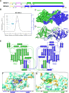
The structure of Tb RESC1–RESC2 heterodimer. (A) Linear representation of RESC1 (dark green) and RESC2 (dark blue). Regions with traced atomic models in solid colour. In yellow the mitochondrial targeting sequence, and in pink the flexibly attached N-terminal domains. Non-resolved linker regions as dashes lines. (B) SEC-MALS experiment with SDS-PAGE of the RESC1–RESC2 sample, indicating a molecular weight of 107 kDa, and confirming the heterodimeric oligomerisation state. (C) The final post-processed EM map with a 3.4 Å resolution, RESC1 is coloured in dark green and RESC2 in dark blue. (D) Cartoon representation of the atomic model built based on the EM map. Coloured as the map. (E) Structure topology diagram of RESC1–RESC2 with secondary structure elements labelled. (FG) Close-up of the dimerisation interfaces, with the β-harpin of RESC2 interacting with a patch in RESC1 in F, and the β-harpin of RESC1 interacting with a cleft in RESC2 in G. Sequence of both β-harpins below, illustrating the different nature of the interaction. The unbuilt region of the RESC1 loop underlined. C-terminal regions are labelled. Surface coloured by molecular lipophilicity, with blue as hydrophilic and yellow as hydrophobic. Residue side chains coloured by heteroatom.
Figure 2.
Figure 2.
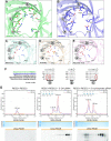
The triphosphate binding fold of Tb RESC1–RESC2. (A–E) Comparison of the chemical environment in the inner triphosphatase tunnels of (A) RESC1, (B) RESC2, (C) Mimivirus capping enzyme (PDB: 2QZE), (D) yeast Cet1p (PDB: 1D8I) and (E) Vaccinia virus capping enzyme (4CKC). Signature sequence motifs (called a, b and c) of the divalent cation-dependent family of RNA triphosphatases are indicated. The bound sulfate ion is shown as sticks in D. Relevant residue side chains are shown by sticks and coloured by heteroatom. (F) Structural sequence alignment of the three linear motifs, which are not conserved in RESC1 and RESC2. Glutamic acids were experimentally identified as fundamental for divalent cation coordination and activity in yeast Cet1p (45,47) annotated by red circles. (G) Analytical size exclusion chromatography of RESC1–RESC2 with gRNA, with or without 5’-triphosphate. Yellow bars indicate the 14 fractions (from 1.1 to 1.8 ml) selected for SDS-PAGE for protein detection and urea–PAGE for RNA detection. Peak shift, increase in 260:280 ratio and co-elution of the protein with the gRNA are only observed for 5’-triphosphate-containing gRNA.
Figure 3.
Figure 3.
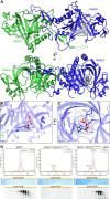
Structure of Tb RESC1–RESC2 bound to gRNA. (A) Two orthogonal views of the cartoon model of RESC1–RESC2 superimposed with the subtraction map between bound and unbound RESC1–RESC2 (in grey), with the GTP shown in sticks and coloured by heteroatom, indicating additional density exclusively inside the triphosphatase tunnel of RESC2. (BC) Two orthogonal views of the triphosphatase tunnel of RESC2, with a GTP fitted in the subtraction map (blue mesh). Lysine and arginine side chains, that coordinate the γ-phosphate of the GTP, are shown in sticks and coloured by heteroatom. (D) Analytical size exclusion chromatography of mutant RESC1–RESC2 and gRNA, together with the SDS-PAGE for protein detection and urea–PAGE for RNA detection. Peak shift and co-elution of the protein with the gRNA is disrupted when key RESC2 residues are mutated to the corresponding residue in RESC1 (K311S and R402D), indicating that only RESC2 is able to bind gRNA through their 5’-triphosphate.

References

    1. Maslov D.A., Opperdoes F.R., Kostygov A.Y., Hashimi H., Lukeš J., Yurchenko V.. Recent advances in trypanosomatid research: genome organization, expression, metabolism, taxonomy and evolution. Parasitology. 2019; 146:1–27. - PubMed
    1. Simpson A.M., Suyama Y., Dewes H., Campbell D.A., Simpson L.. Kinetoplastid mitochondria contain functional tRNAs which are encoded in nuclear DNA and also contain small minicircle and maxicircle transcripts of unknown function. Nucleic. Acids. Res. 1989; 17:5427–5445. - PMC - PubMed
    1. Pollard V.W., Rohrer S.P., Michelotti E.F., Hancock K., Hajduk S.L.. Organization of minicircle genes for guide RNAs in Trypanosoma brucei. Cell. 1990; 63:783–790. - PubMed
    1. Benne R., Van den Burg J., Brakenhoff J.P., Sloof P., Van Boom J.H., Tromp M.C.. Major transcript of the frameshifted coxII gene from trypanosome mitochondria contains four nucleotides that are not encoded in the DNA. Cell. 1986; 46:819–826. - PubMed
    1. Shaw J.M., Feagin J.E., Stuart K., Simpson L.. Editing of kinetoplastid mitochondrial mRNAs by uridine addition and deletion generates conserved amino acid sequences and AUG initiation codons. Cell. 1988; 53:401–411. - PubMed

Publication types

MeSH terms